1 PROTEIN-ENCODING RNA-to-RNA INFORMATION TRANSFER IN MAMMALIAN CELLS:
PRINCIPLES OF RNA-DEPENDENT mRNA AMPLIFICATION.
Vladimir Volloch
Department of Developmental Biology Harvard Dental School
Correspondence: [email protected] [email protected]
ABSTRACT. The transfer of protein-encoding genetic information from DNA to RNA to protein, a process formalized as the “Central Dogma of Molecular Biology”, has undergone a significant evolution since its inception. It was amended to account for the information flow from RNA to DNA, the reverse transcription, and for the information transfer from RNA to RNA, the RNA-dependent RNA synthesis. These processes, both potentially leading to protein production, were initially described only in viral systems, and although RNA-dependent RNA polymerase activity was shown to be present, and RNA-dependent RNA synthesis found to occur, in mammalian cells, its function was presumed to be restricted to regulatory. However, recent results, obtained with multiple mRNA species in several mammalian systems, strongly indicate the occurrence of protein-encoding RNA to RNA information transfer in mammalian cells. It can result in the rapid production of the extraordinary quantities of specific proteins as was seen in cases of terminal cellular differentiation and during cellular deposition of extracellular matrix molecules. A malfunction of this process may be involved in pathologies associated either with the deficiency of a protein normally produced by this mechanism or with the abnormal abundance of a protein or of its C-terminal fragment. It seems to be responsible for some types of familial thalassemia and may underlie the overproduction of beta amyloid in sporadic Alzheimer’s disease. The aim of the present article is to systematize the current knowledge and understanding of this pathway. The outlined framework introduces unexpected features of the mRNA amplification such as its ability to generate polypeptides non-contiguously encoded in the genome, its second Tier, a physiologically occurring intracellular polymerase chain reaction, iPCR, a “Two-Tier Paradox” and RNA “Dark Matter”. RNA-dependent mRNA amplification represents a new mode of genomic protein-encoding information transfer in mammalian cells. Its potential physiological impact is substantial, it appears relevant to multiple pathologies and its understanding opens new venues of therapeutic interference, it suggests powerful novel bioengineering approaches and its further rigorous investigations are highly warranted.
TABLE OF CONTENTS.
1. Introduction. p.3
2. mRNA amplification process may preserve the protein-encoding information
content of a conventional mRNA. p.p.3-7
3. iPCR: physiologically occurring intracellular polymerase chain reaction, the second Tier
of RNA-dependent mRNA amplification. p.p.7-10
4. mRNA amplification process may reduce or change the protein-encoding information content of a conventional mRNA.
Connection to sporadic Alzheimer’s disease. p.p.11-13
5. Two-Tier Paradox: Asymmetry of Outcomes. p.p.13-14
6. mRNA amplification process may enhance protein-encoding information content of a conventional mRNA and generate polypeptides
non-contiguously encoded in the genome. p.p.15-16
7. mRNA amplification may activate dormant protein-encoding information with resulting
polypeptides non-contiguously encoded in the genome. p.p.16-17
8. The RNA end product of mammalian
mRNA amplification is a “Dark Matter”. p.p. 17-19
9. Regulatory aspects of mammalian RNA-dependent
mRNA amplification process. p.p.19-23
10. A definitive proof of the occurrence of mammalian mRNA amplification:
Novel experimental designs. p.p.23-27
11. Significance and directions
of future investigations. p.p.27-28
1. INTRODUCTION
RNA-dependent RNA synthesis and the corresponding enzymatic activity, RNA-dependent RNA polymerase (RdRp), were discovered in studies of mengovirus and polio virus (1-6). Eventually, with the discovery of the RNA-dependent DNA synthesis and the enzyme associated with this process (7, 8), it became clear that among RNA viruses, retroviruses utilize reverse transcriptase for their genomic information transfer whereas almost all other RNA viruses encode and utilize RdRp for this purpose (9, 10). The exceptions are viruses that do not encode their own RNA-dependent RNA polymerase but rely on RdRp activity of either their partners (helper viruses within the same cells) or their hosts. The later category includes plant viroids and hepatitis delta virus, HDV (11). These viruses do not encode an RdRp yet they undergo a robust RNA replication once inside the host cells (9-11). These findings imply that both groups utilize cellular RNA-dependent RNA polymerase activities: viroids in plant cells and HDV in mammalian cells. The presence of RdRp in plant cells is well documented (12-14). The ability of RdRp-deficient HDV to vigorously replicate inside its mammalian hosts establishes the occurrence and functionality of RdRp activity in mammalian cells. Indeed, it became clear that RdRp activity, apparently in a non-conventional form, is constitutively present in most, if not in all, mammalian cells (15-21; see more in section 9 below). Because non-conventional mammalian RdRp activity was shown to produce short transcripts, because of its apparent involvement in RNA interference phenomena, and because double-stranded RNA is known to trigger cellular responses leading to its degradation, it was generally assumed that the function of RdRp activity in mammalian cells is restricted to regulatory. However, at the same time, an RdRp activity (18) capable of generating complete antisense RNA complements of mRNAs, as well as its products (22), were discovered in mammalian cells undergoing terminal differentiation. Moreover, observations of widespread synthesis of antisense RNA initiating at the 3’poly(A) of mRNAs in human cells (17) suggested an extensive cellular utilization of mammalian RdRp activity. These results led to the development of a model of RdRp-facilitated and antisense RNA-mediated amplification of mRNA in mammalian cells (22-25). A major prediction of this model is generation of a chimeric RNA composed of covalently joined antisense and sense strands of the same molecule and uniquely defined by self-priming of the antisense molecule and the extension of its 3’terminus into the sense, mRNA, strand. Recently, this prediction was borne out by the detection of such chimeric RNA molecules in terminally differentiating cells and in cells overproducing the components of the extracellular matrix (23, 24). Moreover, a putative end product of mRNA amplification, which translates into polypeptides indistinguishable from the translation product of genome-originated mRNA, has been identified at levels that are unprecedented for conventional mRNA transcripts (23). Below, we discuss in depth the current understanding of RNA-dependent mammalian mRNA amplification processes, their potentially multiple outcomes, possible regulation, and prospective significance. We also describe in detail novel experimental bioengineering designs suggested by the understanding of the amplification processes, and consider future directions of investigations into mechanisms underlying this powerful pathway of mammalian gene expression.
2. mRNA AMPLIFICATION PROCESS MAY PRESERVE THE PROTEIN-ENCODING INFORMATION CONTENT OF A CONVENTIONAL mRNA.
regular circumstances mammalian RdRp activity lacks a processivity component, and that in special circumstances requiring a substantial overproduction of specific proteins (i.e. hemoglobin in erythroid differentiation, extracellular matrix proteins during connective tissue deposition) or in some pathologies, what is induced is not RdRp activity, but rather its processivity conferring co-factor. A proof of conceptfor the notion of such RdRp co-factor that allows production of complete antisense transcripts is discussed in section 9 below.
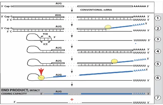
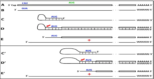
Figure 1. Projected steps in RdRp-facilitated and antisense RNA-mediated amplification of mammalian mRNA with chimeric RNA end product retaining the intact coding capacity of conventional mRNA. Top panel: conventional, genome-originated mRNA molecule. Bottom panel: projected stages of antisense RNA-mediated mRNA amplification. Boxed line – sense strand RNA. Single line – antisense strand RNA. “AUG” – functional translation initiation codon (could be other than “AUG”). “TCE”– 3’-terminal complementary element; “ICE”– internal complementary element, both on the antisense RNA strand. Yellow circle – helicase/modifying activity complex. Blue lines (both single and boxed) – RNA strand modified and separated from its complement by a helicase complex. Red arrowhead – position of cleavage of the chimeric intermediate. Step 1: synthesis of antisense strand; step 2: strand separation; step 3: folding of antisense strand into self-priming configuration; step 4: extension of self-primed antisense RNA; step 5: strand separation; step 6: cleavage of the chimeric intermediate; stage 7: end-products of amplification. Note that chimeric RNA end product retains the intact coding capacity of conventional mRNA.
As for the template eligibility, the only major prerequisite for a potential RNA template appears to be the presence of the poly(A) segment at its 3’ terminus (17, 22, 23). The vast majority of mammalian mRNA species contains 3’-terminal poly(A) segments. The notion that many, or possibly most, of them could be eligible templates for RdRp was suggested in earlier studies (22). Subsequent observations by Kapranov et al. showed a widespread synthesis of antisense RNA initiating, apparently indiscriminately, at the 3’ poly(A) of diverse mRNAs in human cells (17). This, apparently undiscerning, RdRp template eligibility of the bulk of mammalian mRNA species raises questions with regard to mechanisms underlying the manifestly stringent specificity of the mRNA amplification process as seen, for example, in erythropoietic differentiation (22, 23). The specificity of the amplification process appears to be determined at the 3’ terminus of an antisense transcript by its ability or inability to support production of a complementary sense strand molecule (22-25).
The generation of a sense strand on an antisense template occurs via extension of the 3’ terminus of a self-primed antisense template and requires the presence within the antisense transcript of two spatially independent complementary elements whose occurrence in antisense globin molecules was found to be evolutionary conserved across mammalian species (27). One of these is the strictly 3’-Terminal Complementary Element (TCE), the other is the Internal Complementary Element (ICE). These elements (Fig. 1, Step 3) must be complementary to an extent sufficient to form a priming structure but may contain both mismatches and unconventional G/U pairings. Generation of a sense strand also requires the thermodynamic feasibility, enhanced/enabled by the occurrence of these two complementary and topologically compatible elements, of the antisense strand folding into a self-priming configuration. The requirement for terminal localization of the TCE appears to be stringent; an overhang of even a single nucleotide diminishes self-priming (27).
Provided that a self-priming structure is formed, the 3' end of the folded antisense strand is extended by RdRp into a sense-orientation molecule terminating with the poly(A) at the 3'end (Fig. 1, Step 4), thus generating a hairpin-structured chimeric intermediate consisting of covalently joined sense and antisense strands. The double-stranded portion of the resulting structure is separated by a helicase activity invoked above, which mounts the 3'poly(A) of a newly synthesized sense strand component of the chimeric intermediate and proceeds along this strand in the 5' direction modifying the molecule as it advances (Fig. 1, Step 5). When the helicase activity reaches a single stranded portion of the hairpin structure, it, or an associated activity, cleaves the molecule either within the TCE, at a TCE/ICE mismatch, or immediately upstream of the TCE; a cleavage was shown to occur between the 5’ hydroxyl group and the 3’ phosphate (refs. 22, 23; red arrow, Fig. 1, Step 6).
Strand separation, in conjunction with the cleavage, produces two single-stranded molecules (Fig. 1, Step 7) one of which is a chimeric mRNA, the functional mRNA end product of the amplification and the basis for defining this pathway as the "chimeric". The chimeric nature of this end product is due to the presence at its 5’ end of a 3’-terminal segment of the antisense strand consisting, depending on the site of cleavage of the chimeric intermediate, of either the entire TCE or a portion thereof covalently attached, in a 5’ to 3’ orientation, to the 5’-truncated sense strand. This chimeric molecule is modified and 3’polyadenylated; it cannot be further amplified, because its antisense complement would be lacking the TCE, but can be translated into a conventional mRNA-encoded polypeptide (23). Such RNA end product of amplification was putatively identified for murine globin-encoding mRNA and is described in detail and discussed in section 8 below. Figure 1 illustrates the situation whereby the ICE of the antisense strand is located within its segment corresponding to the 5’-untranslated region (5’UTR) of a genome-encoded mRNA. Consequently, the chimeric end product contains the entire protein coding region of a conventional mRNA and can be translated into the original, conventional mRNA-encoded, polypeptide (23). Another single-stranded product of step 7, Figure 1 is the antisense RNA molecule. It is truncated at the 3’terminus and lacks either a portion of or the entire TCE, depending on the position of the cleavage site; its suitability for further amplification is discussed in sections 3 and 9 below. Such antisense beta- globin RNA, cleaved at the three-nucleotide TCE/ICE mismatch (Fig. 4, bottom panel, D), was detected, cloned and sequenced and found to be a predominant variant of globin antisense RNA in murine erythroid cells (22).
just a few days. For this reason, it could be argued that the amplification mechanism might be in restricted use and not relevant to more conventional tissue formation and functions. Recently, however, the evidence for RNA-dependent amplification was obtained for mRNAs encoding all three constituent chains of laminin 111 in a tissue producing extraordinarily large amounts of extracellular matrix proteins, indicating that such an amplification process may operate also during normal development when the production of large quantities of certain polypeptides is required (24).
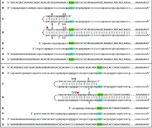
ribosomal RNA, used to generate sequencing libraries. Libraries were sequenced on an Illumina Next500 instrument. Sequencing data were converted into blast databases and analyzed by blasting with chimeric reference sequences. Sequences of interest were extracted from raw data and analyzed. Top panel: 1 laminin sequences; middle panel: 1 laminin sequences; bottom panel: 1 laminin sequences. Uppercase letters – nucleotide sequence of the sense strand; lowercase letters – nucleotide sequence of the antisense strand. Highlighted in green – “AUG” translation initiation codon on the sense strand; highlighted in blue – “uac” complement of translation initiation codon on the antisense strand. In italics and highlighted in grey – detected chimeric fragments. Blue arrows: position of antisense/sense junctions. A: 5’ terminus of conventional laminin mR. B: antisense complement of the 5’ terminus of conventional laminin mRNA. C: folding of the antisense strand into self-priming configuration. D: extension of self-primed antisense strand into sense-oriented sequence. E: projected chimeric junction sequence. F: detected chimeric junction sequence. Note that the priming occurs within the segment of antisense strand corresponding to the 5’UTR of mRNA, thus preserving the coding capacity of amplified mRNA.
Among RNA molecules described above, the best, if not the only, definitive identifier of the occurrence of mRNA amplification is a chimeric junction between sense and antisense components. Although the chimeric end product of amplification would be the most abundant source of the junction sequences, it may not provide the best, or possibly even any, evidence for the occurrence of the amplification. This is because the antisense component could be too short for a definitive identification or even indistinguishable from a sense-oriented sequence. Indeed, if there are no G/U pairings, if a strand-separating activity would cleave at the first mismatch within the TCE or if the TCE would be perfectly complementary to the ICE, the sequence of an antisense component of a chimeric RNA molecule would be identical to and indistinguishable from the sequence of the regular mRNA in the region of interest, and its only characteristic feature would be a 5’ truncation that is insufficient for a definitive identification. The most suitable identifying feature, therefore, appears to be a nucleotide sequence of a yet unmodified portion of non-cleaved chimeric intermediate, containing sufficiently long and unmistakably identifiable sense and antisense junction components. Such chimeric junctions, detected (24) for RNA encoding all three constituent chains of murine laminin 111, and the projected pathways of their generation are presented in Figure 2 above.
3. iPCR: PHYSIOLOGICALLY OCCURRING INTRACELLULAR POLYMERASE CHAIN REACTION, THE SECOND TIER OF RNA-DEPENDENT mRNA AMPLIFICATION.
In addition to chimeric sequences of antisense RNA extended into a sense RNA, described above, the analysis of globin RNA molecules in murine erythroid cells identified sense globin RNA sequences with non-conventionally templated 5’-terminal poly(U) added to the 5’UTR truncated at positions corresponding to potential cleavage sites within the antisense component of the chimeric intermediate (23). What could be the origin of such sequences?
Provided the presence of 3’poly(A) on an RNA molecule is necessary and sufficient for initiation of RNA-dependent RNA synthesis, another mRNA amplification paradigm, constituting the second Tier of RNA-RNA-dependent mRNA amplification, may be considered. If an antisense transcript is polyadenylated at the 3’ end by a known or
a novel cellular poly(A) polymerase, it would become a valid template for RdRp. Since the antisense strand has, by virtue of initiation within the poly(A) of conventional mRNA, a poly(U) stretch at the 5’ end, its transcription by RdRp would result in a sense strand with poly(U) at the 5’ end and poly(A) at the 3’ end, also a legitimate RdRp template. Since strand separation mechanisms are in place and the described sequence of events can occur repeatedly, the process will amount to an intracellular polymerase chain reaction, iPCR. The obvious question regarding such a process is its specificity. If 3’ polyadenylation of an antisense molecule was coupled with the cleavage of the chimeric intermediate, the specificity of iPCR would be equal to that of the initial chimeric amplification round.
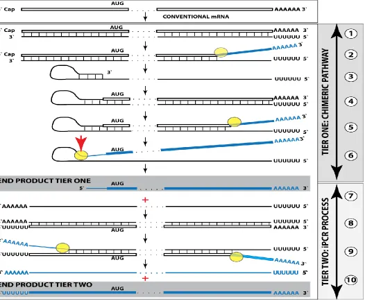
it also contains the 3’-terminal poly(A) (Figure 3, step 7). This molecule constitutes the initial template of a polymerase chain reaction. Indeed, just as in step 1, RdRp activity initiates transcription at the 3’poly(A) and generates the sense strand containing 5’poly(U) and 3’poly(A) (Figure 3, step 8). After strand separation (Figure 3, step 9) there are now two templates, each containing 3’poly(A) and 5’poly(U), and the iPCR is under way. Strand separation and associated nucleotide modification probably commences at 3’poly(A) as soon as it becomes double-stranded (see sections 8 and 9 for more discussion). Therefore, at steady state there would be many more non-modified poly(U)-containing than poly(A)-containing ends and because of problems with analyzing modified RNA (discussed in section 8), their detection would be much more likely.
Conceptually, amplification of a nucleic acid molecule by a polymerase chain reaction necessitates, beside the presence of the building blocks, the occurrence of a template, the priming arrangement for the initial nucleic acid strand and for its complement, a polymerase, and the arrangement for strand separation that doubles the number of template molecules in each cycle. In a conventional PCR reaction, a single-stranded DNA molecule serves as a template, oligonucleotides complementary to the initial DNA strand and to its complement in desired and appropriate positions function as primers (the “forward” and the “return” primers), DNA polymerase of choice extends the 3’ end of a primer, generating a double-stranded molecule, and a thermal treatment separates the strands to enable the next cycle of the chain reaction.
In the cellular iPCR process, an eligible single-stranded RNA molecule acts as a template, the process is driven by an RdRp that generates a complementary strand, the priming arrangements are reflected in the template eligibility requirements that are satisfied by the occurrence of poly (A) segment at the 3’ termini of an RNA template as well as of its transcript/complement, and strand separation is carried out by a helicase activity. When a full-length 5’ poly(U)-containing antisense strand is generated (transcribed by RdRp from a conventional mRNA molecule) and separated from its template (steps 1 and 2, Tier One, Figure 1), it is not an iPCR-eligible template because it lacks the 3’-terminal poly(A) segment. Instead, provided that it contains the 3’-terminal and internal complementary elements, it self-primes its extension into a sense strand molecule; thus generating an intermediate in the chimeric pathway of mRNA amplification. It is the processing of this intermediate that has the potential to produce, in addition to chimeric RNA end product, an iPCR-eligible template. This requires cleavage-coupled 3’ polyadenylation of the antisense strand, which already contains 5’-terminal poly(U) transcribed from the poly(A) of a conventional mRNA progenitor molecule. The presence of the 3’-terminal poly (A) segment would allow the RdRp to initiate and to proceed with the synthesis of a sense strand complement that commences with the 5’-terminal poly(U) and concludes with the 3’-terminal poly(A) segments, also an iPCR-eligible template. The following separation of strands by a helicase activity would enable the next cycle of a polymerase chain reaction. Thus, the key feature underlying the feasibility of iPCR is that both the initial cleavage/polyadenylation-released antisense molecule and its transcript are eligible RdRp templates. Potentially and purely hypothetically, if an antisense RNA transcribed from a conventional mRNA were polyadenylated at the 3’end, it would become an eligible iPCR template. This, however, is highly unlikely because such a process would completely lack specificity. In the Two-Tier amplification process, because the generation of the initial iPCR template is coupled with and enabled by the concluding step of the previous Tier, the specificity of the iPCR process is as stringent as that of the preceding chimeric pathway, which, in turn, is defined by the occurrence within an RdRp transcript of the TCE and the ICE features and by the ability of the initial antisense transcript to self-prime its extension. On the other hand, whether or not the second Tier of amplification, iPCR, occurs does not affect in any way the first Tier, a chimeric pathway. The regulatory aspects of the iPCR amplification pathway are further discussed in section 9 below.
Figure 4 shows detected chimeric junction sequences and 5’-truncated polyurydilated mRNA sequences encoding and globin chains and the projected Two-Tier pathways of their generation. It should be noted that 5’-truncated and polyurydilated sense RNA, the product of Tier Two of amplification process, retains the entire coding information content of conventional mRNA. Such RNA can either serve as a template for further iPCR amplification or be utilized for translation of conventional polypeptide.
be the products of the second stage of the mRNA amplification process, and the extent of 5’ truncations in their genome-encoded portions could reflect the average size of an antisense 3’ truncation at the conclusion of the chimeric pathway, i.e. the distance between the cleavage site within a chimeric intermediate and the 3’ end of an antisense strand.
4. mRNA AMPLIFICATION PROCESS MAY REDUCE OR CHANGE THE PROTEIN-ENCODING INFORMATION CONTENT OF A CONVENTIONAL mRNA. CONNECTION TO SPORADIC ALZHEIMER’S DISEASE.
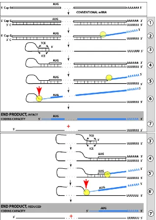
Previous sections discussed a situation where both complementary elements required for an appropriate folding and self-priming of the antisense strand, TCE and ICE, are located within its segment corresponding to the 5’UTR of a conventional genome-encoded mRNA. In such a situation, depicted in steps 3 trough 7 of Figure 5, the
chimeric RNA end product contains the entire protein coding region of a conventional mRNA and can be translated into the original, conventional mRNA-encoded, polypeptide. In the chimeric mRNA amplification pathway, the position of the TCE within the antisense molecule is fixed; it is strictly 3’-terminal. In contrast, the intramolecular location of the ICE is variable, and potentially it can be positioned within a segment of the antisense strand corresponding to the coding portion of an mRNA, a scenario diagrammed in steps 3’ trough 7’ of Figure 5. In this scenario, the chimeric RNA end product would consist of a 3’-terminal segment of the antisense strand (the TCE or its fraction) and a 3’ portion of a conventional mRNA progenitor with a 5’-truncated coding region. In such a case, the translational outcome would be decided by the position of the first functional
(capable of initiation of translation) AUG or other initiation-competent codon. If it were in-frame with the protein-encoding information content of conventional mRNA, translation would result in the C-terminal fragment, CTF, of a conventionally encoded polypeptide.
Fig. 5. RNA-dependent mRNA
amplifica-tion can result in a 5’-truncated molecule encoding C-terminal fragment of a conventionally encoded polypeptide. Boxed line-sense strand RNA. Single line-antisense strand RNA. “AUG”-functional translation initiation codon (could be other than AUG). “TCE”– 3’-terminal complementary element; “ICE”– internal complementary element, both on the antisense RNA strand. Yellow circle – helicase/ modifying activity complex. Blue lines (both single and boxed) – RNA strand modified and separated from its complement by a helicase complex. Red arrow – position of cleavage of the chimeric intermediate. Step
1: synthesis of antisense strand; step 2: strand
separation; step 3: folding of antisense strand
into self-priming configuration; step 4:
extension of self-primed antisense RNA; step
5: strand separation; step 6: cleavage of the
chimeric intermediate; stage 7: end-products
of RNA amplification. Steps 3’-7’ correspond
to steps 3-7. Top panel: Conventional,
genome-transcribed mRNA molecule. Middle
panel: projected stages of RNA-dependent mRNA amplification. “ICE” is located within a segment of antisense RNA corresponding to the 5’UTR of conventional mRNA; the chimeric RNA end product contains the entire coding content of conventional mRNA.
The scenario resulting in a CTF of conventionally encoded polypeptide as the translational outcome of mRNA amplification can be illustrated with the case of beta-amyloid overproduction in sporadic Alzheimer’s disease.
Beta amyloid, Aβ, the peptide associated with and, when overproduced, widely believed to have a pivotal early role in the etiology of Alzheimer’s disease (AD), is normally generated by proteolytic cleavages of a much larger
molecule, β amyloid precursor protein, βAPP (28-36). Two sequential cleavages of βAPP are involved in the
production of Aβ. The first is a cleavage of βAPP by the β secretase enzyme. It occurs between residues 671 and
672 of the βAPP molecule, generating the N-terminus of Aβ. The second cleavage, by γ secretase activity,
generates the C-terminus of Aβ (29-31). In inherited familial cases, constituting about 1% of the AD burden (the rest are sporadic cases) it was shown that the overproduction of Aβ is due to mutations around cleavage sites resulting in an abnormal proteolysis (37, 38). Since the pathological lesions and symptoms in the non-hereditary form of the disease are analogous to those seen in the familial forms, it has been assumed that abnormal proteolytic
processing of βAPP also underlies the pathogenesis of sporadic AD (33-35). It was further assumed, therefore,
that the suppression of APP proteolysis might alleviate the disease (39-41). And, indeed, inhibition of beta secretase rescued functional impairments and actually reversed the disease in animal models bioengineered to imitate familial AD (42-45). However, in several large human clinical trials such inhibition was completely inefficient in sporadic AD (25). This suggested that, in sporadic AD, beta-amyloid could be produced in the amyloid precursor protein-independent manner. But how?
In βAPP mRNA, the Aβ-coding segment is preceded immediately, contiguously and in-frame by the AUG codon normally encoding methionine in position 671 of the APP. If translation were initiated at this position, it would
produce Aβ-containing CTF independently of βAPP. Interestingly, the AUG in question is situated within a
nucleotide context optimal for the initiation of translation (an “A” in position −3 and a “G” in position +4 relative to the “A” of the AUG codon). In fact, of the twenty AUG codons encoding methionine residues in the βAPP mRNA, only the AUG encoding Met671, not even Met1, is located within an optimal translation initiation context. Such favorable positioning of the AUG encoding Met671 was the basis for an early proposal that in AD, Aβ may
be generated independently from βAPP by the internal initiation of translation of the intact βAPP mRNA (46).
The possibility of the internal initiation of translation has been subsequently ruled out by the elegant experiments of Citron and co-investigators (47). There is, however, another possibility of utilization of the AUG in question as a translation initiation codon, namely the generation of a severely 5’-truncated βAPP mRNA in which the AUG
encoding Met671 in the intact mRNA becomes the first AUG codon.
To determine if an mRNA species of interest can potentially be a subject of RNA-dependent mRNA amplification, one needs to assess whether its antisense complement contains both TCE and ICE and is capable of folding into a self-priming configuration. If it were, the position of the ICE would indicate the possible translational outcome. Such an assessment can be conducted in a model experiment where an mRNA of interest serves as a template for synthesis of cDNA and is subsequently removed by RNAse H activity present in a preparation of reverse transcriptase used. If an mRNA were fully transcribed, if complementary elements were present within the antisense strand (cDNA), if one of them were 3’-terminal, and if they were topologically compatible, self-priming and the extension synthesis of a segment of the sense strand would occur. The junction between the antisense and sense components would define the site of self-priming and facilitate identification of the TCE and ICE. Just such an experiment was inadvertently carried out with human βAPP mRNA (48). The results of this experiment, misinterpreted and eventually dismissed by the authors as an artifact (49), indicated the occurrence of topologically compatible TCE and ICE within the antisense strand of βAPP mRNA and defined their sequence as well as the position of self-priming. Based on these results, the TCE/ICE-guided folding of the antisense strand of βAPP mRNA (25; 50-52) can be depicted as shown in Figure 6.
TCE or immediately upstream from it as indicated by the arrow in Fig. 6b. The resulting chimeric RNA end product, shown in Fig. 6c, consists of an antisense segment (TCE or its portion) continued into a sense-orientation molecule. The translational outcome is decided by the first, 5’-most, initiation-competent AUG codon. As shown in Fig. 6b-c, the first AUG codon is located 58 nucleotides downstream from the TCE portion of the chimeric RNA end-product and it is, in fact, the AUG encoding Met671 in the intact APP mRNA! Translation from this position would produce beta amyloid-containing CTF in the APP-independent manner. The major prediction of such a mechanism is a complete inefficiency of beta secretase inhibition in sporadic Alzheimer’s disease. This prediction was, in fact, borne out in several massive stage III clinical trials (25).
Figure 6. Projected topology of RNA-dependent generation of 5’-truncated mRNA encoding beta amyloid-containing C-terminal fragment of human amyloid precursor protein. Lowercase letters- nucleotide sequence of the antisense RNA; uppercase letters- nucleotide sequence of the sense RNA. Double-stranded portions highlighted in yellow: TCE (top) and ICE (bottom) elements of the antisense RNA. Note that the TCE and the ICE are separated by about 2000 nucleotides. “2011-2013”: nucleotide positions on the antisense RNA (starting from the complement of the AUG encoding Met1 of the APP) of the “uac” (highlighted in blue) corresponding to the “AUG” (highlighted in green) encoding Met671 in the APP mRNA. a: TCE/ICE-guided folding of the antisense APP RNA; 3’-terminal “c” corresponds to one of multiple transcription start sites of APP mRNA 149 nucleotides upstream from its AUG initiation codon. (25). b: extension of self-primed antisense RNA into sense RNA and cleavage (red arrow; may also occur at one of the TCE/ICE mismatches), after strand separation, of the chimeric intermediate. c: chimeric RNA end product contains 5’terminal antisense segment extending into severely 5’-truncated APP mRNA. Its translation initiates from the “AUG” (highlighted in green and encoding Met671 in conventional APP mRNA) immediately preceding the beta amyloid-encoding region.
The generation of a CTF of a conventionally encoded polypeptide, described above, requires that the first functional translation initiation codon in the chimeric RNA end product were in-frame with the conventional coding sequence. If it is out-of-frame, but is followed by an open reading frame, ORF, a polypeptide unrelated to a conventionally encoded protein can be produced. If it is out-of-frame and is not followed by an ORF, an abortive translation would result. If there were no functional initiation codon, no translation would occur. An abortive translation or a translational incompetence may appear to be a functional failure of the amplification process, but, as discussed in the next section, even such an apparent malfunction, in terms of the mRNA amplification translational outcome, can potentially be rescued.
5. TWO-TIER PARADOX: ASYMMETRY OF OUTCOMES.
content identical to that of conventional mRNA. In a section 4 scenario above, the ICE is located within a segment of the antisense strand corresponding to the coding region of mRNA and the chimeric amplification pathway Tier results in an RNA molecule encoding a CTF of a conventional polypeptide. In this scenario, as in the previous one (section 3), the chimeric pathway may merge into the second, iPCR Tier of amplification, and one may intuitively think that the iPCR-amplified sense strand would also encode the same CTF as the chimeric RNA end product. This, however, is not the case because of the phenomenon of “Two-Tier Paradox”.
Two Tier Paradox is defined as follows: Regardless of the translational outcome of the chimeric pathway Tier of mRNA amplification – a conventional polypeptide, a CTF, an unrelated polypeptide, and even an abortive translation or translational incompetence – the sense RNA strand produced in the iPCR pathway Tier would always retain the entire coding information of a genome-encoded mRNA and would direct the translation of a polypeptide identical to its conventionally encoded counterpart.
As was mentioned in section 3 above, whether or not the second Tier of amplification, iPCR, occurs, does not affect in any way the first Tier, a chimeric pathway. In turn, the translational outcome of the first, chimeric, Tier does not affect in any way the translational outcome of the second, iPCR, Tier. This is because, in sharp contrast to and irrespective of any of the potentially multiple translational outcomes of the chimeric pathway, there is only one possible translational outcome of the iPCR stage of mRNA amplification - an intact conventional polypeptide. This disparity reflects the asymmetry in the truncation of sense and antisense components of the chimeric intermediate during its cleavage at the conclusion of the Tier One. The source of the asymmetry is that the truncation of molecules in question is determined by two different, spatially independent, elements. The truncation of the sense strand component of the chimeric RNA end product is defined by the position of the ICE whereas the TCE position determines the truncation of the antisense strand. Because the ICE can be anywhere in the antisense RNA molecule, so can the sense strand’s 5’ truncation be, and because the TCE, by definition, can be only 3’-terminal, the antisense always loses only a short segment at its 3’end. This phenomenon is demonstrated in Figure 5. Whereas the sizes of chimeric RNA end products are drastically different in steps 7 and
7’, the sizes of antisense RNA end products in the same steps are identical. The 3’-truncated antisense RNA, if polyadenylated at the conclusion of Tier One, gives rise in Tier Two to poly(U)-containing sense strand truncated at the 5’end by exactly the same extent as the 3’ antisense truncation, which in most, if not all, instances is substantially shorter than a typical 5’UTR of a conventional mammalian mRNA (53). Consequently, because the 5’ poly(U)-containing sense RNA molecule, derived from the 3’ poly(A)-containing antisense protagonist by the iPCR process, retains the bulk of its 5’UTR and the intact protein-encoding portion of a conventional mRNA, regardless of the translational outcome of the Tier One, even if it is an abortive translation, in the Tier Two the translational outcome would always be the intact polypeptide, identical to that attained with a conventional progenitor mRNA. The translational outcome of the Tier Two would match that of the Tier One only when the ICE is positioned within the antisense RNA segment corresponding to the 5’UTR of conventional mRNA (Figures 1 and 3), as seen in the cases of RNA encoding both globin chains (Figure 4) and all three chains of laminin (Figure 2). In other cases, the situation can be quite remarkable. It is conceivable, for example, that the outcome of Tier One is an abortive translation or translational incompetence, yet Tier Two would produce an intact, conventional polypeptide. The same can be said for cases where Tier One results in a CTF of a conventional protein whereas the Tier Two would produce, in parallel, the intact conventionally encoded polypeptide.
6. mRNA AMPLIFICATION PROCESS MAY ENHANCE PROTEIN-CODING INFORMATION CONTENT OF CONVENTIONAL mRNA AND GENERATE POLYPEPTIDES NON-CONTIGUOUSLY ENCODED IN THE GENOME.
The previous sections considered RNA-dependent mRNA amplification scenarios where protein coding information is contained only in the sense RNA strand. In these scenarios, the chimeric RNA end product either retains the entire coding information of a conventional mRNA and is translated from the original translation initiation codon (sections 2 and 3) or looses the 5’-portion of the coding region, and its translation depends on the availability of a functional initiation codon within the remaining portion of the coding region (section 4). There is, however, another possibility. If the TCE of an antisense RNA contains a functional translation initiation codon and if, after the cleavage of the chimeric intermediate, it was retained in the chimeric RNA end product, translation would be initiated from the initiation codon within the antisense portion of the chimeric RNA end product. In such a case, if the folding/self-priming of the antisense RNA occurs within its segment corresponding to the 5’UTR of conventional mRNA and if the initiation codon within the antisense portion of the chimeric RNA end product is in-frame with the mRNA coding sequence, translation will result in a chimeric protein. It will contain, in its entirety, the polypeptide encoded by a conventional mRNA, enhanced at its N-end by additional amino acids encoded by the antisense portion of the chimeric RNAend product and by a segment of the 5’UTR of conventional mRNA. Thus, interestingly, the protein-encoding information content of the chimeric RNA end product of amplification would be non-contiguously encoded in the genome.
If, however, the folding/self priming of the antisense strand occurs within the segment corresponding to the coding region of conventional mRNA, and if the initiation codon within the antisense portion of the chimeric RNA end product is in-frame with the mRNA coding sequence, the translational outcome would be a CTF of the conventional polypeptide, enhanced at its N-end by additional amino acids encoded by the antisense portion of the chimeric RNA end product, also a chimeric protein non-contiguously encoded in the genome. Both variants described above are diagrammatically illustrated in Figure 7.
In this scenario of antisense RNA contributing a functional translation initiation codon to a chimeric RNA end product, if the chimeric pathway was to merge into the iPCR process, the rules of the Two-Tier Paradox described above would fully apply. Regardless of translational outcome of the chimeric Tier of amplification – enhanced conventional polypeptide, enhanced CTF of a conventionally encoded protein, or any other outcome, translational outcome of the second, iPCR Tier of amplification would always be a conventional genome-encoded polypeptide.
7. mRNA AMPLIFICATION MAY ACTIVATE DORMANT PROTEIN-ENCODING INFORMATION WITH RESULTING POLYPEPTIDES NON-CONTIGUOUSLY ENCODED IN THE GENOME.
This section considers a hypothetical yet plausible scenario where an mRNA is translated only under conditions, for example a particular stress, that activate the RNA-dependent mRNA amplification process. It should be emphasized that regardless of the extent of its physiological occurrence, the understanding of such a scenario is important because it could, and actually does as described in section 10 below, suggest very useful experimental and bioengineering designs. In this scenario, RNA encodes a polypeptide but lacks a functional translation initiation codon in-frame with the encoded information. Since its potential to produce a protein is present but unrealized under regular circumstances, it can be considered a silent, or “dormant” mRNA. The potential to direct synthesis of a polypeptide can be realized, and a dormant mRNA activated, by the mRNA amplification process. In the chimeric pathway of mRNA amplification, the chimeric RNA end product contains a 5’-terminal segment contributed by the antisense RNA and not present in the conventional genome encoded RNA molecule. If this additional segment contains a functional translation initiation codon and if this codon were in-frame with the encoded information, the protein-encoding content of a conventional “dormant” mRNA would be activated. The resulting translational outcome would be a chimeric protein consisting of a conventionally encoded polypeptide with the N-end encoded by the antisense RNA. Such a chimeric protein would be, therefore, non-contiguously encoded in the genome. This scenario is diagrammed in Figure 8.
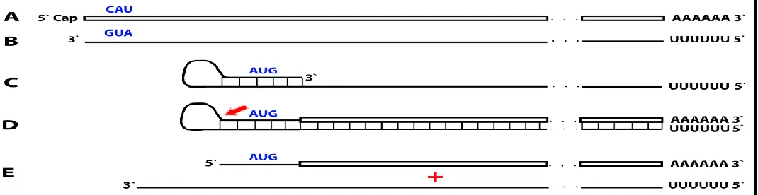
Figure 8. RNA-dependent mRNA amplification process may activate a dormant protein-encoding information and generate polypeptides non-contiguously encoded in the genome. Boxed line: sense strand RNA. Single line: antisense strand RNA. “AUG” (in blue): functional translation initiation codon (could be other than “AUG”). 5’-CAU-3’ (in blue) on the sense RNA: complement of 5’-AUG-3’ on the antisense RNA. Red arrow: position of cleavage of the chimeric intermediate. A: genome-originated “dormant” mRNA containing protein-encoding information but lacking in-frame translation initiation codon. B: antisense complement of conventional mRNA. Note that 3’-GUA-5’(in blue) is the 5’-AUG-3’. C: folding of the antisense strand into self-priming configuration. D: extension of self-primed antisense strand into sense-oriented sequence followed by strand separation and cleavage of the chimeric intermediate. E: end products of RNA-dependent mRNA amplification. Note that translation of the chimeric RNA end product starts from the antisense RNA-originated initiation codon (“AUG”, in blue) and produces a chimeric polypeptide non-contiguously encoded in the genome.
namely that the translational outcome of the iPCR Tier of mRNA amplification is always identical to the outcome of the conventional gene expression, would still remain in force – the resulting sense strand RNA would be translationally dormant, just as the conventional genome-encoded RNA.
8. THE RNA END PRODUCT OF MAMMALIAN mRNA AMPLIFICATION IS A “DARK MATTER”.
The understanding of the RNA end product of the RNA-dependent mRNA amplification process is derived largely from a study of globin mRNA amplification during erythroid differentiation (23). Deficiency of red blood cells in mammals, due to blood loss or induced chemically, by selective degradation of circulating red blood cells with phenylhydrazine injections, stimulates conversion of the spleen into an erythropoietic organ (23; 54-56). This splenic reaction is particularly dramatic in rodents; as shown in Figure 9 (top panel), at seven days post-induction of hemolytic anemia in mice the spleen mass increases nearly 20 fold, and it consists almost entirely of basophilic erythroblasts (23, 56). Electrophoresis of total cytoplasmic RNA from such spleens on denaturing methylmercury hydroxide/agarose gels shows the presence of an increasingly pronounced band that is slightly smaller than globin mRNA (Fig. 9, bottom panel) and whose emergence is associated with the duration of hemolytic treatment and with hemoglobin accumulation (23). Based on the size of this RNA, its kinetics and abundance, and in light of results described below, it was designated pepRNA (putative end product of globin mRNA amplification). Eventually, erythroid cells mature into reticulocytes and are released into the bloodstream; during this erythroblast/reticulocyte transition the cellular pepRNA levels drop sharply (23). The pepRNA band produced no signal upon stringent hybridization of Northern blots with globin-specific probes (22, 23). Since nucleotide modifications can interfere with hybridization properties (57), the nucleoside composition of pepRNA was analyzed (23). In addition to the nucleoside peaks seen in digests of control RNA, two new peaks were evident in the profile of pepRNA (23). Mass-spectrometry produced mass numbers of 304 and 320 for the new peaks and standard numbers for the four conventional peaks (267, 283, 244 and 243 for A, G, U and C, respectively). Together, the new peaks comprised 18%, nearly one fifth, of all nucleoside residues (23). Multiple considerations (23) indicated that the modified nucleotides are adenosine and guanosine with the same size modifying group with a mass of 37 appended to both. Considering that methylmercury interacts with the NH and allowing for the mass requirement, one possible candidate for the modifying group is LiCH2NH2.
produced (23). It was concluded
Figure 9. During induced anemia mouse spleen converts to an erythropoietic organ
and produces new type of RNA. Hemolytic anemia was induced by daily intra-peritoneal injections of 0.1ml of 0.8% neutralized phenylhydrazine hydrochloride. Spleen cells were lysed with non-ionic detergent, nuclei were removed, cytoplasmic fraction was treated with proteinase K in the presence of SDS, and RNA was extracted with phenol/chloroform and treated with DNase. Note that Trizol extraction of whole cell cannot be used because pepRNA separates with the DNA/protein fraction during such extraction (23). Cytoplasmic RNA samples from spleen cells collected at different stages of hemolytic treatment were resolved on denaturing agarose/ methylmercury hydroxide
gels and visualized by ethidium bromide staining. Top panel: Conversion of spleen into
an erythropoietic organ. At seven days post-induction of hemolytic anemia the spleen mass increases almost 20 fold and nearly 90% of spleen cells are erythroid with characteristics of basophilic erythroblasts (22). Upper image: spleen at seven days
post-induction. Lower image: spleen from untreated animal. Bottom panel: Proportion of
pepRNA (putative end product of mRNA amplification) increases with the duration of
hemolytic treatment. Lanes 1-6: number of daily phenylhydrazine injections. Spleens
were collected and processed 24 hours after final injection. Two prominent upper bands
in each lane: 28S and 18S ribosomal RNA. Horizontal arrow: pepRNA. Qualitatively
similar results were obtained in mice with induced hemorrhagic anemia (22).
that modifications interfere both with hybridization and with the primer extension. When oligonucleotide probes were used in RNase H digest mapping, nearly identical maps were obtained for globin mRNA and pepRNA (23). The main differences were that instead of a diffuse poly(A) band with conventional globin RNA, a uniformly short poly(A) was seen at the 3’end with pepRNA whereas at the 5’end fragments of pepRNA were uniformly shorter than corresponding 5’UTR fragments of conventional globin RNA; the truncations did not affect the coding regions. When two, one alpha- and another beta-globin specific probes were used together in digests, a complete cleavage of pepRNA by RNase H was observed, attesting to its homogeneity (23). The end-labeling experiments indicated that at both 5’ and 3’ ends pepRNA terminates with the (OH) group (23). Finally, the notion that pepRNA is a variant of globin mRNA was confirmed by cell-free translation of pepRNA. The resulting polypeptides were indistinguishable from the translation product of conventional globin mRNA (23).
There is very little doubt that pepRNA is a variant of globin mRNA. But is it the projected chimeric RNA end product of the amplification process? Multiple considerations strongly support an affirmative answer. First, the 5’(OH) group of pepRNA indicates that its 5’ terminus is generated by cleavage, just as projected for the chimeric RNA end product. Second, 5’ truncations of pepRNA seen with both alpha-globin probe (about 20-25 nucleotides) and beta-globin probe (about 30-35 nucleotides) match very closely with the sizes projected for 5’ truncations of the chimeric RNA end product (20 nucleotides for chimeric-alpha globin RNA and 36 nucleotides for chimeric beta-globin RNA). Third, the massive cellular levels of pepRNA are unprecedented for and vastly exceed the observed levels of even most abundant conventional mRNA transcripts. In differentiating mammalian erythroid cells, the mass of conventional globin mRNA is about 0.01% of that of ribosomal RNA (58, 59). At its peak, pepRNA constitutes about 15% of ribosomal RNA (ref. 23; Figure 9), a 1500-fold increase and a clear indicator that an amplification process is at work.
If the "visualization" of a nucleic acid species is defined as the determination of its nucleotide sequence, the modified RNA end products of both Tiers of mRNA amplification can be categorized as a "Dark Matter" because they are invisible to the methods of detection, i.e. nucleotide sequencing, based on the reverse transcription of RNA of interest. As was mentioned above, the RNA end product of amplification contains a large proportion of modified nucleotides, presumably "A"s and "G"s, both appended apparently by the same group with a mass of 37 (23). The modifications are apparently introduced on the 3’poly(A)-containing strand during strand-separation process (23); in the case of globin mRNA, on average, about every fifth nucleotide of the amplified end product is modified (23). Apparently, the modified nucleotides are either impassable or create a structure that is impassable for a reverse transcriptase; in related studies, both sequence-specific and random primers were ineffective or extended only by few nucleotides with the modified end product of mRNA amplification as a template, in sharp contrast to their considerable extension with the conventional mRNA template (22, 23). Modified pepRNA showed strong affinity to ribosomal RNA (23). It appears that pepRNA forms complexes with ribosomal RNA where modifying groups are sequestered enabling a codon/anticodon interaction (23). Modified pepRNA appears to be “sticky” and can be lost in storage. The possible regulatory functions of nucleotide modifications in pepRNA are discussed in section 9 below.
In the iPCR stage of mRNA amplification, because the helicase/modifying activity requires the presence of the 3’-terminal poly(A) in a double-stranded configuration to commence strand separation, the 5’ poly(U)-containing mRNA end product remains unmodified until its synthesis is completed. Even after the sense strand RNA product of the iPCR acquires its 3’ poly(A) segment, transcribed from the 5’poly(U) of the antisense strand, its 5’ portion remains unmodified for the limited duration of the strand-separating/modifying activity traversing the length of the molecule. These two processes, the completion of the synthesis and the strand-separation/modification downstream from the region of interest, provide a temporal “window of opportunity” that, apparently, enabled the detection of the yet unmodified 5’-terminal poly(U)-containing segment of the mRNA end product of the iPCR Tier of the mRNA amplification process (Figure 4).
A temporal “window of opportunity” is much smaller with respect to the detection of yet unmodified 3’-terminal poly(A)-containing segment of the antisense strand. At the conclusion of the chimeric pathway, it remains unmodified for probably only a short duration between the cleavage/3’ polyadenylation of the chimeric intermediate and its separation from a probably still nascent 5’poly(U)-containing sense strand being transcribed from it. During the iPCR process this duration could be even shorter, comprised only of the period between completion of the 3’ polyadenylated antisense synthesis and the commencement of the modification starting at its newly added poly (A) component as soon as poly(U) is transcribed. In such a case the modifying activity could be lagging closely behind the polymerase complex. It should be emphasized that the detected, not yet modified, 5’-terminal poly(U)-containing segment of the sense strand product of Tier Two represents probably a very minor subpopulation, with a substantial populations of modified molecules remaining for now, alongside the probably highly abundant end product of the chimeric pathway, a “Dark Matter”.
9. REGULATORY ASPECTS OF MAMMALIAN RNA-dependent mRNA AMPLIFICATION PROCESS. The ubiquitous presence of conventional RdRp was shown in numerous eukaryotic organisms but not in mammals (9). However, the ability of viruses not encoding the RdRp, such as HDV, to robustly replicate in mammalian cells attested to the cellular presence and functionality of this activity (9, 10). The RdRp activity in mammalian cells appears to be non-conventional; two possible candidates for this role are the RNA polymerase II complex or its components (19, 20) and RdRp activity of the TERT complex (21), both ubiquitously present in all cells. Under regular circumstances the RdRp activity in mammalian cells produces only short antisense RNA transcripts. For example, a widespread synthesis of diverse short antisense RNA transcripts initiating at the 3’poly(A) of mRNA was observed in human cells (17). On the other hand, RdRp activity isolated from rabbit reticulocytes (18) was able to produce, in assays, long antisense RNA transcripts. Subsequent studies identified full-length antisense transcripts of globin mRNA in erythroid cells (22). It could be argued that the component responsible for the production of long antisense transcripts in mammalian cells is a processivity conferring co-factor of RdRp activity that is induced under special circumstances when over-production of specific proteins is required (23). The notion of a processivity co-factor is strongly supported by studies of HDV replication in “normal” (i.e. apparently lacking processivity co-factor) mammalian cells. Within the framework of the above considerations, the ability of RdRp-deficient viruses to use RdRp activity of mammalian cell for their replication implies that they should encode a processivity co-factor of cellular RdRp. In case of HDV, it appears to be hepatitis delta antigen HDAg, the only protein encoded by HDV. HDAg is essential (71) both for production of long transcripts by cellular RdRp, and for viral replication. In its absence only short transcripts are generated (71). These observations provide a proof of concept for the notion of RdRp processivity co-factor, central for our understanding of mammalian mRNA amplification. Identification of a cellular homolog of HDAg, DIPA (72,73) suggests directions for a search for the cellular RdRp processivity co-factor.
effected by ribozyme activities present in HDV RNA (71). On the other hand, whereas the nature of initiation of antisense RNA synthesis within 3’poly(A) of conventional mRNA remains to be elucidated, mammalian mRNA amplification involves strand separating/nucleotide modifying activity as well as a chimeric RNA cleaving activity. To understand what is required for the activation of mRNA amplification in mammalian cells, we have to know which of the relevant activities are constitutive and which are inducible. Our current understanding indicates that mammalian RdRp activity is constitutive and its processivity co-factor is inducible; it doesn’t inform us on the status of other activities involved. Elucidation of the nature of all activities involved in RNA-dependent mRNA amplification would, when attained, enable complex and powerful bioengineering designs.
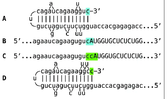
The observed uniformly short 3’poly(A) region of the pepRNA, transcribed from 5’poly(U) of the antisense strand (23) indicates that synthesis of the antisense strand starts within the poly (A) region of the sense strand, in a relatively narrow distance range from the 3’UTR of a conventional mRNA. The manner of antisense RNA initiation remains to be determined. Interestingly, at the 3’terminus of the antisense strand, RdRp appears to be capable of copying the cap”G” of conventional mRNA despite its inverted orientation. The rational for this conclusion is based on the detected antisense/sense junction sequences of chimeric alpha-globin RNA amplification intermediate shown in Figure 4; it is summarized in Figure 10. If the cap “G” is not transcribed, the antisense strand would terminate with the 3’ “c” corresponding to the transcription start site of mRNA (panel A; highlighted in blue). In such a case, antisense RNA folding/self-priming configuration would be as shown in panel
A, and following the extension of self-primed antisense RNA, the antisense/sense junction would consist of the “c/A” (highlighted in blue) as depicted in panel B. The experimental results presented in Fig. 4 are different. They show that the sequence of the antisense/sense junction is, in fact, the “cc/A” (panel C; highlighted in green). Since the genomic sequence upstream of the transcription start site cannot account for the additional 3’-terminal “c” (panel D; highlighted in green) in the antisense strand (23), the remaining possibility is that the “c” in question is a transcript of the cap “G” of the sense strand and that the
antisense folding into a self-priming configuration occurs as shown in panel D. It should be mentioned that conceptually similar results were obtained with chimeric RNA intermediate of murine alpha 1 laminin mRNA amplification (24).
Transcription of conventional mRNA by RdRp activity results in a double-stranded molecule. Separation of strands and associated modifications of one of the strands starts at the poly(A) that is in double-stranded configuration with its poly(U) counterpart. As discussed below, the separation appears to be very rapid. It is possible that a separating/modifying activity mounts the poly(A) segment as soon as the poly(U) is transcribed and lags closely behind the polymerase complex as it progresses. Importantly, during separation of the antisense strand from its conventional RNA template, only the latter is modified. This enables the TCE/ICE interaction and subsequent priming/extension that would be impaired if the antisense strands were modified.
Globin genes contain the “TATA” regulatory element and a fixed transcription start site (TSS). Globin antisense RNA contains both the TCE and the ICE and is eligible for RdRp-mediated amplification. On the other hand,
Figure 10. RNA-dependent RNA polymerase can
transcribe the cap “G” of mRNA. Data shown is adopted from Figure 4, top panel. Uppercase letters – nucleotide sequence of the sense strand; lowercase letters – nucleotide sequence of the antisense strand. “c” highlighted in blue – 3’-terminal nucleotide of the antisense strand corresponding to the transcription start site of mRNA; “cA” highlighted in blue –the projected junction structure in the absence of the cap “G” transcription. “c” highlighted in green – transcript of the cap ”G”; “ccA” highlighted in green – the resulting
junction structure when cap “G” is transcribed. A: projected
self-priming configuration of the antisense strand in the
absence of the cap “G” transcription. B. Projected
nucleotide sequence of the sense/antisense junction in the
absence of the cap “G” transcription. C. Detected nucleotide
sequence of the sense/antisense junction. D. Self-priming
configuration of the antisense strand as defined by
with mRNA transcripts of TATA-less genes where transcription can be initiated from multiple positions, the eligibility for RdRp-mediated amplification appears to be regulated by a shift in the TSS position. For example, genes for all three chains of laminin 111 are TATA-less and have multiple TSS positions (60, 61), most of which are inconsistent with the eventual generation of antisense molecules capable of self-priming within their segments corresponding to the 5’UTRs of mRNA because neither one of the complementary elements on the antisense strand is 3’-terminal. The same is true also for the APP gene discussed in section 4 above. However, a shift in TSS position can make one of the elements 3’-terminal and thus define its eligibility. The concepts of such a regulation are summarized in Figure 11. If the 3’-distant complementary element of the antisense strand is terminal (Figure 11, panel A), it can form a self-priming structure. If, however, both elements are internal (Figure 11, panel B), the downstream shift of the TSS position can make one of them a 3’-terminal and thus enable self-priming. When, on the other hand, the 3’ segment of the antisense strand has no topologically compatible complementary sequences, an upstream shift of the TSS position (Figure 11, panel C) can generate such elements and make the transcript eligible for amplification. The events diagrammed in panels B and C of Figure 11 can also occur in reverse, making previously eligible mRNAs ineligible for the amplification process.
Separation of strands of the chimeric hairpin intermediate occurs as described above for the antisense/conventional mRNA complex. At the conclusion of strand separation,
the chimeric intermediate is cleaved either at or near a TCE/ICE mismatch or at or near the 5’end of the TCE. The cleavage releases the chimeric RNA end product and a 3’-truncated antisense RNA. If cleavage occurs close to the 3’end of the TCE and a substantial portion of the TCE were retained after the cleavage, the self-priming structure could remain stable and be re-used, apparently after 3’-P to 3’-OH phosphatase-mediated conversion, to generate another chimeric intermediate. In such a case, the site of antisense/sense RNA junction would shift upstream from the previous one by the size of the antisense truncation resulting from the initial cleavage. If such a process occurs more than once, multiple antisense truncations are generated and every time the site of the antisense/sense RNA junction is shifted. In fact, such a process was observed with chimeric intermediates of globin mRNA amplification. As can be seen in Fig. 1 of ref. 23, one shift of the antisense/sense junction was observed with -globin RNA and two shifts were seen with -globin RNA. Eventually, the substantial portion of or the entire TCE would be cleaved off the antisense RNA and it could not be re-used. A possible antisense transcript of the chimeric RNA end product would also lack the TCE. The only molecule that can be productively and repeatedly re-used as a template in the chimeric Tier of amplification is a conventional mRNA. Therefore,
for the chimeric amplification pathway to operate, it has to be presumed that conventional mRNA, once utilized and thus modified, can be transcribed by RdRp. Similarly, the iPCR pathway starts with unmodified 3’polyadenylated antisense RNA but apparently utilizes modified RNA templates in the process.
Whereas the occurrence and regulation of the chimeric pathway is entirely independent from the iPCR process, the occurrence of the latter depends on the completion of the former. The occurrence of the iPCR also requires that the last step of the chimeric pathway, a cleavage of the chimeric intermediate, is coupled with the 3’polyadenylation of the truncated antisense RNA strand. Arguably, the iPCR process may vary in a regulated manner in accordance with the needs for the production of a protein encoded by an mRNA being amplified. For
Figure 11. TSS shift as potential regulator of the
eligibility of an mRNA for the amplification process.
Single line: 3’terminus of the antisense strand. Filled grey boxes: sense strand. Filled blue boxes: topologically compatible complementary elements on the antisense
strand. A: one of the complementary elements is
3’-terminal; folding results in a self-priming structure that is
extended into the sense strand. B: both complementary
elements are internal, no self-priming is possible; TSS shift in the downstream direction makes one of the elements 3’-terminal and allows self-priming and extension into the
sense strand. C: there are no complementary elements/no
self-priming; TSS shift in the upstream direction generates 3’-terminal complementary element and thus enables self-priming and extension. Note that processes depicted in
panels B and C can occur in reverse, resulting in a loss,