ISSN Online: 2164-3032 ISSN Print: 2164-3024
DOI: 10.4236/ojrad.2019.94018 Dec. 27, 2019 194 Open Journal of Radiology
Estimation of Thyroid Hormones
Levels & Lung Function in Conventional
Radiotherapy of Breast, Head and Neck
Cancer’s
Mohammed A. Ali Omer
1,2, Abdulrahman A. S. Alsayyari
1, Abdullah Mohammed Aldokhail
1,
Nasraldeen Alnaeem M. Alkhidir
11Department of Radiologic Technology, College of Applied Medical Science, Qassim University, Buraidah, KSA 2College of Medical Radiologic Sciences, Sudan University of Science and Technology, Khartoum, Sudan
Abstract
Consequences of breast, head and neck cancers (HNC) radiotherapy are common among developing country patients; hence the aim of this work was to assess the impact of HNC (nasopharyngeal, laryngeal and hypopharyngeal and parotid) radiotherapy in thyroid and lungs functions. The data (tumor dose, dose histogram) has been retrieved from treatment planning system (TPS) and the thyroid hormones (T3, T4 and TSH) level pre/post radiotherapy
was measured by radioimmunoassay (RIA) technique. Ages (18 - 55 for HNC and 20 - 65 for breast cancer) derived from PACS and respiratory rate (RR) assessed by counting the number of breathing/minutes. The analyzed data using Excel showed that: the impact of HNC without parotid and supraclavi-cular irradiation was significant (P = 0.00) reduction on T3 & T4, and
in-creasing TSH hormones relative to applied tumor dose. The average doses (2.8, 30, 32, 33 and 20.5 Gy) received by thyroid gland from irradiation of parotid, larynx, breast, hypopharynx and nasopharynx respectively reduced T4/T3 hormones to 125.9/0.8, 109/0.6, 67.8/0.4, 33.9/0.3 and 105.8/0.7
respec-tively and increased TSH to 4.5, 6.3, 8.1, 11.5 and 0.65 mU/l respecrespec-tively. The RR increased significantly (P = 0.05) from 19.1 ± 3.6 to 22.1 ± 3.4 in average due to tangential fields irradiation of breast. Conclusion addressing that: conventional radiotherapy for HNC & breast induce a significant reduction in thyroid hormones and increment of RR.
Keywords
Breast, Head, Neck, Radiotherapy, Thyroid, Lungs
How to cite this paper: Ali Omer, M.A., Alsayyari, A.A.S., Aldokhail, A.M. and Alk-hidir, N.A.M. (2019) Estimation of Thyroid Hormones Levels & Lung Function in Con-ventional Radiotherapy of Breast, Head and Neck Cancer’s. Open Journal of Radiology, 9, 194-205.
https://doi.org/10.4236/ojrad.2019.94018
Received: December 9, 2019 Accepted: December 24, 2019 Published: December 27, 2019
Copyright © 2019 by author(s) and Scientific Research Publishing Inc. This work is licensed under the Creative Commons Attribution International License (CC BY 4.0).
DOI: 10.4236/ojrad.2019.94018 195 Open Journal of Radiology
1. Introduction
The malignant tumors originated in head and neck representing 9th most com-mon cancer and threatening diseases where being disseminated over the world which striking throat, larynx, hypopharynx, nasopharynx, nose, sinuses, parotid and mouth [1] that denoted by head and neck cancer (HNC). The annual inci-dence had been estimated between 400,000 - 600,000 and mortality rate between 223,000 - 300,000 deaths/year [2]. Regionally, these malignancies represented the most top ten in South East Asia and India [3] [4]. Among these cancers; naso-pharyngeal carcinoma (NPC) showed a rising rate in Asia, Middle East, North Africa and peaked in Southern China [5]. In Saudi Arabia, NPC has been de-creased by a factor of 2.9% annually since 1990 with mortality value of 0.68 deaths per 10,000 men in 2013, which was higher compared among women as 0.33 per 10,000 women [6]. It ranked at 18 with an incidence of 1.7% based on international classification of diseases 2018 [7]. The encouraging aspect for con-sidering NPC is its consequences, late discovery among Saudis’ population (70% of patients presented with stage III and IV) [6] [8] and the induced hypothy-roidism due to conventional radiation therapy.
In Sudan, NPC represented 6% of all cancer cases based on Sudan Cancer Re-gistry (SCR) records, and 7% based on Radiation Isotope Center-Khartoum (RICK) and having a male to female ratio of 2.6:1 respectively [8]. The recent Sudan cancer registry (2018) showed that: NPC ranked at 17 and the incidence as 1.8%. The patients with NPC presented for medication with advanced stages as 68%, 65.97%, and 15.58% for stage IV, III, and II respectively [9]. Also, La-ryngeal cancer is ranked at the 14th most common cancer worldwide and it is the most common cancer among HNC [8]. In Sudan during 2014-2016, Laryn-geal cancer represents 95.1% as common among Sudanese male as well as re-ported by other scholars [10] [11] [12]. In Saudi Arabia, it ranked at 25 with an incidence of 0.63% in 2018 based on international classification of diseases 2018 [7]. Hypopharyngeal carcinoma (HPoPC) (carcinoma originated at pharyngoe-sophageal junction (postcricoid), pyriform fossa, and posterior pharyngeal wall) has been encountered in radiation centers in Saudi Arabia and represented 0.2%. The involved anatomical parts showed 28.3% and 19.9% and 52.9%, for post-cricoid, pyriform fossa and both sites respectively. It has a survival rate of 16% - 25% irrespective to treatment models such as surgery and radiation ther-apy [13]. And it ranked at 29 with an incidence of 0.21% based on international classification of diseases 2018 [7]. While in Sudan, it ranked at 27 with an inci-dence of 0.44%. However, it has been increasing in western regions of Sudan (Dilling, Kadogli and Nuba Mountains) and predominantly among the ages of 15 - 19 years and one at 50 - 54 years with male to female ratio of 2.6:1. The common presented cases for treatment represented stage II (15.58%) and III (65.97%) [9].
DOI: 10.4236/ojrad.2019.94018 196 Open Journal of Radiology While Ibrahim et al.[17] predicted that: the incidence percent of breast cancer will increase over the next few decades as the population grows and ages. Com-paring that with Sudan; breast cancer has been representing as top (ranked at 1) most common cancer among Sudanese female with incidence of 22.1%. It has been managed by different treatment modalities such as surgery, radiotherapy, chemotherapy and hormonal therapy [18]. The radiotherapy (RT) is commonly given to eradicate the residual microscopic cancer cells after operation or for palliation or adjuvant treatment, hence commonly accompanied by radiation sickening extended from mild to severe side effects. The side effects under focus in this research are the physiological effects of thyroid gland and the lung due to external radiotherapy of the HNC and the breast cancers. Thyroid dysfunction commonly develops after ionizing radiation therapy at therapeutic doses 225 - 4300 cGy [19]. Also, in the same realm, Bonato etal.[20] have reported that: the hypothyroidism is very common in survivors of childhood cancer treated with external beam radiation. In the study carried out by Laway etal. [21]; they showed that: the primary hypothyroidism often develops as a result of radiotherapy to the cervical region in therapeutic doses (30 - 70 Gy) in patients with head and neck cancer and the mean time for development of hypothyroidism was 4.5 months. Relative to radiation effect in the lungs that maintaining the level of oxygen and carbon dioxide in arterial blood, some scholars [22] [23] [24] revealed that: breast cancer irradiation leading to pneumonitis of Grade I (16%) and II (4.9%) after 6 months of RT course. And more over: Lopez etal.[25] showed a reduc-tion in lung diffusing capacity for carbon monoxide (DLCO) among the majori-ty of the patients after RT with modern techniques which furtherly affecting the mean normal respiratory rate (MRR) which is 19 breath/ minute [26].
Therefore, the main focus of this work is on the accompanied hypothyroidism and respiratory incompetence due to HNC & breast irradiation respectively. And the general objective of this study is to correlate the level of thyroid hor-mones (T3, T4, TSH) with the applied prescribed carcinocidal radiation dose,
de-termine the hormones level at specific committed radiation dose in addition to RR after Nasopharyngeal, hypo-pharyngeal, laryngeal, parotid and breast carci-noma with respect to applied conventional radiotherapy technique.
2. Method
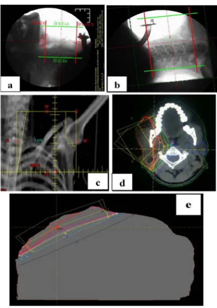
DOI: 10.4236/ojrad.2019.94018 197 Open Journal of Radiology by using simulator and pentogram shown in Figure 1. The radiation dose re-ceived by thyroid gland retrieved from the dose histogram obtained by PlanW- Treatment Planning System for Radiotherapy (Version 3.0.2017) with its quality controlled verified based on Benedick etal.[27] and IAEA [28] method. The ap-plied prescribed carcinocidal doses were 4500 - 5250 cGy for breast cancer, 6000 - 6500 cGy for nasopharyngeal, laryngeal/hypo-pharyngeal and Parotid carcino-mas. The RT machine used for irradiation was 60 co-teletherapy machine with conventional collimator.
Thyroid hormones: Tri/tetra iodothyronine and Thyroid Stimulating Hor-mone (T3, T4, TSH) have been measured from the withdrawal blood samples as 15 cc from each patient using disposable syringe and collected in three dry glass tubes (5 cc/tube for each patient) with anticoagulant and immediately centri-fuged at 2000 rpm (rotation per minutes) for 5 minutes and the amount of sepa-rated serum assessed by using Radio-immunoassay kits (DPC, USA) on Gam-mamatic II gamma counter (Contron, Switzerland) to determine the amount of T3, T4 and TSH for all cases pre/post radiotherapy course, and as well the WBC and RBCs that stored in patient’s file in PACS.
[image:4.595.263.483.335.647.2]DOI: 10.4236/ojrad.2019.94018 198 Open Journal of Radiology
3. Results
The results present the scattered plot correlation for received dose in cGy versus thyroid hormone level for irradiated HNC and breast (Figures 1-5), while Fig-ure 6 shows the average dose received by thyroid gland from each irradiated case versus the level of thyroid hormones (T3, T4, and TSH). Figure 7 presents the
correlation of respiratory rate (RR) versus ages of patient’s pre/post radiotherapy course.
4. Discussion
[image:5.595.246.503.283.443.2]Up to date, the conventional radiotherapy has been a common and main tech-nique to eradicate the residual cancer cells post operation, and further could be given as pre-operation (enhancing operation technique) or palliative treatment in developing countries. However, the consequences due to irradiation still
Figure 2. The correlation between the applied prescribed carcinocidal radiation dose for nasopharyngeal carcinoma and the relative thyroid hormones level as a reduction impact on T3 and T4 and increasing impact on TSH levels.
Figure 3. The applied prescribed carcinocidal radiation doses for Laryngeal carcinoma irradiation and the relative thyroid hormonal level as a reduction impact on T3 and T4 and increasing impact on TSH levels.
y = -0.002x + 13.72 R² = 0.708 y = -0.181x + 1219
R² = 0.708
y = 0.001x - 9.603 R² = 0.821
0.1 1 10 100 1000
5900 6000 6100 6200 6300 6400 6500 6600
H
orm
on
e
lev
el
in
n
m
/l
Tumor dose in cGy
T3-Nasophx T4-Nasophx TSH-Naphx
y = -0.000x + 5.043 R² = 0.746
y = -0.100x + 684.4 R² = 0.602
y = 0.040x - 236.4 R² = 0.662
0.1 1 10 100
6000 6100 6200 6300 6400 6500 6600
H
or
m
on
e
lev
el
in
m
U
/l
Applied dose in cGy
[image:5.595.231.517.499.683.2]DOI: 10.4236/ojrad.2019.94018 199 Open Journal of Radiology
[image:6.595.251.498.288.453.2]Figure 4. The applied prescribed carcinocidal radiation doses for Hypopharyngeal carcinoma irradiation and relative thyroid hormonal level as a reduction impact on T3 and T4 and increasing impact on TSH levels.
Figure 5. The applied prescribed carcinocidal radiation doses for supraclavicular field of (targeting cervical, supra/infra clavicle and axillary lymph nodes) breast carcinoma irradiation and relative thyroid hormonal levels as a reduction impact on T3 and T4 and increasing impact on TSH levels.
Figure 6. The applied prescribed carcinocidal radiation doses for Parotid carcinoma irradiation and the relative thyroid hormonal level as a reduction impact on T3
and T4 and increasing impact on TSH levels.
y = -0.001x + 8.488 R² = 0.546 y = -0.16x + 1063.
R² = 0.626
y = 0.024x - 143.2 R² = 0.646
0.1 1 10 100 1000
6000 6100 6200 6300 6400 6500 6600
H or m on e lev el in m U /l
Applied dose in cGy
T3-Hypop T4-Hypop TSH-Hypo
y = -0.002x + 13.52 R² = 0.648 y = -0.070x + 408.0
R² = 0.441
y = 0.003x - 10.82 R² = 0.539
0.1 1 10 100 1000
4000 4200 4400 4600 4800 5000 5200 5400
Le ve l o f H or m on es in n m /L
Applied dose in CGy
T3-Breast T4-Breast TSH-Brest
y = -0.008x + 56.70 R² = 0.774
y = -0.267x + 1778. R² = 0.632
y = 0.007x - 44.97 R² = 0.604 0.1
1 10 100 1000
6000 6100 6200 6300 6400 6500 6600
H or m on e lev el in n m /l
Applied dose in CGy
Parotid T3
[image:6.595.252.498.522.682.2]DOI: 10.4236/ojrad.2019.94018 200 Open Journal of Radiology
Figure 7. The average dose received by thyroid gland from each irradiated case (Parotid, Larynx, Breast, Hypopharynx and Nasopharynx) and the relative level of thyroid hormones (T3, T4, and TSH).
prominent and turning the scholar’s attention. Figures 2-6 show the correlation between the applied tumor dose for HNC and the relative influenced hormonal levels in logarithmic scale. In which thyroid hormones T4 and T3 decreased sig-nificantly (R2 = 0.7 and 0.8) in a linear form following the radiation dose incre-ment as correlated in equations: y = −0.1816x + 1219 and y = −0.0021x + 13.724 respectively and TSH increased significantly (R2 = 0.8) following the radiation dose increment as correlated in equation: y = 0.0016x − 9.6036, for nasopharyn-geal irradiation. For larynnasopharyn-geal irradiation, the correlations were fitted to equa-tions: y = −0.1008x + 684.42 and y = 0.0402x − 236.43 (T4 and T3) and y =
−0.0008x + 5.0432 (TSH). For hypopharyngeal irradiation, the correlations were fitted to equations: y = −0.16x + 1063.3 and y = 0.0243x − 143.22 (T4 and T3) and y = −0.0013x + 8.4887 (TSH). For supraclavicular of breast irradiation, the correlations were fitted to equations: y = −0.0708x + 408.05 and y = 0.003x − 10.825 (T4 and T3) and y = −0.0024x + 13.527 (TSH). For parotid gland
irradia-tion, the correlations were fitted to equations: y = −0.2677x + 1778.9 and y = −0.0087x + 56.702 (T4 and T3) and y = 0.0076x − 44.97 (TSH), where x refers to
radiation dose in cGy and y refers to the level of hormones in nmol/l for all cor-related equations. The t-test has been carried out for the level of hormones pre/post irradiation with references to normal levels (T3: 0.8 - 3.0 nmol/L, T4: 50 - 150 nmol/L, TSH: 0.4 - 4.0 mu/L) which was showed significant impact at P = 0.00, for nasopharyngeal, larynx, hypopharynx and P = 0.05 for breast and paro-tid gland irradiation. The reduction of hormones could be ascribed to damage or inhibition of the active follicular epithelium and reduce the number of function-al follicles; or it may reduce the vascular permeability or may trigger immuno-logic reactions leading to histoimmuno-logical changes, as reported by Mizukami et al. [29]. Also, Jung etal., [30] showed that: numerous numbers of abnormal small follicles were observed in the thyroid tissues of rats which were surrounded by cuboidal or columnar epithelium on days 4 and 7 after irradiation and inflam-matory cells were observed in the inter-follicular areas. While Chougule and
0 1 10 100 1000
Parot 2.8 Larynx 30 Breast 22 Hypo 33 Nasop 20.5
0.8 0.6
0.4 0.3 0.7
125.9 109.0
67.8
33.9
105.8
4.47 6.31 8.13
11.48
0.65
H
or
m
one
le
ve
l i
n nm
/L
DOI: 10.4236/ojrad.2019.94018 201 Open Journal of Radiology Kochar [31] found that the levels of T4 & T3were decreased significantly (p <
0.001, p < 0.005 after irradiation. In previous studies, scholars [32] [33] [34] stated that: the HNC irradiation results in hypothyroidism in at least 50% of pa-tients. The increment of TSH could be ascribed to a reduced level of T4 and T3 in the circulating blood that triggers and stimulate the production of TSH by the pituitary gland.
In supraclavicular irradiated field for breast, despite the radiation field was partially encompassing the thyroid gland compared with the cases of HNC however the impact was so significant due to large penumbra profile of the 60Co source of teletherapy machine as well as inadequate shielding during radiothe-rapy session which furtherly led to shoulder joint stiffness for some patients.
The radiation dose received by thyroid gland Figure 7, due to Nasopharynx, parotid, larynx, breast and hypopharynx irradiation were 20.5, 2.8, 30, 22 and 33 Gy respectively that reducing significantly (p = 0.00 − 0.05) the thyroid hormone T3 to (0.7, 0.8, 0.6, 0.4, 0.3) and T4 to (105.8, 125.9, 109, 67.8, 33.9) and TSH to
(0.7, 4.5, 6.3, 8.1, 11.5) respectively. Such results agreed with study done by Rei-nertsen etal.[35] and Smith etal.[36] where they noticed about 18% and 14% of the patients developed post radiotherapy hypothyroidism respectively due to 31 Gy average dose.
Based on the literature highlighted by Johansen etal.[37]; Ahmed etal. [38] and Chaurasia et al. [39]; the impact of irradiation in thyroid depends on vo-lume of the thyroid, applied dose for cancer case, fields distribution, planning method, applied technique and the type of radiotherapy machine in addition to gender and the age. The general HNC irradiation has been noticed to cause hy-pothyroidism among 48% of patients underwent radiotherapy course [40]. Ac-cording to such findings, the applied radiotherapy technique and the number of radiation fields distribution have to be verified seriously with usage of modern treatment planning systems with multi-leave collimators facilities and the worth to be recommended is the usage of intensity modulated radiation therapy (IMRT) as it gives modular radiation intensity concise with the tumor density and thickness in addition to precise shaping of the radiation field relative to tu-mor extension and sparing the adjacent vital organs.
DOI: 10.4236/ojrad.2019.94018 202 Open Journal of Radiology
Figure 8. Shows the scattered plot to correlate between age in years and respiratory rate for irradiated patients (Ca. breast tangential fields) pre and after RT course, tumor dose 4500 5000 cGy.
5. Conclusion
The conclusion addressing that: conventional radiotherapy for HNC & breast induce a significant reduction in thyroid hormones and increment of RR, lead-ing to the necessity of uslead-ing IMRT.
Acknowledgements
The authors gratefully acknowledge Qassim University represented by the Dean-ship of Scientific Research, on the material support for this research under the number [CAMS 1-2018-1-14-S-3555] during 1440 H/2018 AD.
Conflicts of Interest
The authors declare no conflicts of interest regarding the publication of this pa-per.
References
[1] Ferlay, J., Soerjomataram, I., Ervik, M., Dikshit, R., Eser, S., Mathers, C., Rebelo, M., Parkin, D.M., Forman, D. and Bray, F. (2013) GLOBOCAN 2012 v1.0, Cancer Inci-dence and Mortality Worldwide: IARC Cancer Base No. 11 [Internet]. International Agency for Research on Cancer, Lyon.
[2] Chaturvedi, A.K., Anderson, W.F., Lortet-Tieulent, J., etal. (2013) Worldwide Trends in Incidence Rates for Oral Cavity and Oropharyngeal Cancers. Journal of Clinical Oncology, 31, 4550-4559.https://doi.org/10.1200/JCO.2013.50.3870
[3] Jemal, A., Bray, F., Center, M.M., etal. (2011) Global Cancer Statistics. CA: A Cancer Journal for Clinicians, 61, 69-90.https://doi.org/10.3322/caac.20107
[4] Krishna Rao, S.V., Mejia, G., Roberts-Thomson, K., etal. (2013) Epidemiology of Oral Cancer in Asia in the Past Decade: An Update (2000-2012). Asian Pacific Journal of Cancer Prevention, 14, 5567-5677.https://doi.org/10.7314/APJCP.2013.14.10.5567
[5] Chang, E.T. and Adami, H.-O. (2006) The Enigmatic Epidemiology of Nasopha-ryngeal Carcinoma. Cancer Epidemiology, Biomarkers & Prevention, 15, 1765-1777. https://doi.org/10.1158/1055-9965.EPI-06-0353
y = 0.207x + 8.385 R² = 0.864 y = 0.201x + 13.66
R² = 0.896
0 5 10 15 20 25 30 35
20 30 40 50 60 70 80
R
ep
irat
ory rat
e
Ages in years
[image:9.595.231.524.65.227.2]DOI: 10.4236/ojrad.2019.94018 203 Open Journal of Radiology
[6] Alotaibi, A.D., Ahmed, H.G. and Elasbali, A.M. (2019) Nasopharyngeal Cancer in Saudi Arabia: Epidemiology and Possible Risk Factors. Journal of Oncological Sciences, 5, 23-30.https://doi.org/10.1016/j.jons.2019.01.002
[7] Al-Shahrani, Z.S., Al-Rawaji, A.I., Al-Madouj, A.N. and Hayder, M.S. (2017) Saudi Cancer Registry—Cancer Incidence Report Saudi Arabia 2014. Saudi Health Coun-cil-KSA.
[8] Zhang, L., Zhao, C., Ghimire, B., etal. (2010) The Role of Concurrent Chemoradi-otherapy in the Treatment of Locoregionally Advanced Nasopharyngeal Carcinoma among Endemic Population: A Meta-Analysis of the Phase III Randomized Trials. BMC Cancer, 10, 558.https://doi.org/10.1186/1471-2407-10-558
[9] Abdullah, N.E., Adam, A.A.M., Khalifa, E.H., El Hassan, L.A.M., Ibrahim, M.E., Hamad, K.M. and El Hassan, A.M. (2011) Nasopharyngeal Cancer in Sudan: Epi-demiology, Clinical and Histological Characteristics. Clinical Medicine Insights: Ear, Nose and Throat, 4, 5-11.https://doi.org/10.4137/CMENT.S5825
[10] Villanueva-Reyes, A., Strand, E., Nazario, C.M. and Irizarry-Ramirez, M. (2008) Can-cer of the Larynx in Puerto Rico. Puerto Rico Health Sciences Journal, 27, 196-203. [11] Ahmed, S., Abdelrhman, S., Taha, M., Malik, M. and El Naseh, W. (2018) Etiology and Clinical Presentation of Laryngeal Cancer in Sudanese Patients. International Journal of Otolaryngology, 5, 3.https://doi.org/10.13188/2380-0569.1000022
[12] Jones, T.M., De, M., Foran, B., Harrington, K. and Mortimore, S. (2016) Laryngeal Cancer: United Kingdom National Multidisciplinary Guidelines. The Journal of La-ryngology & Otology, 130, S75-S82.https://doi.org/10.1017/S0022215116000487
[13] Kajanti, M. and Manty, A. (1990) Carcinoma of the Hypopharynx. A Retrospective Analysis of the Treatment Result Over a 25-Year Period. Acta Otologica, 29, 903-907. [14] Mahasin, Z. and Khan, B. (1996) Hypopharyngeal Carcinoma: King Faisal Specialist
Hospital and Research Centre Experience. Annals of Saudi Medicine, 16, 539-544. https://doi.org/10.5144/0256-4947.1996.539
[15] Alotaibi, R.M., Rezk, H.R., Juliana, C.I. and Guure, C. (2018) Breast Cancer Mortal-ity in Saudi Arabia: Modelling Observed and Unobserved Factors. PLoS ONE, 13, e0206148.https://doi.org/10.1371/journal.pone.0206148
[16] Ayadi, W., Khabir, A., Hadhri-Guiga, B., etal. (2010) North African and Southeast Asian Nasopharyngeal Carcinomas: Between the Resemblance and the Dissem-blance. Bulletin du Cancer, 97, 475-482.https://doi.org/10.1684/bdc.2010.1090
[17] Nguyen-Van, D., Ernberg, I., Phan-Thi Phi, P., Tran-Thi, C. and Hu, L. (2008) Eps-tein-Barr Virus Genetic Variation in Vietnamese Patients with Nasopharyngeal Carcinoma: Full-Length Analysis of LMP1. Virus Gene, 37, 273-281.
https://doi.org/10.1007/s11262-008-0262-9
[18] Ibrahim, E.M., Zeeneldin, A.A., Sadiq, B.B. and Ezzat, A.A. (2008) The Present and the Future of Breast Cancer Burden in the Kingdom of Saudi Arabia. Medical On-cology, 25, 387-393.https://doi.org/10.1007/s12032-008-9051-5
[19] Elamin, A., Ibrahim, M.E., Abuidris, D., Mohamed, K.E.H. and Mohammed, S.I. (2015) Part I: Cancer in Sudan—Burden, Distribution, and Trends Breast, Gyneco-logical, and Prostate Cancers. Cancer Medicine, 4, 447-456.
https://doi.org/10.1002/cam4.378
[20] Fuks, Z., Glatstein, E., et al. (1976) Long-Term Effects of External Radiation on the Pituitary and Thyroid Glands. Cancer, 37, 1152-1161.
DOI: 10.4236/ojrad.2019.94018 204 Open Journal of Radiology
[21] Bonato, C., Severino, R.F. and Elnecave, R.H. (2008) Reduced Thyroid Volume and Hypothyroidism in Survivors of Childhood Cancer Treated with Radiotherapy. Journal of Pediatric Endocrinology and Metabolism, 21, 943-949.
https://doi.org/10.1515/JPEM.2008.21.10.943
[22] Laway, B.A., Shafi, K.M., Majid, S., Lone, M.M., Afroz, F., Khan, S. and Roohi, R. (2012) Incidence of Primary Hypothyroidism in Patients Exposed to Therapeutic External Beam Radiation, Where Radiation Portals Include a Part or Whole of the Thyroid Gland. Indian Journal of Endocrinology and Metabolism, 16, 329-331. [23] Jaén, J., Vázquez, G., Alonso, E., León, A., Guerrero, R. and Almansa, J.F. (2006)
Changes in Pulmonary Function after Incidental Lung Irradiation for Breast Can-cer: A Prospective Study. International Journal of Radiation Oncology, Biology, Physics, 65, 1381-1388.https://doi.org/10.1016/j.ijrobp.2006.03.008
[24] Hernberg, M., Virkkunen, P., Maasilta, P., Keyriläinen, J., Blomqvist, C., Bergh, J., etal. (2002) Pulmonary Toxicity after Radiotherapy in Primary Breast Cancer Pa-tients: Results from a Randomized Chemotherapy Study. International Journal of Radiation Oncology, Biology, Physics, 52, 128-136.
https://doi.org/10.1016/S0360-3016(01)01760-6
[25] Lopez Guerra, J.L., Gomez, D.R., Zhuang, Y., Levy, L.B., Eapen, G., Liu, H.M., Mo-han, R., Komaki, R., Cox, J.D. and Liao, Z.X. (2012) Changes in Pulmonary Func-tion after Three-Dimensional Conformal RadiaFunc-tion Therapy, Intensity-Modulated Radiation Therapy, or Proton Beam Therapy for Non-Small Cell Lung Cancer. In-ternational Journal of Radiation Oncology, Biology, Physics, 83, e537-e543. https://doi.org/10.1016/j.ijrobp.2012.01.019
[26] Subbe, C.P., Davies, R.G., Williams, E., etal. (2003) Effect of Introducing the Mod-ified Early Warning Score on Clinical Outcomes, Cardio-Pulmonary Arrests and Intensive Care Utilisation in Acute Medical Admissions. Anaesthesia, 58, 797-802. https://doi.org/10.1046/j.1365-2044.2003.03258.x
[27] Benedick, F., Doppke, K., Hunt, M., Kutcher, G., Starkschall, G., Stern, R. and Van Dyke, J. (1998) AAPM Radiation Therapy Committee TG53: Quality Assurance Program for Radiotherapy Treatment Planning. Medical Physics, 25, 1773-1836.
https://doi.org/10.1118/1.598373
[28] IAEA (2004) Commissioning and Quality Assurance of Computerized Planning
Systems for Radiation Treatment of Cancer. TRS 430, Austria.
[29] Mizukami, Y., Michigishi, T., Nonomura, A., Hashimoto, T., Noguchi, M., Ohmura, K. and Matsubara, F. (1992) Histologic Changes in Graves’ Thyroid Gland after 131I
Therapy for Hyperthyroidism. Acta Pathologica Japonica, 42, 419-426. https://doi.org/10.1111/j.1440-1827.1992.tb03247.x
[30] Jung, J.H., Jung, J., Kim, S.K., Woo, S.H., Kang, K.M., Jeong, B.-K., Jung, M.H., Kim, J.H. and Hahm, J.R. (2014) Alpha Lipoic Acid Atteuates Radiation-Induced Thyroid Injury in Rats. PLoS ONE, 9, e112253.
https://doi.org/10.1371/journal.pone.0112253
[31] Chougule, A. and Kochar, B. (2011) Thyroid Dysfunction Following Therapeutic External Radiation to Head and Neck Cancer. Asian Pacific Journal of Cancer Pre-vention, 12, 443-445.
[32] Garcia-Serra, A., Amdur, R.J., Morris, C.G., Mazzaferri, E. and Mendenhall, W.M. (2005) Thyroid Function Should Be Monitored Following Radiotherapy to the Low Neck. American Journal of Clinical Oncology, 28, 255-258.
https://doi.org/10.1097/01.coc.0000145985.64640.ac
Receiv-DOI: 10.4236/ojrad.2019.94018 205 Open Journal of Radiology
ing Neck Irradiation for Cancer. Cancer, 55, 1190-1194.
https://doi.org/10.1002/1097-0142(19850315)55:6<1190::AID-CNCR2820550609>3. 0.CO;2-6
[34] Khoo, V.S., Liew, K.H., Crennan, E.C., D’Costa, I.M. and Quong, G. (1998) Thyroid Dysfunction after Mantle Irradiation of Hodgkin’s Disease Patients. Australasian Radiology, 42, 52-57.https://doi.org/10.1111/j.1440-1673.1998.tb00565.x
[35] Reinertsen, K.V., Cvancarova, M., Wist, E., Bjøro, T., Dahl, A.A., Danielsen, T. and Fosså, S.D. (2009) Thyroid Function in Women after Multimodal Treatment for Breast Cancer Stage II/III: Comparison with Controls from a Population Sample. International Journal of Radiation Oncology, Biology, Physics, 75, 764-770. https://doi.org/10.1016/j.ijrobp.2008.11.037
[36] Smith, G.L., Smith, B.D., Giordano, S.H., Shih, Y.C., Woodward, W.A., Strom, E.A., Perkins, G.H., Tereffe, W., Yu, T.K. and Buchholz, T.A. (2008) Risk of Hypothy-roidism in Older Breast Cancer Patients Treated with Radiation. Cancer, 112, 1371-1379.https://doi.org/10.1002/cncr.23307
[37] Johansen, S., Reinertsen, K.V., Knutstad, K., Olsen, D.R. and Fosså, S.D. (2011) Dose Distribution in the Thyroid Gland Following Radiation Therapy of Breast Cancer: A Retrospective Study. Radiation Oncology, 6, 68.
https://doi.org/10.1186/1748-717X-6-68
[38] Ahmed, Z., Khan, M.A., Haq, A., Attaullag, S. and ur Rehman, Z. (2009) Effect of Race, Gender and Age on Thyroid and Thyroid Stimulating Hormone Levels in Northwest Frontier Province, Pakistan. Journal of Ayub Medical College Abbotta-bad, 21, 21-24.
[39] Chaurasia, P., Modi, B., Mangukiya, S., Jadav, P. and Shah, R. (2011) Variation of Thyroid Hormones Level among People of Different Age, Gender and Seasons, Pi-paria, Gujarat. National Journal of Medical Research, 1, 57-59.
[40] Mercado, G., Adelstein, D.J., Saxton, J.P., Secic, M., Larto, M.A. and Lavertu, P. (2001) Hypothyroidism: A Frequent Event after Radiotherapy and after Radiothe-rapy with ChemotheRadiothe-rapy for Patients with Head and Neck Carcinoma. Cancer, 92, 2892-2897.
https://doi.org/10.1002/1097-0142(20011201)92:11<2892::AID-CNCR10134>3.0.C O;2-T
[41] Hong, W., Earnest, A., Sultana, P., etal. (2013) How Accurate Are Vital Signs in Predicting Clinical Outcomes in Critically Ill Emergency Department Patients. Eu-ropean Journal of Emergency Medicine, 20, 27-32.
https://doi.org/10.1097/MEJ.0b013e32834fdcf3
[42] Goldman, B.U., Anderson, M., Wennberg, B. and Lind, P. (2014) Radiation Pneu-monitis and Pulmonary Function with Lung Dose-Volume Constraints in Breast Cancer Irradiation. Journal of Radiotherapy in Practice, 13, 211-217.