organic papers
o1582
Liuet al. C10H10O2 doi:10.1107/S1600536806010440 Acta Cryst.(2006). E62, o1582–o1583
Acta Crystallographica Section E Structure Reports
Online
ISSN 1600-5368
1,4-Diacetylbenzene
Shan Liu,aZhen-Yi Wu,aNing
Shan,bDan-Dan Wangaand
Hong-Jun Zhua*
aDepartment of Applied Chemistry, College of
Science, Nanjing University of Technology, Nanjing 210009, People’s Republic of China, andbInstitute of Chemical and Engineering
Sciences, The Singapore Government Agency for Science, Technology and Research, 627833, Singapore
Correspondence e-mail: [email protected]
Key indicators
Single-crystal X-ray study
T= 180 K
Mean(C–C) = 0.002 A˚
Rfactor = 0.069
wRfactor = 0.195
Data-to-parameter ratio = 20.7
For details of how these key indicators were automatically derived from the article, see http://journals.iucr.org/e.
Received 6 March 2006 Accepted 22 March 2006
#2006 International Union of Crystallography
All rights reserved
The asymmetric unit of the title compound, C10H10O2,
contains two half-molecules, each molecules being located on a centre of inversion. The dihedral angle between the planes of the two independent molecules is 39.67 (4).
Comment
1,4-Diacetylbenzene is used in polymerization reactions to obtain polymers of quinoline derivatives (Bracke, 1969; Imai
et al., 1975). We report here the crystal structure of the title compound, (I).
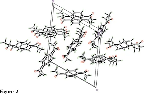
The asymmetric unit of (I) contains two half-molecules, each molecule being located on a centre of inversion (Fig. 1). Selected geometric parameters are listed in Table 1. Corres-ponding bond lengths and angles in the two independent molecules agree with each other. Both the molecules are essentially planar; the dihedral angle between the planes of the two molecules is 39.67 (4). No–stacking interactions
or hydrogen bonds are observed in the crystal structure (Fig. 2).
Experimental
p-Ethylacetophenone (14.825 g, 0.1 mol) was added dropwise to a mixture of magnesium oxide (10.0 g, 0.25 mol), water (258 ml), concentrated nitric acid (34 ml, 0.53 mol) and potassium permanga-nate (39.5 g, 0.25 mol), with stirring at 332–334 K. The mixture was stirred further at 331–335 K for 4.5 h and then cooled, filtered by vacuum and dried. The powdered residue was dissolved in hot benzene (300 ml) to extract the crude product. This was slurried with cold anhydrous diethyl ether (70 ml), filtered, and dried to obtain white crystals of the title compound (13.3 g). Single crystals suitable for X-ray diffraction were obtained by dissolving the compound (2.0 g, 12.3 mmol) in benzene (25 ml) and allowing the solution to evaporate at room temperature for about 15 d.
Crystal data
C10H10O2
Mr= 162.18
Monoclinic,P21=n a= 12.699 (3) A˚
b= 5.4604 (11) A˚
c= 12.950 (3) A˚
= 114.07 (3) V= 819.9 (4) A˚3
Z= 4
Dx= 1.314 Mg m
3 MoKradiation Cell parameters from 2627
reflections
= 1.7–31.1
= 0.09 mm1
Data collection
Bruker SMART CCD area-detector diffractometer
’and!scans
Absorption correction: multi-scan (SADABS; Sheldrick, 2002)
Tmin= 0.979,Tmax= 0.992 6624 measured reflections
2297 independent reflections 1979 reflections withI> 2(I)
Rint= 0.024
max= 31.3
h=18!18
k=7!6
l=16!17
Refinement
Refinement onF2
R[F2> 2(F2)] = 0.069
wR(F2) = 0.195
S= 1.16 2297 reflections 111 parameters
H-atom parameters constrained
w= 1/[2(F
o2) + (0.0962P)2 + 0.1961P]
whereP= (Fo2+ 2Fc2)/3 (/)max= 0.001
max= 0.29 e A˚
3
min=0.27 e A˚
[image:2.610.314.564.71.248.2]3
Table 1
Selected geometric parameters (A˚ ,).
O1—C4 1.221 (2) O2—C9 1.218 (2)
O1—C4—C1 119.89 (15) O1—C4—C5 121.29 (14)
O2—C9—C10 121.12 (15) O2—C9—C6 120.23 (15)
H atoms were placed in idealized positions and constrained to ride on their parent atoms, with C—H distances of 0.95 (aromatic) or 0.97(methyl) A˚ , and withUiso(H) = 1.2 or 1.5(methyl) timesUeq(C).
Data collection:SMART(Bruker, 2001); cell refinement:SAINT
(Bruker, 2001); data reduction: SAINT; program(s) used to solve structure: SHELXS97(Sheldrick, 1997); program(s) used to refine structure: SHELXL97 (Sheldrick, 1997); molecular graphics:
SHELXTL (Bruker, 2001); software used to prepare material for publication:SHELXTL.
References
Bracke, W. (1969).Macromolecules,2, 286–289.
Bruker (2001).SMART,SAINTandSHELXTL. Bruker AXS Inc., Madison, Wisconsin, USA.
Imai, Y., Johnson, E. F., Katto, T., Katto, T., Kurihara, M. & Stille, J. K. (1975).
J. Polym. Sci.13, 2233–2249.
Sheldrick, G. M. (1997). SHELXS97 and SHELXL97. University of Go¨ttingen, Germany.
Sheldrick, G. M. (2002).SADABS. University of Go¨ttingen, Germany.
Figure 1
The asymmetric unit of (I), showing the atom-numbering scheme. Displacement ellipsoids are drawn at the 50% probability level. Atoms labeled with the suffixes A and B are generated by the symmetry operations (1x,y,z) and (2x,y,z), respectively.
Figure 2
[image:2.610.315.563.318.482.2]supporting information
sup-1 Acta Cryst. (2006). E62, o1582–o1583
supporting information
Acta Cryst. (2006). E62, o1582–o1583 [https://doi.org/10.1107/S1600536806010440]
1,4-Diacetylbenzene
Shan Liu, Zhen-Yi Wu, Ning Shan, Dan-Dan Wang and Hong-Jun Zhu
1,4-Diacetylbenzene
Crystal data
C10H10O2 Mr = 162.18 Monoclinic, P21/n
Hall symbol: -P 2yn
a = 12.699 (3) Å
b = 5.4604 (11) Å
c = 12.950 (3) Å
β = 114.07 (3)°
V = 819.9 (4) Å3 Z = 4
F(000) = 344
Dx = 1.314 Mg m−3 Dm = 1.314 Mg m−3
Dm measured by not measured
Melting point: 113(1) K Mo Kα radiation, λ = 0.71073 Å Cell parameters from 2627 reflections
θ = 1.7–31.1°
µ = 0.09 mm−1 T = 180 K Prism, colourless 0.24 × 0.15 × 0.09 mm
Data collection
Bruker SMART CCD area-detector diffractometer
Radiation source: fine-focus sealed tube Graphite monochromator
Detector resolution: 28.5714 pixels mm-1 φ and ω scans
Absorption correction: multi-scan (SADABS; Sheldrick, 2002)
Tmin = 0.979, Tmax = 0.992
6624 measured reflections 2297 independent reflections 1979 reflections with I > 2σ(I)
Rint = 0.024
θmax = 31.3°, θmin = 1.9° h = −18→18
k = −7→6
l = −16→17
Refinement
Refinement on F2
Least-squares matrix: full
R[F2 > 2σ(F2)] = 0.069 wR(F2) = 0.195 S = 1.16 2297 reflections 111 parameters 0 restraints
Primary atom site location: structure-invariant direct methods
Secondary atom site location: difference Fourier map
Hydrogen site location: inferred from neighbouring sites
H-atom parameters constrained
w = 1/[σ2(F
o2) + (0.0962P)2 + 0.1961P]
where P = (Fo2 + 2Fc2)/3
(Δ/σ)max = 0.001
Δρmax = 0.29 e Å−3
Special details
Geometry. All e.s.d.'s (except the e.s.d. in the dihedral angle between two l.s. planes) are estimated using the full covariance matrix. The cell e.s.d.'s are taken into account individually in the estimation of e.s.d.'s in distances, angles and torsion angles; correlations between e.s.d.'s in cell parameters are only used when they are defined by crystal symmetry. An approximate (isotropic) treatment of cell e.s.d.'s is used for estimating e.s.d.'s involving l.s. planes.
Refinement. Refinement of F2 against ALL reflections. The weighted R-factor wR and goodness of fit S are based on F2,
conventional R-factors R are based on F, with F set to zero for negative F2. The threshold expression of F2 > σ(F2) is used
only for calculating R-factors(gt) etc. and is not relevant to the choice of reflections for refinement. R-factors based on F2
are statistically about twice as large as those based on F, and R- factors based on ALL data will be even larger.
Fractional atomic coordinates and isotropic or equivalent isotropic displacement parameters (Å2)
x y z Uiso*/Ueq
O1 0.80092 (10) 0.0913 (2) 0.19525 (10) 0.0411 (3)
C1 0.61872 (12) −0.0341 (3) 0.06097 (12) 0.0296 (3)
O2 0.80864 (11) 0.1179 (2) −0.29558 (10) 0.0436 (4)
C7 1.02083 (14) −0.1997 (3) −0.05480 (13) 0.0330 (4)
H7 1.0352 −0.3370 −0.0923 0.040*
C2 0.56419 (13) 0.1697 (3) 0.08133 (13) 0.0323 (4)
H2 0.6078 0.2862 0.1368 0.039*
C6 0.93984 (12) −0.0249 (3) −0.11728 (13) 0.0307 (4)
C4 0.74619 (13) −0.0645 (3) 0.12672 (13) 0.0321 (4)
C3 0.55359 (13) −0.2037 (3) −0.02102 (13) 0.0325 (4)
H3 0.5901 −0.3431 −0.0356 0.039*
C9 0.87567 (13) −0.0430 (3) −0.24330 (13) 0.0337 (4)
C5 0.80422 (14) −0.2857 (3) 0.10517 (15) 0.0380 (4)
H5A 0.8870 −0.2795 0.1535 0.057*
H5B 0.7927 −0.2883 0.0256 0.057*
H5C 0.7709 −0.4340 0.1223 0.057*
C10 0.89811 (15) −0.2588 (4) −0.30297 (14) 0.0404 (4)
H10A 0.8498 −0.2467 −0.3842 0.061*
H10B 0.8796 −0.4101 −0.2732 0.061*
H10C 0.9796 −0.2606 −0.2905 0.061*
C8 0.91933 (13) 0.1758 (3) −0.06135 (13) 0.0327 (4)
H8 0.8643 0.2961 −0.1029 0.039*
Atomic displacement parameters (Å2)
U11 U22 U33 U12 U13 U23
O1 0.0345 (6) 0.0419 (7) 0.0406 (7) −0.0040 (5) 0.0087 (5) −0.0049 (5)
C1 0.0306 (7) 0.0301 (8) 0.0282 (7) −0.0004 (5) 0.0121 (6) 0.0030 (5)
O2 0.0499 (7) 0.0420 (7) 0.0338 (6) 0.0051 (5) 0.0117 (5) 0.0036 (5)
C7 0.0346 (7) 0.0314 (8) 0.0333 (8) 0.0011 (6) 0.0140 (6) −0.0040 (6)
supporting information
sup-3 Acta Cryst. (2006). E62, o1582–o1583
C10 0.0443 (9) 0.0452 (10) 0.0322 (8) −0.0009 (7) 0.0160 (7) −0.0063 (7)
C8 0.0325 (7) 0.0315 (8) 0.0324 (8) 0.0025 (6) 0.0115 (6) 0.0009 (6)
Geometric parameters (Å, º)
O1—C4 1.221 (2) C4—C5 1.500 (2)
C1—C2 1.392 (2) C3—C2ii 1.389 (2)
C1—C3 1.397 (2) C3—H3 0.95
C1—C4 1.500 (2) C9—C10 1.499 (2)
O2—C9 1.218 (2) C5—H5A 0.98
C7—C8i 1.387 (2) C5—H5B 0.98
C7—C6 1.394 (2) C5—H5C 0.98
C7—H7 0.95 C10—H10A 0.98
C2—C3ii 1.389 (2) C10—H10B 0.98
C2—H2 0.95 C10—H10C 0.98
C6—C8 1.395 (2) C8—C7i 1.387 (2)
C6—C9 1.501 (2) C8—H8 0.95
C2—C1—C3 119.20 (14) O2—C9—C10 121.12 (15)
C2—C1—C4 119.08 (14) O2—C9—C6 120.23 (15)
C3—C1—C4 121.71 (14) C10—C9—C6 118.63 (14)
C8i—C7—C6 120.85 (14) C4—C5—H5A 109.5
C8i—C7—H7 119.6 C4—C5—H5B 109.5
C6—C7—H7 119.6 H5A—C5—H5B 109.5
C3ii—C2—C1 120.34 (14) C4—C5—H5C 109.5
C3ii—C2—H2 119.8 H5A—C5—H5C 109.5
C1—C2—H2 119.8 H5B—C5—H5C 109.5
C7—C6—C8 119.07 (14) C9—C10—H10A 109.5
C7—C6—C9 122.07 (14) C9—C10—H10B 109.5
C8—C6—C9 118.85 (14) H10A—C10—H10B 109.5
O1—C4—C1 119.89 (15) C9—C10—H10C 109.5
O1—C4—C5 121.29 (14) H10A—C10—H10C 109.5
C1—C4—C5 118.82 (13) H10B—C10—H10C 109.5
C2ii—C3—C1 120.46 (14) C7i—C8—C6 120.08 (14)
C2ii—C3—H3 119.8 C7i—C8—H8 120.0
C1—C3—H3 119.8 C6—C8—H8 120.0