RASA1-dependent cellular export of collagen IV
controls blood and lymphatic vascular
development
Di Chen, … , Philip E. Lapinski, Philip D. King
J Clin Invest.
2019;
129(9)
:3545-3561.
https://doi.org/10.1172/JCI124917
.
Combined germline and somatic second-hit inactivating mutations of the
RASA1
gene,
which encodes a negative regulator of the Ras signaling pathway, cause blood and
lymphatic vascular lesions in the human autosomal-dominant vascular disorder capillary
malformation–arteriovenous malformation (CM-AVM). How
RASA1
mutations in endothelial
cells (ECs) result in vascular lesions in CM-AVM is unknown. Here, using different murine
models of RASA1 deficiency, we found that RASA1 was essential for the survival of ECs
during developmental angiogenesis, in which primitive vascular plexuses are remodeled
into hierarchical vascular networks. RASA1 was required for EC survival during
developmental angiogenesis, because it was necessary for export of collagen IV from ECs
and deposition in vascular basement membranes. In the absence of RASA1, dysregulated
Ras/MAPK signal transduction in ECs resulted in impaired folding of collagen IV and its
retention in the endoplasmic reticulum (ER), leading to EC death. Remarkably, the chemical
chaperone 4-phenylbutyric acid and small-molecule inhibitors of MAPK and 2-oxoglutarate–
dependent collagen IV–modifying enzymes rescued ER retention of collagen IV and EC
apoptosis and resulted in normal developmental angiogenesis. These findings have
important implications for a better understanding of the molecular pathogenesis of CM-AVM
and possible means of treatment.
Research Article
Angiogenesis
Vascular biology
Find the latest version:
Introduction
Capillary malformation–arteriovenous malformation (CM-AVM) is an autosomal-dominant inherited vascular disease that is char-acterized by 1 or more cutaneous CMs together with fast-flow vas-cular lesions in one-third of patients (1–3). Fast-flow lesions, which include AVMs and arteriovenous fistulas (AFs), occur in different anatomical locations and can be life threatening. Lymphatic vessel (LV) abnormalities that result in lymphedema, chylothorax, and chylous ascites have also been identified in a minority of patients with CM-AVM (2–7). In the majority of CM-AVM cases, blood ves-sel (BV) and LV lesions are present at birth, although they can also develop throughout childhood and up to early adulthood.
Inactivating germline mutations of the RASA1 gene are respon-sible for approximately 50% of CM-AVM cases (1–3). RASA1 encodes p120 Ras GTPase-activating protein (p120 RasGAP or RASA1), a negative regulator of the Ras small GTP-binding protein that promotes cell growth, proliferation, and differentiation (8–10). In quiescent cells, Ras exists predominantly in an inactive GDP-bound state. Growth factors promote the conversion of Ras to an active GTP-bound state that results in the triggering of downstream signaling pathways including the MAPK and PI3K pathways that drive cellular responses. RASA1 inhibits Ras signal transduction by augmenting the ability of Ras to hydrolyze bound GTP, resulting in its conversion to the inactive GDP-bound form (8). Vascular lesions in patients with CM-AVM with germline RASA1 mutations arise as a
consequence of somatic inactivating mutation of the inherited WT RASA1 allele in endothelial cells (ECs) or their precursors (6, 11). Loss of RASA1 in these ECs would be expected to result in dysregu-lated Ras signal transduction that could drive lesion development.
Recently, it has been shown that inactivating germline muta-tions of EPHB4, which encodes the ephrin receptor B4, are respon-sible for the majority of CM-AVM cases that are not explained by the mutation of RASA1 (12). Accordingly, CM-AVM resulting from RASA1 mutation has been renamed CM-AVM1, and CM-AVM resulting from EPHB4 mutation has been named CM-AVM2. Clinically, CM-AVM1 and CM-AVM2 are almost indistinguish-able except for the additional occurrence of telangiectasias in CM-AVM2 (12). These findings raise the possibility that lesion development in CM-AVM results from loss of an EPHB4/RASA1 negative-regulatory axis in ECs, in which EPHB4 serves to recruit RASA1 to the inner leaflet of the cell membrane, allowing its jux-taposition to Ras-GTP (12, 13). It is likely that second-hit muta-tions of EPHB4 are required for the development of lesions in CM-AVM2, although this has yet to be demonstrated
Studies of genetically engineered mutant mice have the poten-tial to provide information on the pathogenesis of diseases such as CM-AVM that could not otherwise be obtained from human stud-ies alone. Concerning RASA1 and CM-AVM1, constitutive loss of Rasa1 in mice results in mid-gestation lethality at E10.5 as a conse-quence of impaired vascular development (14, 15). Developmental angiogenesis, in which primitive vascular plexuses are remodeled into hierarchical vascular networks, is abnormal in these embry-os. This is evident in the yolk sac, for example, where ECs initial-ly assemble into a vascular plexus but then fail to organize into a vascular network that supplies blood to the developing embryo. Some defects in vasculogenesis are also evident in RASA1-defi-disorder capillary malformation–arteriovenous malformation (CM-AVM). How RASA1 mutations in endothelial cells (ECs)
result in vascular lesions in CM-AVM is unknown. Here, using different murine models of RASA1 deficiency, we found that RASA1 was essential for the survival of ECs during developmental angiogenesis, in which primitive vascular plexuses are remodeled into hierarchical vascular networks. RASA1 was required for EC survival during developmental angiogenesis, because it was necessary for export of collagen IV from ECs and deposition in vascular basement membranes. In the absence of RASA1, dysregulated Ras/MAPK signal transduction in ECs resulted in impaired folding of collagen IV and its retention in the endoplasmic reticulum (ER), leading to EC death. Remarkably, the chemical chaperone 4-phenylbutyric acid and small-molecule inhibitors of MAPK and 2-oxoglutarate–dependent collagen IV–modifying enzymes rescued ER retention of collagen IV and EC apoptosis and resulted in normal developmental angiogenesis. These findings have important implications for a better understanding of the molecular pathogenesis of CM-AVM and possible means of treatment.
Conflict of interest: The authors have declared that no conflict of interest exists.
Copyright: © 2019, American Society for Clinical Investigation.
Submitted: September 13, 2018; Accepted: June 6, 2019; Published: August 5, 2019.
Results
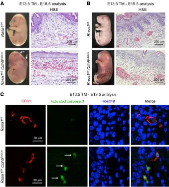
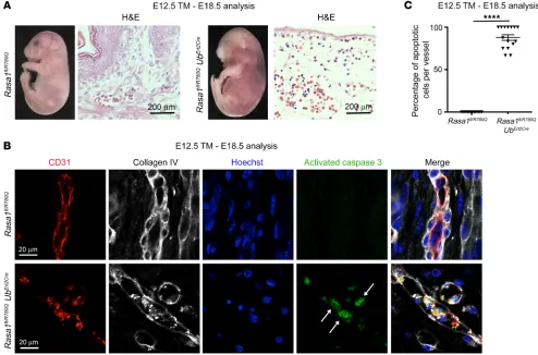
Global disruption of Rasa1 during developmental angiogenesis results in hemorrhage, edema, and EC apoptosis. To examine the influence of global RASA1 loss upon developmental angiogenesis, pregnant Rasa1fl/fl mice carrying Rasa1fl/fl and Rasa1fl/fl UbErt2Cre embryos at the E12.5–E14.5 stage were administered tamoxifen (TM). We found that administration of TM to Rasa1fl/fl UbErt2Cre embryos at this time point resulted in visible cutaneous hemorrhage and an edematous appearance at E18.5 to E19.5 (Table 1 and Figure 1A). Histological analysis of embryos revealed extravasated erythro-cytes in skin associated with damaged cutaneous BVs and a vastly reduced number of cutaneous LVs (Figure 1A). We did not observe the same phenotypes in Rasa1fl/fl UbErt2Cre embryos that were not administered TM (Supplemental Figure 1A). Administration of TM to Rasa1fl/fl UbErt2Cre embryos at E15.5 and later also did not result in hemorrhage or other spontaneous embryonic BV abnormalities, although TM administration at this embryonic stage does result in failed LV valve development, as we reported previously (17).
Failed LV valve development in embryos administered TM at E15.5 is explained by apoptosis of LV endothelial cells (LECs) in developing LV valve leaflets (17). Therefore, we asked wheth-er disruption of Rasa1 in Rasa1fl/fl UbErt2Cre embryos before E15.5 induced apoptosis of BV endothelial cells (BECs) and LECs in BV and LV walls. As revealed by immunostaining for activat-ed caspase 3, we identifiactivat-ed apoptotic BECs in the vast majority of cutaneous BVs of Rasa1fl/fl UbErt2Cre embryos that were admin-istered TM between E12.5 and E14.5, as determined at E18.5 to cient embryos. In contrast to this, in adult mice, induced global
disruption of Rasa1 does not result in any spontaneous BV abnor-malities (16). Instead, the mice develop LV hyperplasia and leak-age that result in chylous ascites and chylothorax (16). Recently, we demonstrated that RASA1 is essential for the development and maintenance of valves in collecting LVs, which accounts for LV leakage in the absence of RASA1 (17).
[image:3.585.37.544.84.365.2]To further understand the role of RASA1 in the BV and LV systems and how its loss may contribute to the vascular pheno-types observed in CM-AVM1, in the current study, we examined the influence of embryonic loss of RASA1 after E10.5. By E10.5, vasculogenesis is largely complete, and the remainder of vascular development is devoted to remodeling of the vascular network by angiogenic processes (18). RASA1 was found to be essential for continued vascular development during this period by promot-ing the survival of ECs. Unexpectedly, the prosurvival function of RASA1 in ECs during developmental angiogenesis could be explained on the grounds that RASA1 is required for the proper folding and export from ECs and vascular smooth muscle cells (VSMCs) of collagen IV, a major constituent of vascular basement membranes (BMs). We further show that RASA1 is required for normal retinal angiogenesis in newborns and pathological angio-genesis in adults and that this is again most likely explained by its role in the export of collagen IV for deposition in the BM. These findings reveal a previously unappreciated role for RASA1 in vas-cular biology and are of relevance to the understanding of the pathogenesis and treatment of CM-AVM.
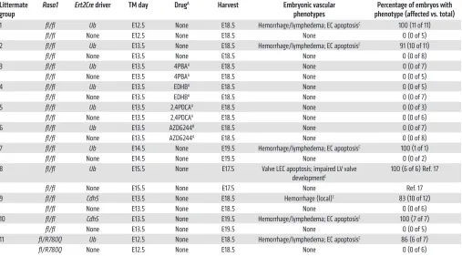
Table 1. Embryonic disruption of Rasa1
Littermate
group Rasa1 Ert2Cre driver TM day Drug
A Harvest Embryonic vascular
phenotypes phenotype (affected vs. total)Percentage of embryos with 1 fl/fl Ub E12.5 None E18.5 Hemorrhage/lymphedema; EC apoptosisC 100 (11 of 11)
fl/fl None E12.5 None E18.5 None 0 (0 of 5)
2 fl/fl Ub E13.5 None E18.5 Hemorrhage/lymphedema; EC apoptosisC 91 (10 of 11)
fl/fl None E13.5 None E18.5 None 0 (0 of 8)
3 fl/fl Ub E13.5 4PBAA E18.5 None 0 (0 of 7)
fl/fl None E13.5 4PBAA E18.5 None 0 (0 of 5)
4 fl/fl Ub E13.5 EDHBA E18.5 None 0 (0 of 5)
fl/fl None E13.5 EDHBA E18.5 None 0 (0 of 7)
5 fl/fl Ub E13.5 2,4PDCAA E18.5 None 0 (0 of 3)
fl/fl None E13.5 2,4PDCAA E18.5 None 0 (0 of 6)
6 fl/fl Ub E13.5 AZD6244B E18.5 None 0 (0 of 7)
fl/fl None E13.5 AZD6244A E18.5 None 0 (0 of 8)
7 fl/fl Ub E14.5 None E19.5 Hemorrhage/lymphedema; EC apoptosisC 100 (1 of 1)
fl/fl None E14.5 None E19.5 None 0 (0 of 2)
8 fl/fl Ub E15.5 None E17.5 Valve LEC apoptosis; impaired LV valve developmentC
100 (6 of 6) Ref. 17
fl/fl None E15.5 None E17.5 None Ref. 17
9 fl/fl Cdh5 E13.5 None E18.5 Hemorrhage (local)C 83 (10 of 12)
fl/fl None E13.5 None E18.5 None 0 (0 of 6)
10 fl/fl Cdh5 E13.5 None E19.5 Hemorrhage/lymphedema; EC apoptosisC 100 (7 of 7)
fl/fl None E13.5 None E19.5 None 0 (0 of 5)
11 fl/R780Q Ub E12.5 None E18.5 Hemorrhage/lymphedema; EC apoptosisC 86 (6 of 7)
fl/R780Q None E12.5 None E18.5 None 0 (0 of 6)
ed, Rasa1fl/fl Cdh5Ert2Cre embryos, which was confirmed by histo-logical analysis (Table 1, Figure 2A and Supplemental Figure 3A). However, hemorrhage was more localized in Rasa1fl/fl Cdh5Ert2Cre embryos than in Rasa1fl/fl UbErt2Cre embryos at this time point (com-pare with Figure 1A). In contrast, at E19.5, we observed extensive hemorrhage and edema in Rasa1fl/fl Cdh5Ert2Cre embryos (Table 1 and Figure 2B). Furthermore, apoptotic BECs were readily observed in BVs of Rasa1fl/fl Cdh5Ert2Cre embryos at E19.5 (Figure 2C) but not at E18.5 (see below). As shown by real-time quantitative PCR (qPCR) analysis of sorted skin BECs from TM-treated Rasa1fl/fl UbErt2Cre and Rasa1fl/fl Cdh5Ert2Cre embryos, deletion efficiency of the Rasa1 gene in BECs was comparable using the 2 different types of ErtCre driv-ers at E18.5 (Supplemental Figure 4). Therefore, differences in the time of onset of the phenotypes cannot be explained by differenc-es in Rasa1 gene deletion efficiency. In conclusion, disruption of Rasa1 within ECs during developmental angiogenesis was suffi-cient for the development of vascular abnormalities, including EC E19.5 (Figure 1, B and C) but not in Rasa1fl/fl UbErt2Cre embryos that
did not receive TM (Supplemental Figure 1B). Likewise, within the few LVs that could be identified in these embryos at these time points, we frequently observed apoptotic LECs (Supplemen-tal Figure 2). Apoptosis of BECs and LECs, therefore, is likely to contribute to hemorrhage and edema in Rasa1fl/fl UbErt2Cre embryos administered TM between E12.5 and E14.5.
Disruption of Rasa1 specifically within ECs is sufficient for EC apoptosis during developmental angiogenesis. To determine whether the vascular abnormalities observed in Rasa1fl/fl UbErt2Cre embryos treated with TM between E12.5 and E14.5 were a consequence of loss of RASA1 within ECs themselves, we performed similar experiments using an EC-specific Cdh5Ert2Cre driver (19). Pregnant Rasa1fl/fl mice carrying Rasa1fl/fl and Rasa1fl/fl Cdh5Ert2Cre embryos were administered TM at E13.5, and the embryos were harvested at E18.5 or E19.5, i.e., 5 or 6 days later, respectively. At E18.5, we observed cutaneous hemorrhage in TM-treated, but not
untreat-Figure 1. Hemorrhage, edema, and EC apoptosis following global disruption of Rasa1 during developmental angiogenesis. (A)TM was administered to
littermate Rasa1fl/fl and Rasa1fl/fl UbErt2Cre embryos at E13.5, and embryos were harvested at E18.5. Rasa1fl/fl UbErt2Cre embryos show extensive cutaneous
hem-orrhage that was confirmed by staining of skin sections with H&E. Sections were additionally stained with antibodies against CD31 and LYVE-1 to identify BVs and LVs, respectively. Note the abundant BVs and LVs in skin of control Rasa1fl/fl embryos (separate representative fields are shown) and damaged BVs
and the absence of LVs in skin of Rasa1fl/fl UbErt2Cre embryos (separate fields show areas with and without extravasated autofluorescent erythrocytes in
yel-low). Scale bars: 200 μm and 40 μm. (B) TM was administered to littermate Rasa1fl/fl and Rasa1fl/fl UbErt2Cre embryos at E12.5, and embryos were harvested
at E18.5. Skin sections were stained with Hoechst and antibodies against CD31 and activated caspase 3. Note the activated caspase 3 (arrows) surrounding fragmented nuclei of apoptotic BECs of Rasa1fl/fl UbErt2Cre embryos. Scale bars: 20 μm. (C) Quantitation of BEC apoptosis in skin BVs of Rasa1fl/fl and Rasa1fl/fl UbErt2Cre embryos administered TM at E12.5 and harvested at E18.5. Data show the mean ± 1 SEM of the percentage of activated caspase 3+ BECs per BV (n =
[image:4.585.56.523.55.406.2]To determine whether intracellular collagen IV accumula-tion was a consequence of or independent of BEC apoptosis, we stained skin sections of E18.5 Rasa1fl/fl Cdh5Ert2Cre embryos administered TM at E13.5 with collagen IV antibodies (Figure 3). BEC apoptosis was observed infrequently in Rasa1fl/fl Cdh5Ert2Cre embryos at this time point (Supplemental Figure 6). Nonetheless, BECs in these embryos (but not BECs in E18.5 Rasa1fl/fl Cdh5Ert2Cre embryos that were not treated with TM) showed intracellular accumulation of collagen IV associated with a reduced density of collagen IV in the BM (Figure 3 and Supplemental Figures 3 and 6). Therefore, intracellular accumulation of collagen IV occurred independently of BEC apoptosis. In contrast to collagen IV, lami-nin α 4 was deposited normally in BV BMs in Rasa1fl/fl Cdh5Ert2Cre embryos at E18.5 (Supplemental Figure 7). These findings indi-cate that impaired export of collagen IV from BECs is a contrib-uting factor to the paucity of collagen IV in BV BMs, independent of BEC apoptosis. We also observed less intensity of collagen IV staining in BMs of LVs in TM-treated E18.5 Rasa1fl/fl Cdh5Ert2Cre embryos compared with controls (Supplemental Figure 8). Fur-thermore, this was associated with intracellular accumulation of collagen IV within LECs (Supplemental Figure 8). Therefore, as with BVs, intracellular accumulation of collagen IV in LECs prob-ably contributes to the reduced density of collagen IV in LV BM following loss of RASA1.
apoptosis, although the full manifestation of vascular phenotypes was slightly delayed.
[image:5.585.37.376.54.429.2]Accumulation of collagen IV within BECs of embryos with induced EC-specific RASA1-deficiency. Since induced loss of RASA1 during developmental angiogenesis results in hemorrhage, we also exam-ined the integrity of vascular BMs. Vascular BMs are predominant-ly composed of collagen IV and laminins that are produced mostpredominant-ly by ECs but also VSMCs during developmental angiogenesis (20). Apoptotic death of BECs would be expected to result in reduced deposition of BM that would result in reduced barrier function that could contribute to hemorrhage. To examine this, we stained skin sections from Rasa1fl/fl and Rasa1fl/fl UbErt2Cre embryos that had been administered TM between E12.5 and E14.5 (or not) with antibod-ies against collagen IV (Supplemental Figure 1C and Supplemen-tal Figure 5). As predicted, at E18.5 to 19.5, BV BMs in TM-treated Rasa1fl/fl UbErt2Cre embryos stained less intensely with collagen IV antibodies than did BV BMs in Rasa1fl/fl littermates (Supplemental Figure 5). In addition, in TM-treated Rasa1fl/fl UbErt2Cre embryos, the collagen IV staining of BMs was frequently discontinuous. Unex-pected, however, was the finding that BECs in TM-treated Rasa1fl/fl UbErt2Cre embryos frequently contained intracellular accumula-tions of collagen IV in discrete foci (Supplemental Figure 5). These abnormalities of collagen IV distribution were not observed in the absence of TM treatment (Supplemental Figure 1C).
Figure 2. BV abnormalities following
disruption of Rasa1 specifically within ECs
during developmental angiogenesis. TM
was administered to littermate Rasa1fl/fl and Rasa1fl/fl Cdh5Ert2Cre embryos at E13.5. (A and
B) Embryos were harvested at E18.5 or E19.5, and skin sections were stained with H&E. Note the localized hemorrhage in Rasa1fl/fl Cdh5Ert2Cre embryos at E18.5 (arrows) and the
more extensive hemorrhage and edema at E19.5. Note the extravasated erythrocytes in skin sections from E18.5 and E19.5 embryos. The section from the E18.5 Rasa1fl/fl Cdh5Ert2Cre
embryo is from an area of skin with visible hemorrhage. Scale bars: 200 μm. (C) Embryos were harvested at E19.5, and skin sections were stained with Hoechst and antibodies against CD31 and activated caspase 3. Note the apoptotic BECs in Rasa1fl/fl Cdh5Ert2Cre
Mechanism of EC death upon loss of RASA1 during developmen-tal angiogenesis. Blocked export of collagen IV from vascular cells could contribute to EC death during developmental angiogenesis in 2 distinct ways. First, blocked export could result in detachment or failed attachment of ECs to the vascular BM, thereby resulting in apoptotic death by anoikis (23). In this regard, we frequent-ly observed ECs with accumulated collagen IV in the process of detachment from the underlying BM following induced loss of RASA1 (Supplemental Figure 9). Furthermore, the notion that anoikis contributes to EC death in the absence of RASA1 is sup-ported by the observation of an earlier EC apoptotic response in TM-treated Rasa1fl/fl UbErt2Cre compared with Rasa1fl/fl Cdh5Ert2Cre embryos (Figure 1, Figure 2, and Table 1). Vascular BM collagen IV is synthesized b oth by ECs and VSMCs (20). In TM-treated Rasa1fl/fl UbErt2Cre embryos, collagen IV export from both types of cells could be affected, resulting in less collagen IV deposition in BM compared with TM-treated Rasa1fl/fl Cdh5Ert2Cre embryos, in which export of collagen IV only from ECs would be affected. To address this possibility, we examined whether VSMCs in Rasa1fl/fl UbErt2Cre embryos also accumulated collagen IV following adminis-tration of TM. As predicted, intracellular accumulation of collagen IV was readily identified in the VSMCs of these embryos (Supple-mental Figure 10). Furthermore, apoptotic VSMCs were occasion-ally identified (Supplemental Figure 11).
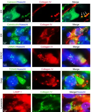
[image:6.585.101.492.65.372.2]Collagen IV is retained within the endoplasmic reticulum of ECs of embryos with induced EC-specific RASA1-deficiency. Newly syn-thesized collagen IV in the endoplasmic reticulum (ER) is pack-aged into coat protein II–coated (COPII-coated) vesicles that deliver collagen IV to the Golgi apparatus via the ER Golgi inter-mediate compartment (ERGIC). From the Golgi apparatus, colla-gen IV is further packaged into secretory vesicles for export to the extracellular space (21). Potentially, therefore, intracellular accu-mulation of collagen IV in RASA1-deficient ECs could be a result of retention in any of the ER, ERGIC, or the Golgi itself. It is also theoretically possible that intracellular collagen IV reflects not impaired secretion, but ingestion of collagen IV through an endo-cytic process. To examine this, skin sections from E18.5 Rasa1fl/fl Cdh5Ert2Cre embryos treated with TM at E13.5 were costained with antibodies against collagen IV and organelle-specific anti-bodies (Figure 4). We observed no colocalization of collagen IV with ERGIC, Golgi, or lysosomal markers. In contrast, both of 2 different ER markers colocalized with collagen IV. Calnexin, a transmembrane ER chaperone that is highly restricted to the ER, encircled discrete collagen IV punctae (22). In contrast, calretic-ulin, an ER lumenal chaperone, was coincident with the majority of collagen IV punctae (Figure 4). Thus, intracellular collagen IV accumulation in RASA1-deficient ECs is explained by impaired export of collagen IV from the ER.
Figure 3. EC-specific disruption of Rasa1 during developmental angiogenesis results in retention of collagen IV within BECs. TM was administered to
littermate Rasa1fl/fl and Rasa1fl/fl Cdh5Ert2Cre embryos at E13.5. Embryos were harvested at E18.5 and skin sections were stained with Hoechst and antibodies
A second mechanism through which accumulated intracellular collagen IV could induce EC apoptosis is through induction of ER stress, resulting in an unfolded protein response (UPR) (24, 25). The purpose of the UPR is to assist the cell with the folding of unfold-ed and misfoldunfold-ed proteins in the ER. However, in circumstances in which there remains an excess of unfolded protein, the UPR triggers apoptosis. In humans and mice, point-mutated collagen IV variants induce cell apoptosis via this mechanism (26–29). In addi-tion, in mice deficient in the TANGO1 protein that is involved in export of collagen IV from the ER and in mice that are deficient in the hsp47 chaperone that assists with collagen IV folding, accumu-lating WT collagen IV in the ER induces a UPR and BEC apoptosis (30, 31). The principal sensor of unfolded protein in the ER is BIP (also known as GRP78), whose expression is increased during the course of a UPR. Therefore, to determine whether a UPR is induced in BECs upon loss of RASA1 during developmental angiogenesis, we examined BiP expression. We found that the amounts of BiP were sharply increased in ECs of E18.5 Rasa1fl/fl Cdh5Ert2Cre embry-os treated with TM at E13.5 compared with ECs of E18.5 Rasa1fl/fl controls and ECs of E18.5 Rasa1fl/fl Cdh5Ert2Cre embryos not treated with TM (Figure 5 and Supplemental Figure 12). These findings are consistent with the induction of a UPR in ECs upon loss of RASA1.
The chemical chaperone 4-phenylbutyrate rescues blood vascular phenotypes in embryos with RASA1-deficiency. ER retention of collagen IV in RASA1-defcient BECs could be a direct con-sequence of impaired collagen IV folding in the ER or may instead be a result of altered expres-sion or function of proteins involved in COPII- mediated protein secretion (32–34). To address this, we tested whether a chemical chaperone, 4-phenylbutyrate (4PBA), could ameliorate vascular phenotypes that result from loss of RASA1. Previously, it was demonstrated that 4PBA rescued the blocked export of misfolded point–mutant collagen IV variants from human and mouse ECs in vitro and reversed intracere-bral hemorrhage in mouse models that express these mutants (27, 35). Pregnant Rasa1fl/fl mice carrying Rasa1fl/fl and Rasa1fl/fl UbErt2Cre embryos were administered TM with 4PBA at E13.5 followed by 4PBA every day thereafter until embryo harvesting at E18.5. Administra-tion of 4PBA in these experiments completely rescued EC export of collagen IV, EC apoptosis, and blood vascu-lar hemorrhage (Table 1 and Figure 6, compare with Figure 1). In contrast, 4PBA had no influence on vascular development when administered alone to embryos in the absence of TM (Supplemen-tal Figure 13). As determined by real-time qPCR of tail genomic DNA, 4PBA did not affect the ability of TM to disrupt the Rasa1 gene in Rasa1fl/fl UbErt2Cre embryos (Supplemental Figure 14). RASA1 siRNA–mediated knockdown of RASA1 in human umbilical vein endothelial cells (HUVECs) also resulted in intracellular accumu-lation of collagen IV, which could be rescued by 4PBA treatment (Figure 7). These findings provide strong evidence that blocked export of collagen IV from RASA1-deficient ECs is a consequence of impaired collagen IV folding in the ER rather than a defect in COPII-mediated secretion.
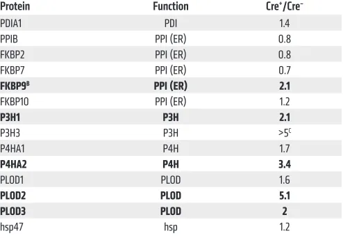
Loss of RASA1 during developmental angiogenesis results in an increased abundance of collagen IV–modifying enzymes in ECs. Het-erotrimerization of 2 collagen IV α-1 monomers and 1 alpha-2 monomer and folding to form the mature protomer in the ER (collagen α-1 and α-2 are the predominant forms of collagen IV in ECs) is a complex process that is regulated by different collagen IV–modifying enzymes and molecular chaperones that include protein disulfide isomerase A1 (PDIA1), peptidyl proline
isomer-Figure 4. Collagen IV is trapped within the ER of BECs
following disruption of Rasa1 during developmental
angiogenesis. Skin sections from E18.5 Rasa1fl/fl Cdh5Ert2Cre embryos administered TM at E13.5 were
[image:7.585.36.350.56.442.2]ases (PPIs), proline-4 and proline-3 hydroxylases (P4HA1–3 and P3H1–3, respectively), lysine hydroxylases (LH1–3) also known as procollagen lysine, 2-oxoglutarate 5-dioxygenase enzymes (PLOD1–3) and hsp47. Potentially, therefore, an increased or decreased abundance of collagen IV–modifying enzymes or
[image:8.585.85.506.62.214.2]chap-erones in RASA1-deficient BECs could affect collagen IV folding (36–40). To examine whether loss of RASA1 results in changes in the amounts of collagen IV–modifying enzymes or chaper-ones in embryonic BECs during developmental angiogenesis, we performed proteomic analyses. Pregnant Rasa1fl/fl mice carrying
Figure 5. Increased amounts of BIP in BECs following disruption of Rasa1 during developmental angiogenesis. Skin sections from E18.5 Rasa1fl/fl Cdh5Ert2Cre
embryos administered TM at E13.5 were stained with Hoechst and antibodies against CD31 and BIP. Note the increase in BIP in BECs from the Rasa1fl/fl Cdh5Ert2Cre embryos, indicative of an ongoing UPR (arrows). Scale bars: 20 μm.
Figure 6. Rescue of developmental angiogenesis defects with the chemical chaperone 4PBA in mice with induced RASA1 deficiency. TM was
admin-istered to littermate Rasa1fl/fl and Rasa1fl/fl UbErt2Cre embryos at E13.5. 4PBA was coadministered with TM and was also administered to embryos on
con-secutive days thereafter until embryo harvesting at E18.5. (A) Gross appearance of embryos. Note absence of hemorrhage and edema in Rasa1fl/fl UbErt2Cre
[image:8.585.66.525.359.670.2]notion that impaired export of collagen IV is not a result of defects in the COPII secretion mechanism.
Inhibitors of 2-oxoglutarate–dependent oxygenases rescue blood vascular phenotypes in embryos with induced RASA1-deficiency. P4HA2, P3H1, PLOD2, and PLOD3 all belong to the same family of enzymes known as 2-oxoglutarate–dependent (2OG-depen-dent) oxygenases, so called because of their dependency on 2OG for catalysis (41). Consequently, drugs are available that generical-ly inhibit all members of this famigenerical-ly. One such drug is the catechol ethyl-3,4-dihydroxybenzoic acid (EDHB), which has been used in vitro and in vivo to block the activity of collagen proline and lysine hydroxylases (41, 42). Therefore, to determine whether increased abundance of any or all of these enzymes is responsible for col-lagen IV accumulation in BECs during developmental angiogen-esis, we examined the ability of EDHB to rescue blocked collagen IV export and vascular phenotypes in induced RASA1-deficient embryos. Pregnant Rasa1fl/fl mice carrying Rasa1fl/fl and Rasa1fl/fl UbErt2Cre embryos were administered TM at E13.5 together with Rasa1fl/fl and Rasa1fl/fl UbErt2Cre embryos were given TM at E14.5,
[image:9.585.93.484.57.413.2]and the embryos were harvested at E18.5, i.e., prior to BEC apop-tosis and hemorrhage (Table 1). Subsequently, BECs from skin of individual embryos were purified, pooled according to geno-type, and lysed. Tryptic digests of lysates were then analyzed by liquid chromatography tandem mass spectrometry (LC-MS/MS). With this approach, we obtained data on the relative abundance of nearly 4000 BEC proteins. Of these, approximately 250 pro-teins were increased by at least 2-fold, and 200 were decreased by at least 2-fold in RASA1-deficient BECs compared with control BECs (Supplemental Table 1). Strikingly, several of the enzymes involved in collagen IV posttranslational modification were increased in abundance, including FKBP9, P3H1, P4HA2, LH2/ PLOD2, and LH3/PLOD3 (Table 2). In contrast, other ER-res-ident proteins implicated in collagen folding, including PDIA1 and hsp47, showed no or only modest changes in abundance. Fur-thermore, no significant changes in the abundance of any COPII secretory pathway proteins were apparent, consistent with the
Figure 7. RASA1 knockdown in HUVECs results in collagen IV accumulation in the ER that can be rescued by 4PBA. HUVECs were transfected with control
responsible for BV phenotypes upon loss of RASA1 during devel-opmental angiogenesis. To address this, Rasa1fl/fl mice carrying Rasa1fl/fl and Rasa1fl/fl UbErt2Cre embryos were administered TM at E13.5 together with a MAPK pathway inhibitor (AZD6244) (43, 44) or a PI3K inhibitor (PX-866) (45), either of which was also admin-istered to mice on each of the 2 days following the TM injection. As assessed at E18.5, the MAPK pathway inhibitor rescued the block-age of collblock-agen IV export from BECs and prevented development of the hemorrhaging in Rasa1fl/fl UbErt2Cre embryos that was observed after TM treatment alone (Table 1 and Figure 10, compare with Figure 1). AZD6244 did not affect TM-induced Rasa1 gene dele-tion efficiency in Rasa1fl/fl UbErt2Cre embryos (Supplemental Figure 14), and, by itself, AZD6244 did not induce any vascular abnormal-ities (Supplemental Figure 18). In contrast, the PI3K inhibitor was unable to rescue BEC export of collagen IV or apoptosis, and exten-sive cutaneous hemorrhage was evident at E18.5 (data not shown). These findings show that dysregulated Ras/MAPK signaling rather than dysregulated Ras/PI3K signaling drives BV phenotypes upon loss of RASA1 during developmental angiogenesis.
RASA1 is required for normal retinal angiogenesis in newborns. No spontaneous BV abnormalities have been noted in mice in which the Rasa1 gene is disrupted after E15.5 (16, 17). We hypothe-size that this is because the majority of the collagen IV in vascular BM is deposited during developmental angiogenesis. Collagen IV is recognized to be one of the most stable proteins in the animal kingdom (38). Thus, in postnatal life, a continued high rate of collagen IV synthesis would be unnecessary for BECs to remain attached to BM. Nonetheless, in situations where de novo depo-sition of BM is required, abnormalities of BV function might be expected in RASA1-deficient mice. Two such situations are reti-nal angiogenesis in newborns and pathological angiogenesis in adult mice. To examine retinal angiogenesis, we administered TM to littermate Rasa1fl/fl UbErt2Cre and Rasa1fl/R780Q UbErt2Cre mice and Cre-negative controls at P1 and examined the retinal vascula-ture at P4. The extent of new vessel growth in TM-treated Rasa1fl/fl UbErt2Cre and Rasa1fl/R780Q UbErt2Cre mice was significantly less than in Rasa1fl/fl controls as assessed by the number of vessel branch points and the percentage of coverage of the retina with BECs (Figure 11, A and D). Furthermore, the number of BEC filopodia, which are a feature of sprouting angiogenesis, at the periphery of the vascular coverage area was reduced in the Rasa1fl/fl UbErt2Cre and Rasa1fl/R780Q UbErt2Cre mice (Figure 11, B and D). Intracellular accumulation of collagen IV could be detected in retinal BECs of Rasa1fl/fl UbErt2Cre and Rasa1fl/R780Q UbErt2Cre mice but not Rasa1fl/fl control mice at P4 (Figure 11C). In addition, in Rasa1fl/fl UbErt2Cre and Rasa1fl/R780Q EDHB, which was additionally administered to mice every day
thereafter until embryos were harvested, at E18.5. Administra-tion of EDHB in these experiments completely rescued EC export of collagen IV, EC apoptosis, and blood vascular hemorrhage (Table 1 and Figure 8, compare with Figure 1). In contrast, when administered to embryos in the absence of TM, EDHB did not affect vascular development (Supplemental Figure 15). As with 4PBA, EDHB did not affect Rasa1 gene disruption induced by TM in Rasa1fl/fl UbErt2Cre embryos (Supplemental Figure 14). The same results were obtained using another generic 2OG-dependent oxy-genase inhibitor, 2,4 pyridinedicarboxylic acid (2,4PDCA) (Table 1, Supplemental Figure 14 and Supplemental Figure 16). These findings are consistent with observations from a model in which loss of RASA1 in BECs during developmental angiogenesis results in increased amounts of collagen IV–modifying, 2OG-dependent oxygenases in BECs that account for collagen IV retention in the ER and downstream vascular phenotypes.
Dysregulated Ras/MAPK signaling is responsible for the develop-ment of BV phenotypes following RASA1 loss during developdevelop-mental angiogenesis. RASA1 may participate in certain signaling path-ways independently of its ability to regulate Ras (10). Therefore, to address whether vascular phenotypes that result from induced loss of RASA1 during developmental angiogenesis result from dysregulated Ras signaling or to perturbation of a distinct signal-ing pathway, we examined Rasa1fl/R780Q UbErt2Cre embryos that we have described previously (15, 17). Administration of TM to these embryos resulted in the expression of Rasa1R780Q alone, which encodes a catalytically inactive form of RASA1, in which all puta-tive alternaputa-tive functions of RASA1 are predicted to remain intact. Pregnant Rasa1fl/fl mice carrying Rasa1fl/fl and Rasa1fl/R780Q embryos with and without UbErt2Cre were administered TM at E12.5, and the embryos were harvested at E18.5. Like the Rasa1fl/fl UbErt2Cre embryos (Figure 1), Rasa1fl/R780Q UbErt2Cre embryos showed extensive
P4HA1 P4H 1.7
P4HA2 P4H 3.4
PLOD1 PLOD 1.6
PLOD2 PLOD 5.1
PLOD3 PLOD 2
hsp47 hsp 1.2
AAbundance of all detectable known collagen IV–modifying enzymes
and chaperones are shown; Bproteins with 2-fold or greater changes in
abundance are indicated with bold; CP3H3 was undetectable in control
[image:10.585.42.289.97.268.2]tumors from control mice, indicating that reduced tumor growth in the former was a result of impaired BV tumor angiogenesis (Supplemental Figure 21C). Further analysis of BVs in tumors from Rasa1fl/fl UbErt2Cre mice revealed intracellular accumulation of collagen IV in BECs and BEC apoptosis (Supplemental Figure 21C and Supplemental Figure 22). Thus, blocked export of collagen IV from BECs and BEC apoptosis likely account for an impaired pathological angiogenesis response in the absence of RASA1.
To examine this further, we switched to a B16 melanoma model (47). B16 cells grow more rapidly than do ID8 cells in vivo, thus per-mitting a more ready analysis of the effect of drugs such as 4PBA that promote collagen IV folding. B16 cells were injected into the flanks of littermate TM-treated Rasa1fl/fl and Rasa1fl/fl UbErt2Cre mice. Some mice were also injected with 4BPA at the same time that tumor cells were injected, and additional 4PBA was administered to mice every day thereafter. After 13 days, we assessed tumor growth and angiogenesis (Figure 12). As with ID8 tumor growth, we found that B16 growth was inhibited in TM-treated Rasa1fl/fl UbErt2Cre mice compared with Rasa1fl/fl controls (Figure 12, A and B). In addition, the reduced growth of B16 tumors was also associated with impaired intratumoral BV angiogenesis and collagen IV accu-mulation in BECs (Figure 12, C–E). 4PBA restored the growth of UbErt2Cre retinas, the number of “empty sleeves” that comprised a
thin tube of collagen IV with no BECs was substantially increased compared with controls (Figure 11, C and D). This latter obser-vation is consistent with impaired deposition of collagen IV into BM and BEC death during retinal angiogenesis in the absence of catalytically active RASA1. We also observed decreased angiogen-esis in retinas of Rasa1fl/fl Cdh5Ert2Cre mice administered TM at P1 and analyzed at P6 (Supplemental Figure 19), confirming an EC- intrinsic role for RASA1 in retinal angiogenesis. In Rasa1fl/fl UbErt2Cre mice administered TM at P3, we observed areas of hemorrhage in retinas at P10 (Supplemental Figure 20).
[image:11.585.65.507.54.383.2]Rasa1 is required for pathological angiogenesis in adult mice. To examine pathological angiogenesis, we initially used an ID8 ovarian tumor model (46). Adult littermate female Rasa1fl/fl and Rasa1fl/fl UbErt2Cre mice were administered TM and 1 week later were injected in the flanks with ID8 tumor cells. Growth of injected ID8 tumor cells in female recipients is strictly dependent on host BV angiogenesis (46). Six weeks after injection, we found that the ID8 tumors were substantially smaller in Rasa1fl/fl UbErt2Cre mice compared with those in controls (Supplemental Figure 21, A and B). Upon histological analysis, the density of BVs in tumors from Rasa1fl/fl UbErt2Cre mice was found to be substantially less than in
Figure 8. Rescue of developmental angiogenesis defects in mice with induced RASA1 deficiency using the 2OG-dependent oxygenase inhibitor EDHB.
TM was administered to littermate Rasa1fl/fl and Rasa1fl/fl UbErt2Cre embryos at E13.5. EDHB was coadministered with TM and was also administered to
embryos on consecutive days thereafter until embryo harvesting at E18.5. (A) Gross appearance of embryos. Note the absence of hemorrhage and edema in the Rasa1fl/fl UbErt2Cre embryos, which was confirmed by H&E staining of skin sections. Scale bars: 200 μm. (B) Skin sections were stained with Hoechst
impaired protein folding. Further mechanistic studies indicated that loss of RASA1 within ECs leads to dysregulated Ras/MAPK signaling, which results in an increased abundance of several ER-resident enzymes that carry out posttranslational modifica-tions of collagen IV that are known to regulate folding and ER export of this protein (36, 37). Most notable among these are P3H, P4H, and PLOD enzymes, of which there are 3 different isoforms each in mammals. Of the 7 isoforms of a total of 9 that could be detected in BECs by LC-MS/MS, all were increased in abundance in RASA1-deficient BECs. Increased abundance of these enzymes could lead to excessive posttranslational modification of colla-gen IV that would explain impaired folding and ER retention of this protein (36–40). Consistent with this, generic inhibitors of this class of enzymes, EDHB and 2,4PDCA, rescued ER collagen IV retention, EC apoptosis, and hemorrhage in induced RASA1- deficient mice. This finding demonstrates that, although the abundance of numerous other proteins was also altered by more than 2-fold in RASA1-deficient BECs (Supplemental Table 1), an increased abundance of these collagen IV–modifying enzymes specifically is responsible for the development of vascular pheno-types in the absence of RASA1.
B16 cells in TM-treated Rasa1fl/fl UbErt2Cre recipients, and this was associated with normal export of collagen IV from BECs and BV angiogenesis (Figure 12). In contrast, 4PBA had no influence on B16 cell growth in TM-treated Rasa1fl/fl mice (Figure 12) or when administered alone to Rasa1fl/fl and Rasa1fl/fl UbErt2Cre mice that had not previously been injected with TM (Supplemental Figure 23). These findings are consistent with the notion that impaired patho-logical angiogenesis and tumor growth in RASA1-deficient adult mice is also a consequence of an inability of BECs to export colla-gen IV for deposition in newly forming BMs.
Discussion
[image:12.585.44.538.57.383.2]In this study we show that RASA1 has what we believe to be a pre-viously unappreciated critical function in the export of collagen IV from ECs during developmental angiogenesis. In the absence of RASA1, collagen IV is retained in the ER of ECs, leading to their apoptotic death as a result of ER stress and anoikis. The chemical chaperone 4PBA rescued ER retention of collagen IV, EC apop-tosis, and BV hemorrhage in induced RASA1-deficient embryos. This finding strongly supports the notion that retention of col-lagen IV in the ER in the absence of RASA1 is a consequence of
Figure 9. BV abnormalities in embryos induced to express RASA1 R780Q alone during developmental angiogenesis. TM was administered to
litter-mate Rasa1fl/R780Q and Rasa1fl/R780Q UbErt2Cre embryos at E12.5 and embryos were harvested at E18.5. (A) Gross appearance of embryos. Note the cutaneous
hemorrhage in Rasa1fl/R780Q UbErt2Cre embryos, which was confirmed by H&E staining of skin sections. Scale bars: 200 μm. (B) Skin sections were stained with
Hoechst and antibodies against collagen IV, CD31, and activated caspase 3. Note the discontinuous distribution of collagen IV in BV BM, the accumulation of collagen IV in BECs, and the presence of activated caspase 3 in BEC nuclei (arrows) of Rasa1fl/R780Q UbErt2Cre embryos. Scale bars: 20 μm. (C) Quantitation
of BEC apoptosis in skin BVs of Rasa1fl/R780Q and Rasa1fl/R780 UbErt2Cre embryos administered TM at E12.5 and harvested at E18.5. Data represent the mean ± 1
all 3 classes of collagen-modifying enzymes or an increase in the abundance of select enzymes remains to be determined. The col-lagenous domains of collagen IV contain multiple repeats of the sequence of Gly-Xaa-Yaa (where Xaa and Yaa are any amino acid). P4Hs hydroxylate prolines in the Yaa position, whereas P3Hs sub-sequently hydroxylate prolines in the Xaa position of Gly-Xaa-4-hydroxyPro. PLODs hydroxylate lysine residues in the Yaa position of Gly-Xaa-Yaa, and PLOD3 additionally catalyzes glycosylation of hydroxylysine to form galactosylhydroxylysine or galactosylgluco-sylhydroxylysine. Proline 3 hydroxylation is known to destabilize the collagen triple helix, and, thus, excessive proline 3 hydrox-ylation of collagen IV is probably an important contributor to impaired folding (38–40). In contrast, proline 4 hydroxylation pro-motes electrostatic interactions between collagen IV monomers, and lysine hydroxylation and glycosylation are required for colla-gen IV secretion (36–38). Nonetheless, an increased abundance of P4Hs and PLODs could lead to excessive modification that could also have a negative impact on folding of collagen protomers.
Vasculogenesis commonly results in the formation of a vas-cular plexus that comprises a primitive vasvas-cular network with presumptive arterial inputs and venous outputs. Subsequently, Dysregulated Ras/MAPK signaling could result in an
increased abundance of collagen IV–modifying enzymes as a result of increased gene transcription. MAPK signaling modu-lates the activity of several different transcription factor complex-es including activator protein 1 (AP1) complexcomplex-es and ternary com-plex factors (TCFs) with the potential to modify the transcription of collagen IV–modifying genes (48). Alternatively, dysregulated MAPK signaling could affect the abundance of these enzymes at a posttranslational level, for example, by phosphorylation of sub-strates that affect protein stability. To distinguish between these possibilities, we examined mRNA levels of 2 select enzymes, Plod2 and P3h1, in sorted skin BECs from TM-treated Rasa1fl/fl and Rasa1fl/fl UbErt2Cre embryos (Supplemental Figure 24). We found that the levels of Plod2 mRNA were significantly increased in Rasa1fl/fl UbErt2Cre BECs, which was consistent with increased Plod2 transcription. In contrast, P3h1 mRNA levels were not sig-nificantly increased in Rasa1fl/fl UbErt2Cre BECs, suggesting that loss of RASA1 in BECs results in an increased abundance of P3H1 through a posttranslational mechanism.
[image:13.585.97.479.56.388.2]Whether impaired folding of collagen IV in RASA1-defi-cient ECs is a result of a collective increase in the abundance of
Figure 10. An inhibitor of MAPK signaling blocks the development of BV abnormalities resulting from induced loss of RASA1 during developmental
angiogenesis. TM was administered to littermate Rasa1fl/fl and Rasa1fl/fl UbErt2Cre embryos at E13.5. The MAPK pathway inhibitor AZD6244 was
coadmin-istered with TM and was also admincoadmin-istered to embryos on the following 2 days afterward. Embryos were harvested at E18.5. (A) Gross appearance of embryos. Note the absence of hemorrhage and edema in the Rasa1fl/fl UbErt2Cre embryos, which was confirmed by H&E staining of skin sections. Scale bars:
vascular plexuses (49). Thus, in EPHB4-deficient embryos and in embryos of mice deficient in the EPHB4 ligand ephrin B2, arteries form near-direct connections with veins through large-diameter vessels, as observed in CM-AVM (50, 51). Similarly, in RASA1- deficient embryos, the same defect in angiogenic remodeling of vascular plexuses has also been noted (14). In light of these obser-vations, findings in this study are directly relevant to an under-angiogenic processes that include fusion, intussusception,
[image:14.585.58.542.54.529.2]regres-sion, and sprouting angiogenesis remodel the plexus to yield a hierarchical network of arteries, arterioles, venules, and veins connected by smaller-diameter capillaries (18). Previous studies of global EPHB4-deficient and RASA1-deficient mice have pro-vided evidence that AVMs and AFs in CM-AVM arise as a con-sequence of impaired angiogenic remodeling of these primitive
Figure 11. Impaired retinal angiogenesis in neonatal mice with induced RASA1 R780Q and mice with induced RASA1 deficiency. TM was administered
to littermate Rasa1fl/fl, Rasa1fl/R780QUbErt2Cre, and Rasa1fl/fl UbErt2Cre mice at P1, and retinas were harvested at P4. (A–C) Retinas were stained with isolectin
B4 (IB4) to identify BVs and anti–collagen IV (C). (A and B) Representative low-power (A) and high-power (B) images of IB4 staining are shown. Asterisks indicate filopodia at the vascular front. (C) High-power images (left) show collagen IV accumulation in BECs from Rasa1fl/R780QUbErt2Cre and Rasa1fl/fl UbErt2Cre
retinas (arrows), and lower-power images (right) illustrate empty collagen IV sleeves in Rasa1fl/R780Q UbErt2Cre and Rasa1fl/fl UbErt2Cre retinas (arrows). (D) Graphs
show the mean ± 1 SEM of the number of branch points from veins (n = 6 retinas/genotype), the percentage of coverage of retinas with BECs per field (n = 5–7 retinas/genotype), the number of filopodia per vascular field (n = 7–10 retinas/genotype), and the number of empty collagen sleeves per field (n = 5–8 retinas/genotype). **P < 0.01, ***P < 0.001, and ****P < 0.0001, by 1-way ANOVA test with Dunnett’s multiple comparisons post-hoc test. Scale bars: 100
loss of RASA1 in mice at any point after birth is consistent with a model in which a continued high rate of collagen IV synthe-sis by BECs is not necessary for BV function. Exceptions to this would include retinal angiogenesis in newborn mice and patho-logical angiogenesis in adult mice, in which EC synthesis of col-lagen IV and deposition in BMs would be necessary. Accordingly, we show here that RASA1 is required for both processes, and, at least for pathological angiogenesis, our evidence indicates that impaired angiogenesis is, again, consequent to an inability of ECs to export collagen IV. Earlier studies of pathological angio-genesis and retinal angioangio-genesis in mice indicated that miR-132– mediated downregulation of Rasa1 mRNA in ECs is required in order for Ras activation and angiogenesis to proceed (54, 55). The current studies do not contradict this notion but instead explore the consequences of complete and permanent loss of RASA1 in ECs. miR-132–mediated downregulation of Rasa1 during normal angiogenesis was probably not complete and would be expected to be transient. In contrast, genetic disruption of Rasa1 would result in chronic uncontrolled activation of Ras in ECs, with distinct downstream consequences. In this regard, it is of note that mice that express constitutively active H-Ras (resistant to RasGAP-mediated inactivation) in ECs develop brain vascular malformations and hemorrhagic stroke (56). Moreover, somatic activating mutations in K-Ras have been identified in the major-ity of brain AVMs in humans (57). To our knowledge, the effect in mice of induced loss of RASA1 upon retinal angiogenesis and pathological angiogenesis caused by actively growing tumors had not previously been examined. The current studies, therefore, are the first to our knowledge to address this question.
Notably, RASA1 was also required for the maintenance of LEC numbers in LV valves in adult mice. We propose that the loss of LECs in adult mouse valves upon RASA1 loss reflects a requirement for valve leaflet LECs to engage in a high rate of collagen IV syn-thesis in order to remain attached to leaflets that encounter higher shear stress forces than LV or BV wall ECs (52, 53). Indeed, shear stress has previously been shown to induce collagen IV expression in ECs (58). Whether RASA1 is also required for the maintenance of BECs in venous valves that would encounter similarly high sheer stress forces remains to be determined.
Methods
For a full description of the Methods, see Supplemental Methods. Mice. Rasa1fl/fl and Rasa1fl/R780Q mice with and without UbErt2Cre
transgenes have been described previously (15–17, 59). Cdh5Ert2Cre mice
were obtained from Cancer Research UK. Rasa1fl/fl Cdh5Ert2Cre mice
standing of the pathogenesis of CM-AVM. Acquisition of somatic second-hit mutations in RASA1 in ECs or their precursors at the time of or prior to vasculogenesis, respectively, could potentially result in the loss of RASA1 in the majority of ECs in an individual primitive vessel within a vascular plexus. Consequently, sprouting angiogenesis and potentially other forms of angiogenesis from that vessel would be blocked, as these events would require de novo synthesis of collagen IV by ECs in these vessels and deposition of collagen IV in newly forming BM. Thus, an inability of ECs in these vessels to export collagen IV during angiogenic remodeling could account for the development of AVMs and AFs in CM-AVM. Alternatively, acquisition of second-hit mutations in RASA1 in ECs later in development, after remodeling of vascular plexuses, could result in CM, again as a consequence of blocked export of collagen IV. In either scenario, the ability of chemical chaperones and inhibitors of 2OG-dependent oxygenases to rescue impaired developmental angiogenesis in the absence of RASA1 suggests possible means of prevention of vascular lesions in human embry-os and patients with inherited RASA1 mutations. Whether such drugs would be effective in the treatment of existing vascular lesions in CM-AVM is far less certain. ECs of CM-AVM lesions would not be expected to be engaged in a high rate synthesis of collagen IV, and, in the absence of angiogenic stimuli, intracellu-lar accumulation of collagen IV in ECs of lesions would not be pre-dicted, nor would it be expected to contribute to lesion pathology. To confirm this, we examined collagen IV accumulation in CM and AVM lesions of multiple patients with CM-AVM1, including 2 patients in whom somatic inactivating second-hit RASA1 muta-tions had been identified in lesional tissue. Intracellular collagen IV accumulation was not apparent in ECs of theses lesions, again, as predicted (Supplemental Figure 25).
LV abnormalities have also been reported in CM-AVM and may also be explained by impaired angiogenic remodeling of primitive LV plexuses as a consequence of LEC retention of colla-gen IV (2–7). In addition, RASA1 is required for the development of LV valves (17). Therefore, second-hit mutations of RASA1, if they were to affect a sufficient number of valve-forming LECs at a site of valvulogenesis, could affect leaflet development in that vessel, against collagen IV and CD31. Representative images are shown. Note the accumulation of collagen IV in tumor BECs from Rasa1fl/fl UbErt2Cre mice
JT and PN provided CM-AVM1 tissue samples. PN assisted with interpretation of the findings. The manuscript was written by PDK.
Acknowledgments
This work was supported by NIH grant HL120888 (to PDK). Project consultation and mass spectrometric data analysis was performed at the Proteomics and Peptide Synthesis Core of the University of Michigan by Henriette A. Remmer. The experimental processing was contracted with MS Bioworks LLC.
Address correspondence to: Philip D. King, Department of Microbiology and Immunology, University of Michigan Medical School, 6606 Med Sci II, 1150 West Medical Center Drive, Ann Arbor, Michigan 48109-5620, USA. Phone: 734.615.9073; Email: [email protected].
were generated through cross-breeding. All mice were on a mixed 129S6/SvEv and C57BL/6 genetic background.
Statistics. Statistical analysis was performed using Student’s 1-sample and 2-sample t tests or 1-way ANOVA with Dunnett’s multi-ple comparisons post hoc test. A P value of less than 0.05 was consid-ered statistically significant.
Study approval. All experiments performed with mice were in com-pliance with University of Michigan guidelines and were approved by the IACUC of the University of Michigan. All work performed with CM-AVM1 tissue samples was approved by the IRBs at the University of Michigan, Stanford University, and the Medical College of Wisconsin.
Author contributions
DC, PEL, and PDK contributed to the design of the studies. DC performed the majority of experiments, with assistance from PEL.
1. Eerola I, et al. Capillary malformation-arteriove-nous malformation, a new clinical and genetic disorder caused by RASA1 mutations. Am J Hum
Genet. 2003;73(6):1240–1249.
2. Revencu N, et al. RASA1 mutations and associ-ated phenotypes in 68 families with capillary malformation-arteriovenous malformation. Hum
Mutat. 2013;34(12):1632–1641.
3. Revencu N, et al. Parkes Weber syndrome, vein of Galen aneurysmal malformation, and other fast-flow vascular anomalies are caused by RASA1 mutations. Hum Mutat. 2008;29(7):959–965. 4. Burrows PE, et al. Lymphatic abnormalities
are associated with RASA1 gene mutations in mouse and man. Proc Natl Acad Sci U S A. 2013;110(21):8621–8626.
5. de Wijn RS, Oduber CE, Breugem CC, Alders M, Hennekam RC, van der Horst CM. Phenotypic variability in a family with capillary malforma-tions caused by a mutation in the RASA1 gene.
Eur J Med Genet. 2012;55(3):191–195.
6. Macmurdo CF, et al. RASA1 somatic mutation and variable expressivity in capillary malformation/ arteriovenous malformation (CM/AVM) syn-drome. Am J Med Genet A. 2016;170(6):1450–1454. 7. Sevick-Muraca EM, King PD. Lymphatic vessel
abnormalities arising from disorders of Ras signal transduction. Trends Cardiovasc Med. 2014;24(3):121–127.
8. Buday L, Downward J. Many faces of Ras activa-tion. Biochim Biophys Acta. 2008;1786(2):178–187. 9. Wennerberg K, Rossman KL, Der CJ. The Ras
superfamily at a glance. J Cell Sci. 2005;118(Pt 5):843–846.
10. King PD, Lubeck BA, Lapinski PE. Nonredundant functions for Ras GTPase-activating proteins in tissue homeostasis. Sci Signal. 2013;6(264):re1. 11. Lapinski PE, Doosti A, Salato V, North P, Burrows
PE, King PD. Somatic second hit mutation of RASA1 in vascular endothelial cells in capillary malformation-arteriovenous malformation. Eur J
Med Genet. 2018;61(1):11–16.
12. Amyere M, et al. Germline loss-of-function muta-tions in EPHB4 cause a second form of capillary malformation-arteriovenous malformation (CM-AVM2) deregulating RAS-MAPK signaling.
Circulation. 2017;136(11):1037–1048.
13. Kawasaki J, et al. RASA1 functions in EPHB4
sig-naling pathway to suppress endothelial mTORC1 activity. J Clin Invest. 2014;124(6):2774–2784. 14. Henkemeyer M, et al. Vascular system
defects and neuronal apoptosis in mice lack-ing ras GTPase-activatlack-ing protein. Nature. 1995;377(6551):695–701.
15. Lubeck BA, et al. Blood vascular abnormalities in Rasa1(R780Q) knockin mice: implications for the pathogenesis of capillary malforma-tion-arteriovenous malformation. Am J Pathol. 2014;184(12):3163–3169.
16. Lapinski PE, et al. RASA1 maintains the lymphat-ic vasculature in a quiescent functional state in mice. J Clin Invest. 2012;122(2):733–747. 17. Lapinski PE, et al. RASA1 regulates the function
of lymphatic vessel valves in mice. J Clin Invest. 2017;127(7):2569–2585.
18. Udan RS, Culver JC, Dickinson ME. Understand-ing vascular development. Wiley Interdiscip Rev
Dev Biol. 2013;2(3):327–346.
19. Wang Y, et al. Ephrin-B2 controls VEGF-induced angiogenesis and lymphangiogenesis. Nature. 2010;465(7297):483–486.
20. Glentis A, Gurchenkov V, Matic Vignjevic D. Assembly, heterogeneity, and breaching of the basement membranes. Cell Adh Migr. 2014;8(3):236–245.
21. Malhotra V, Erlmann P. The pathway of collagen secretion. Annu Rev Cell Dev Biol. 2015;31:109–124. 22. Butler J, Watson HR, Lee AG, Schuppe HJ, East
JM. Retrieval from the ER-golgi intermediate compartment is key to the targeting of c-termi-nally anchored ER-resident proteins. J Cell
Bio-chem. 2011;112(12):3543–3548.
23. Michel JB. Anoikis in the cardiovascular system: known and unknown extracellular mediators.
Arte-rioscler Thromb Vasc Biol. 2003;23(12):2146–2154.
24. Kim I, Xu W, Reed JC. Cell death and endoplas-mic reticulum stress: disease relevance and therapeutic opportunities. Nat Rev Drug Discov. 2008;7(12):1013–1030.
25. Oslowski CM, Urano F. The binary switch between life and death of endoplasmic reticu-lum-stressed beta cells. Curr Opin Endocrinol
Diabetes Obes. 2010;17(2):107–112.
26. Guiraud S, et al. HANAC Col4a1 Mutation in mice leads to skeletal muscle alterations due to a primary vascular defect. Am J Pathol.
2017;187(3):505–516.
27. Jeanne M, Jorgensen J, Gould DB. Molecular and genetic analyses of collagen type IV mutant mouse models of spontaneous intracerebral hemorrhage identify mechanisms for stroke pre-vention. Circulation. 2015;131(18):1555–1565. 28. Jeanne M, et al. COL4A2 mutations impair
COL4A1 and COL4A2 secretion and cause hemor-rhagic stroke. Am J Hum Genet. 2012;90(1):91–101. 29. Weng YC, et al. COL4A1 mutations in patients
with sporadic late-onset intracerebral hemor-rhage. Ann Neurol. 2012;71(4):470–477. 30. Marutani T, Yamamoto A, Nagai N, Kubota H,
Nagata K. Accumulation of type IV collagen in dilated ER leads to apoptosis in Hsp47-knockout mouse embryos via induction of CHOP. J Cell Sci. 2004;117(Pt 24):5913–5922.
31. Wilson DG, et al. Global defects in collagen secretion in a Mia3/TANGO1 knockout mouse.
J Cell Biol. 2011;193(5):935–951.
32. Saito K, Katada T. Mechanisms for exporting large-sized cargoes from the endoplasmic reticu-lum. Cell Mol Life Sci. 2015;72(19):3709–3720. 33. Unlu G, Levic DS, Melville DB, Knapik EW.
Trafficking mechanisms of extracellular matrix macromolecules: insights from vertebrate devel-opment and human diseases. Int J Biochem Cell
Biol. 2014;47:57–67.
34. Melville DB, et al. The feelgood mutation in zebra-fish dysregulates COPII-dependent secretion of select extracellular matrix proteins in skeletal mor-phogenesis. Dis Model Mech. 2011;4(6):763–776. 35. Kuo DS, et al. Allelic heterogeneity contributes
to variability in ocular dysgenesis, myopathy and brain malformations caused by Col4a1 and Col4a2 mutations. Hum Mol Genet. 2014;23(7):1709–1722. 36. Chioran A, Duncan S, Catalano A, Brown
TJ, Ringuette MJ. Collagen IV trafficking: the inside-out and beyond story. Dev Biol. 2017;431(2):124–133.
37. Ishikawa Y, Bächinger HP. A molecular ensemble in the rER for procollagen maturation. Biochim
Biophys Acta. 2013;1833(11):2479–2491.
38. Shoulders MD, Raines RT. Collagen structure and stability. Annu Rev Biochem. 2009;78:929–958. 39. Mizuno K, Hayashi T, Peyton DH, Bachinger HP.
2008;14(12):1351–1356.
44. Pratilas CA, Solit DB. Targeting the mitogen- activated protein kinase pathway: physiological feedback and drug response. Clin Cancer Res. 2010;16(13):3329–3334.
45. Courtney KD, Corcoran RB, Engelman JA. The PI3K pathway as drug target in human cancer.
J Clin Oncol. 2010;28(6):1075–1083.
46. Su F, et al. Apolipoprotein A-I (apoA-I) and apoA-I mimetic peptides inhibit tumor development in a mouse model of ovarian cancer. Proc Natl Acad
Genes Dev. 1999;13(3):295–306.
51. Gerety SS, Wang HU, Chen ZF, Anderson DJ. Symmetrical mutant phenotypes of the receptor EphB4 and its specific transmembrane ligand ephrin-B2 in cardiovascular development. Mol
Cell. 1999;4(3):403–414.
52. Bazigou E, Makinen T. Flow control in our ves-sels: vascular valves make sure there is no way back. Cell Mol Life Sci. 2013;70(6):1055–1066. 53. Bazigou E, Wilson JT, Moore JE. Primary and
sec-ondary lymphatic valve development: molecular,
mutations in arteriovenous malformations of the brain. N Engl J Med. 2018;378(3):250–261. 58. Yamane T, et al. Laminar high shear stress
up-regulates type IV collagen synthesis and down-regulates MMP-2 secretion in endothe-lium. A quantitative analysis. Cell Tissue Res. 2010;340(3):471–479.