Genetic diversity of the
Cucumber green mottle mosaic virus
and the development of RT-LAMP assay for its detection
Beata Hasiów-Jaroszewska*, Daria Budzyńska, Natasza Borodynko-Filas
Department of Virologyand Bacteriology, Institute of Plant Protection-NationalResearch Institute, Poznań, Poland
*Corresponding author: [email protected]
Citation: Hasiów-Jaroszewska B., Budzyńska D., Borodynko-Filas N. (2019): Genetic diversity of the Cucumber green mottle mosaic virus and the development of RT-LAMP assay for its detection. Plant Protect. Sci., 55: 1–7.
Abstract: To analyse the genetic diversity of the Cucumber green mottle mosaic virus (CGMMV) population in Po-land and to establish the phylogenetic relationships between the Polish and other isolates described to date, 91 iso-lates were collected from cucumber plants. The analysis, based on coat protein (CP) gene, revealed the presence of two phylogenetic groups: one consisting of the Polish isolates collected in 2017 and those originated mainly from Asia region and the second including the Polish isolates collected in 2016 and the others from European countries. The sensitive, specific, and rapid one-step loop-mediated isothermal amplification assay was developed for the early detection of genetically diverse CGMMV isolates in seeds and plant material.
Keywords: CGMMV; cucumber; genetic variability; identification; LAMP method
Cucumber green mottle mosaic virus (CGMMV) belongs to thegenus Tobamovirus within the family
Tombusviridae. The virus was first identified in the United Kingdom in 1935 (Ainsworth 1935) and since then it has been reported in Europe, Asia, North America, and Australia (Antignus et al. 1990; Kim
et al. 2003; Li et al. 2015; Tesoriero et al. 2016). CGMMV causes severe yield and quality losses in
Cucurbitaceae crops. Recently, several outbreaks of CGMMV have been observed in greenhouse cu-cumber in Poland (Borodynko-Filas et al. 2017). The infected plants were often stunted and displayed symptoms of green mottle and mosaic symptoms. The fruits were reduced in size and number, and did not have the market value. CGMMV is one of the economically important seed-transmitted viruses. The spread of CGMMV over long distances and its introduction into new areas is correlated with the increased exchange of cucurbit crops. Therefore, the effective, sensitive, specific, and rapid
detec-tion methods are required to identify the virus and prevent its further spread within the crop.
The virus genome consists of positive-sense single-stranded RNA of approximately 6.4 kb. It contains four open reading frames (ORFs 1-4), encoding two viral replicases, movement and coat protein. Sev-eral sequence-based detection methods, such as reverse transcription polymerase chain reaction (RT-PCR) or real-time RT-PCR methods have been applied for the detection of CGMMV (Hongyun
et al. 2008; Tian et al. 2014); however, they require expensive equipment and are time-consuming. Re-cently, reverse transcription loop-mediated iso-thermal amplification assay has been developed for CGMMV detection (Li et al. 2013). However, we were not able to detect all the Polish isolates us-ing the proposed primer pairs probably due to the genetic diversity in the virus population. RT-LAMP has several advantages over the conventional RT-PCR assay; it amplifies a target sequence with high
ficity under isothermal conditions (63–65°C), and the reaction can be carried out with a simple heater. Moreover, a large amount of DNA (10–30 µg/25 µl) can be synthesised in a short time (15–60 min) while maintaining high specificity (Mori et al. 2006). The results of amplification can be observed directly under UV light by adding fluorescent dyes intercalating to DNA, eliminating the need for gel electrophoresis and greatly reducing the time required for result analysis. Altogether, it makes the LAMP technique a rapid, reliable, and cost-effective method for patho-gen detection.
In the present study, we analysed the genetic vari-ability of the CGMMV population from cucum-ber in Poland and developed a rapid and sensitive RT-LAMP assay for the detection of a wide range of CGMMV isolates.
MATERIAL AND METHODS
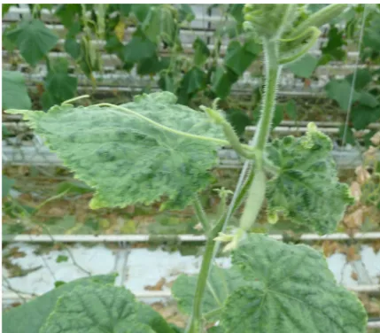
Virus source and RNA isolation. During 2016–2017, 105 samples of cucumber (Cucumis sativus) plants of the following varieties: Medora, Mewa, Me-len, Mirabelle, and Kybria were collected from greenhouses located in the Kujawsko-Pomorskie, Wielkopolskie and Mazowieckie regions. Symptoms of growth reduction, mottled areas of light and dark green and mosaics were observed on the leaves and fruits (Figure 1). No significant differences in symp-toms induced on plants were noticed between the CGMMV isolates collected in 2016 and 2017 in Po-land. The total RNA was extracted using an RNeasy
Plant Mini Kit (Qiagen, Hilde, Germany) accord-ing to the manufacturer’s instructions. The RNA concentration and quality were measured using an ND-1000 Spectrophotometer (Thermo Fisher Sci-entific, Waltham, USA) and adjusted to 500 ng/µl.
RT-PCR reaction. In order to amplify the region encompassing the CP gene, CGMMV-F5370 and CGMMV-R6390 primers were used (Tian et al.
2014). RT-PCR was performed using a Transcriptor One-Step RT-PCR Kit (Roche, Basel, Switzerland) with each 50 µl reaction mixture containing: 1 µl of RNA, 10 µl of 5X reaction buffer, 0.4 µM of each primer, 1 µl Transcriptor Enzyme Mix, and 34 µl of water. Reverse transcription was performed at 50°C during 30 minutes. Initial denaturation was performed at 94°C for 10 s, followed by 35 cycles of denaturation at 94°C for 10 s, annealing at 50°C for 30 s, and extension at 68°C for 1 minute. A final extension at 68°C for 5 min was also performed. The RT-PCR products were separated on 1% agarose gel stained with Midori Green (NIPPON Genetics, Düren, Germany). The RT-PCR products obtained of ca. 1 000 bp were ligated into a TOPO® TA cloning vec-tor (Thermo Fisher Scientific, USA) and transformed into One Shot TOP10 Escherichia coli (Thermo Fisher Scientific, USA), according to the manufacturer’s instructions. Plasmid DNAs were further isolated and subjected to sequencing using M13F/R primers (Genomed, Warsaw, Poland). The compilation and editing of sequences of gene encoding coat protein were performed using BioEdit v7.2.5 (Hall 1999). The samples were also screened for the presence of the Cucumber mosaic virus (Bashir et al. 2006),
Zucchini yellow mosaic virus using ZY-1/ZY-2 prim-ers (Thomson et al. 1995), and Watermelon mosaic virus using WMVF/R primers (Sharifi et al. 2008).
Phylogenetic analysis. The phylogenetic rela-tionships between CGMMV isolates were analysed using the CP sequences of CGMMV isolates ob-tained in this study and 31 others retrieved from the GenBank database. The multiple sequence align-ments were performed using ClustalW implemented in BioEdit software (Hall 1999). A comparison between the Polish and other CGMMV isolates was carried out at both the nucleotide and ami-no acid levels. Prior to phylogenetic reconstruc-tion, recombination events were analysed using the RDP, GENECONV, Chimaera, MaxChi, Boot- Scan, SiScan, and 3Seq methods implemented in the Recombination Detection Program v4 (RDP) with default settings (Martin et al. 2015). Recombina-Figure 1. Symptoms induced by CGMMV-1 isolate:
[image:2.595.63.278.537.725.2]tion events were considered as significant if four or more methods had a consensus P-value < 0.05 in ad-dition to phylogenetic evidence of recombination. A maximum-likelihood tree was constructed based on nucleotide alignments using MEGA v6.0.5 (Tamura
et al. 2013). An appropriate nucleotide substitution model was determined using the ModelTest program implemented in MEGA6. Bootstrap values were calculated using 1.000 random replicates.
Development of the RT-LAMP assay. RT-LAMP primers were designed using PrimerExplorer V5 software (http://primerexplorer.jp/elamp 4.0.0/index. html) based on the aligned CP nucleotide sequences of the different CGMMV isolates. RNA was extracted from healthy and infected cucumber leaf tissues us-ing the RNeasy Plant Mini Kit (Qiagen, Germany) according to the manufacturer’s instructions. The RNA concentration and quality were measured us-ing an ND-1000 Spectrophotometer (Thermo Fisher
Scientific, USA) and adjusted to 500 ng/µl. Each 25 µl LAMP reaction consisted of 2 µl of each of FIP 5' GCGTAAAAACCCGCATCTGGGA-CCCT-GTCTGCGTTACCCT 3' nt 5950-5971; nt 5905–5922 [F1c–F2], and BIP 5' TCAACGGTCCTGTGTTGAG-GC-TATTGCGCGTATCCGTGGA 3' nt 5977–5997; nt 6021–6039 [B1c–B2] primers (10 µM), 0.5 µl of each of F3 5' AGCGGGAAGAGATTCTTTCC 3' nt 5879–5898 and B3 5' GCAGTCGTAGGATTGCTAGG 3' nt 6060–6079 primers (10 µM), 15 µl Isothermal Mastermix (ISO-001t) (OptiGene, Horsham, UK), 1 µl of the target RNA and 0.25 µl (1 u/µl) of LAMP reverse transcriptase (Novazym, Poznan, Poland). The specificity of the RT-LAMP assay was evaluated by adding an RNA sample of Tobacco mosaic virus
[image:3.595.66.364.352.749.2](TMV, representing the genus Tobamovirus) to the set. The mixture was incubated at 63°C for 30 min in a thermoblock (Biometra, Göttingen, Germany). The RT-LAMP products were analysed through
electrophoresis in 1.5% agarose gels containing Mi-dori Green and direct visual inspection of the so-lution colour under UV light after the addition of 3 µl EvaGreen®Dye (Biotium, Fremont, USA). The experiment was also carried out using Isothermal Mastermix ISO-001 (OptiGene, Horsham, UK) in a LightCycler® 480 (Roche, Basel, Switzerland). The amplification was monitored in real-time condi-tions, and fluorescence data were obtained on the FAM channel (excitation at 470 nm and detection at 510 nm) in 30 minutes.
The sensitivity of the RT-LAMP method was com-pared to the RT-PCR technique using a series of 10-fold dilutions of total RNA (adjusted to 1000 ng/µl) of GCMMV1 isolate as templates. The reactions were performed as described above. The detection limit was verified through electrophoresis separation on 1.5% agarose gel.
RESULTS
Virus detection. Among 105 samples tested, the presence of CGMMV was confirmed in 91 (86.6%). In 5 samples, the presence of CMV was noticed, whereas in the remaining 9 symptomatic samples none of the tested viruses was identified. In 2016, all samples origi-nated from the Wielkopolska region (40 samples/100% infected), whereas in 2017, the incidence of CGMMV in the Wielkopolska region (35/33 infected samples) and the Mazowieckie region (20/18 infected samples) was 95 and 90%, respectively. The results suggested that the virus is widespread in Poland.
Genetic diversity of the CGMMV population. In 2016, the presence of CGMMV was confirmed
in 40 of the tested samples (Borodynko-Filas
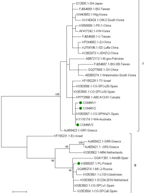
[image:4.595.63.524.564.698.2]et al. 2017). The sequence analysis based on the CP coding region revealed 100% identity among the collected CGMMV isolates, which suggests a common source of infection. The sequence of one representative was deposited in the GenBank data-base under accession no. KX589257. No remarkable molecular diversity was found among the 51 CGMMV isolates collected in 2017, which shared 99.5–100% identity with the CP gene. The sequences of three isolates that represent particular genetic variants (CGMMV1-3) were deposited in GenBank under accession Nos MH492014–16, respectively. The Kimura 2+G model of nucleotide substitution was estimated as the most appropriate using MEGA 6 and chosen for the construction of phylogenetic trees. Phylogenetic analysis based on CP sequences of the Polish isolates and corresponding sequences from GenBank revealed that CGMMV isolates can be divided into two groups. The majority of the isolates (24/35) clustered in group I whereas the remain-ing 11 isolates were located in group II. The Polish isolates collected in 2016 grouped together with European ones (group II), whereas those collected in 2017 clustered in group I where the majority of the isolates originated from Asia region (Figure 2). The isolates representing two phylogenetic groups shared 91.7–92.1% nucleotide sequence identity. Interest-ingly, the analysis based on amino acid sequences revealed that the majority of the point mutations were silent and the isolates from separate groups shared 95–100% CP amino acid sequence identity. Within the Polish isolates the amino acid identity of CP ranged from 99.3% to 100%. No recombination events were detected among the analysed sequences.
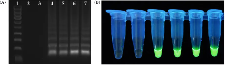
Figure 3. Detection of CGMMV isolates by RT-LAMP: (A) Analysis of RT-LAMP products on agarose gel; lane 1 – HyperLadderTM 100 bp (Bioline, London, UK); lane 2 – negative control (water); lane 3 – negative control (TMV); lane
4–7 – CGMMV isolates and (B) Visual detection of RT-LAMP products using EvaGreen® Dye (Biotium, Fremont,
USA), tubes correspond to gel lanes (2–7) in panel A
Development of RT-LAMP assay. The success of LAMP amplification depends on the specificity of the designed primer sets. The primers used in this study were selected based upon the multiple sequence alignment of different CGMMV isolates. For an RT-LAMP assay four CGMMV isolates representing different genetic variants were chosen (CGMMV-PL collected in 2016 and CGMMV1-3 collected in 2017). Our primers can detect a large part of the CGMMV variants representing two phylogenetic groups. The amplified products for CGMMV presented as ladder-like bands were observed for samples infected with all CGMMV isolates, but not in the virus-free samples or in those with the RNA of TMV(Figure 3A). The results obtained by the visual detection methods cor-related with the agarose gel electrophoresis results, and the green colour was observed in UV light after adding EvaGreen dye to the CGMMV infected sam-ples, whereas no reaction was observed in negative and TMV-infected samples (Figure 3B). Amplification curves in real-time reactions using a LightCycler® 480 (Roche, Switzerland) were observed only in the samples with RNA of CGMMV (Figure 4). The sensitivity test showed that RT-LAMP was capable
of detecting CGMMV in 10 fg/µl of the total RNA. Our results showed that the detection sensitivity of the RT-LAMP assay was 100 times higher than that of conventional RT-PCR (Figure 5).
DISCUSSION
[image:5.595.70.459.93.221.2]Since its first detection in 1935, CGMMV has achieved global distribution and become a major threat to cucurbit industries worldwide. Among the main cucurbit-infecting tobamoviruses, CGMMV is the most economically important and currently considered as a serious peril in the production of cucumbers, melons, watermelons, gherkins, and pumpkins. The ability of CGMMV to spread locally or over long distances is related with its transmis-sion through infected seeds. An adverse effect of the global seed trade is re-elected in disease outbreaks in new growing areas, countries, and continents. CGMMV is a new emerging Tobamovirus in Poland, and since its first detection in 2016, its presence has been reported in different regions of the country. The analysis of the genetic diversity of CGMMV in
Figure 5. Comparison of sensitivity of RT-LAMP and RT-PCR methods using CGMMV specific primers. The obtained bands are of expected size: (A) Detection limit of the RT-LAMP assay using total RNA isolated from plants infected by CGMMV1 isolate; lane 1 – HyperLadderTM 100 bp (Bioline, London, UK); lane 2 – negative control (water); lanes
3–12 – serial tenfold dilutions of RNA (1 000 ng/µl–1 fg/µl) and (B) Detection limit of the RT-PCR assay using total RNA isolated from plants infected by CGMMV1 isolate; lane 1 – HyperLadderTM 100 bp (Bioline, London, UK); lane 2
[image:5.595.70.533.563.672.2]– negative control (water); lanes 3–10 – serial tenfold dilutions of RNA (1000 ng/µl–100 fg/µl)
Figure 4. Real-time monitoring of RT-LAMP in LightCycler® 480
Poland revealed that the Polish isolates clustered in two separate phylogenetic groups, thereby indicating different sources of their origin. In the first group, the majority of the isolates originated from Asia, whereas the second group consisted of the European isolates. Interestingly, in 2016 in the Wielkopolska region, only the isolates representing group II were found, whereas in 2017, they were replaced by iso-lates from group I. A similar situation was observed in Spain where two major clusters of CGMMV iso-lates were observed: one cluster included 14 isoiso-lates collected between 2013 and 2014, and the second grouped 12 isolates that were mostly collected in 2015 (Crespo et al. 2017). The Polish isolates col-lected in 2016 were closely related to those found in Spain in 2013–2014 and clustered together with European isolates. The Polish isolates collected in 2017 grouped with the Spanish isolates collected in 2015 and others originated mainly from Asia. The comparison of the amino acid sequences of the Polish and Spanish isolates revealed 100% identity. It may suggest their common source of origin. CGMMV can spread readily by contact because its virions are stable and can remain infectious on a contaminated surface for long periods (Dombrovsky et al. 2017). It has been shown that CGMMV can be transmitted during grafting and pruning procedures, by overwin-ter contaminated soil, irrigation waoverwin-ter, soil mix, and dirty and contaminated surfaces. CGMMV spreads over long distances mostly via contaminated cucurbit seeds or infected cucurbit seedlings (Dombrovsky et al. 2017). The majority of the reports indicated that the incidence of vertical transmission of the virus in the progeny seedlings was rather low (Wu et al.
2011). Nevertheless, even extremely low frequencies of seed transmission can result in severe epidemics (Simmons & Munkvold 2014). It is very likely that, due to the commercial exchange of CGMMV infected seed and/or plant material, similar genetic variants can be found in different geographic regions. The co-existence of different Asian and European CGMMV isolates in Poland was noticed, which suggests the dispersion of the virus through the international seed trade. Cucurbit seed production in the Indian subcon-tinent or Southeast Asia and international seed trade may explain, for example, the presence in the Polish greenhouses of the genetic variants of CGMMV that have been previously observed in Asia. To prevent their introduction via seed and further spreading, the early detection and identification of virus infec-tion are required. Accurate identificainfec-tion and rapid
detection of the CGMMV are the first steps in the successful management of disease. In this study, we developed an RT-LAMP assay, which can be routinely used for the detection of different genetic variants of CGMMV. The development of RT-LAMP assays that could be widely applied for the detection of a wide range of viral isolates requires the knowledge of gene sequences, careful primer design, and assay optimisation. Taking into account the genetic diver-sity of the CGMMV population, the primers for an RT-LAMP assay were designed based on the align-ment of CP sequences instead of a single sequence as it has been done in the previously published reports (Li et al. 2013). The RT-LAMP developed here was capable of the detection of phylogenetically different CGMMV in less than one hour. Next, the RT-LAMP assay was 100 times more sensitive compared to the conventional RT-PCR. It makes it a very good tool that can be routinely used by phytosanitary services for the detection and identification of viral infection in seed and plant material.
References
Ainsworth G.C. (1935): Mosaic diseases of the cucumber. Annals of Applied Biology, 22: 55–67.
Antignus Y., Pearlsman M., Rachel B.Y., Cohen S. (1990): Occurrence of a variant of cucumber green mottle mosaic virus in Israel. Phytoparasitica, 18: 50–56.
Bashir N.S., Kalhor M.R., Zarghani S.N. (2006): Detection, differentiation and phylogenetic analysis of Cucumber mosaic virus isolates from cucurbits in the northwest region of Iran. Virus Genes, 32: 277–288.
Borodynko-Filas N., Minicka J., Hasiów-Jaroszewska B. (2017): The occurrence of Cucumber green mottle mosaic virus infecting greenhouse cucumber in Poland. Plant Disease, 101: 1336.
Crespo O., Janssen D., García C., Ruiz L. (2017): Biological and molecular diversity of Cucumber green mottle mosaic virus in Spain. Plant Disease, 101: 997–984.
Dombrovsky A., Tran-Nguyen L.T.T., Jones R.A.C. (2017).
Cucumber green mottle mosaic virus: Rapidly increasing global distribution, etiology, epidemiology, and manage-ment. Annual Review of Phytopathology, 55: 231–256. Hall T.A. (1999): BioEdit: a user-friendly biological sequence
alignment editor and analysis program for Windows 95/98/ NT. Nucleic Acids Symposium Series, 41: 95–98. Hongyun C., Wenjun Z., Qinsheng G., Qing C., Shiming
Kim S.M., Lee J.M., Yim K.O., Oh M.H., Park J.W., Kim J.H. (2003): Nucleotide sequences of two Korean isolates of Cucumber green mottle mosaic virus. Molecules and Cells, 16: 407–412.
Li J., Wei Q., Liu Y., Tan X., Zhang W., Wu J., Charimbu M.K., Hu B., Cheng Z., Yu C., Tao X. (2013): One-step reverse transcription loop-mediated isothermal am-plification for the rapid detection of Cucumber green mottle mosaic virus. Journal of Virological Methods, 193: 583–588.
Li R., Zheng Y., Fei Z., Ling K.S. (2015): First complete genome sequence of an emerging Cucumber green mottle mosaic virus isolate in North America. Genome Announcements, 3: e00452–15. doi: 10.1128/genomeA.00452-15
Martin D.P., Murrell B., Golden M., Khoosal A., Muhire B. (2015): RDP4: Detection and analysis of recombination patterns in virus genomes. Virus Evolution, 1: 1–5. Mori Y., Hirano T., Notomi T. (2006): Sequence specific
visual detection of LAMP reactions by addition of cati-onic polymers. BMC Biotechnology, 6: 3.
Sharifi M., Massumi H., Heydarnejad J., Pour A.H., Shaaba-nian M., Rahimian H. (2008): Analysis of the biological and molecular variability of Watermelon mosaic virus
isolates from Iran. Virus Genes, 37: 304–313.
Simmons H.E., Munkvold G.P. (2014): Seed transmission in the Potyviridae. In: Gullino M.L., Munkvold G. (eds): Global Perspectives on the Health of Seeds and Plant
Propagation Material. Plant Pathology in the 21st
Cen-tury – Contributions to the 9th International Congress,
Vol. 6. Dordrecht, Springer: 3–15.
Tamura K., Stecher G., Peterson D., Filipski A., Kumar S. (2013): MEGA6: Molecular evolutionary genetics analy-sis version 6.0. Molecular Biology and Evolution, 30: 2725–2729.
Tesoriero L.A., Chambers G., Srivastava M., Smith S., Conde B., Trannguyen L.T.T. (2016): First report of Cucumber green mottle mosaic virus in Australia. Australasian Plant Disease Notes, 11 (1). doi: 10.1007/s13314-015-0186-x Thomson K.G., Dietzgen R.G., Gibbs A.J., Tang Y.C.,
Liesack W., Teakle D.S., Stackebrandt E. (1995): Identi-fication of Zucchini yellow mosaic potyvirus by RT-PCR and analysis of sequence variability. Journal of Virological Methods, 55: 83–96.
Tian T., Posis K., Maroon-Lango C.J., Mavrodieva V., Haymes S., Pitman T.L., Falk B.W. (2014): First report of Cucumber green mottle mosaicvirus on melon in the United States. Plant Disease, 98: 1163.
Wu H., Qin B., Chen H., Peng B., Cai J., Gu Q. (2011): The rate of seed contamination and transmission of Cucum-ber green mottle mosaic virus in watermelon and melon. Scientia Agricultura Sinica, 44: 527–532.