2-Methyl-4-nitrophenol
Sheng Bi,aYong-Zhong Wu,bYi-Xin Zhou,aJian-Guo Tangaand Cheng Guoa*
a
College of Science, Nanjing University of Technology, Xinmofan Road No. 5 Nanjing, Nanjing 210009, People’s Republic of China, andbDepartment of Applied Chemistry, Nanjing College of Chemical Technology, Geguan Road No. 625 Dachang District Nanjing, Nanjing 210048, People’s Republic of China Correspondence e-mail: [email protected]
Received 14 May 2009; accepted 18 May 2009
Key indicators: single-crystal X-ray study;T= 294 K; mean(C–C) = 0.005 A˚; Rfactor = 0.054;wRfactor = 0.181; data-to-parameter ratio = 12.2.
The molecule of the title compound, C7H7NO3, is nearly
planar [maximum deviation 0.112 (3) A˚ for one of the notro O atoms]. In the crystal structure, intermolecular O—H O and C—H O interactions link the molecules into a three-dimensional network.
Related literature
For a related structure, see: Ahmed & Ashwini (2004). For bond-length data, see: Allenet al.(1987).
Experimental
Crystal data
C7H7NO3
Mr= 153.14
Monoclinic,P21=n a= 5.6210 (11) A˚
c= 14.300 (3) A˚
= 100.71 (3)
V= 690.4 (2) A˚3
Z= 4
= 0.12 mm
T= 294 K
0.300.200.10 mm
Data collection
Enraf–Nonius CAD-4 diffractometer
Absorption correction: scan (Northet al., 1968)
Tmin= 0.966,Tmax= 0.988 1378 measured reflections
1245 independent reflections 870 reflections withI> 2(I)
Rint= 0.027 3 standard reflections
frequency: 120 min intensity decay: 1%
Refinement
R[F2> 2(F2)] = 0.054
wR(F2) = 0.181
S= 1.01 1245 reflections
102 parameters
H-atom parameters constrained
max= 0.25 e A˚ 3
min=0.24 e A˚ 3
Table 1
Hydrogen-bond geometry (A˚ ,).
D—H A D—H H A D A D—H A
O3—H3A O2i 0.82 2.10 2.770 (4) 138 C7—H7C O1ii
0.96 2.57 3.505 (5) 165
Symmetry codes: (i)x;y1;z; (ii)x1;yþ1;zþ2.
Data collection: CAD-4 Software (Enraf–Nonius, 1989); cell refinement: CAD-4 Software; data reduction: XCAD4 (Harms & Wocadlo, 1995); program(s) used to solve structure: SHELXS97 (Sheldrick, 2008); program(s) used to refine structure:SHELXL97 (Sheldrick, 2008); molecular graphics: ORTEP-3 for Windows (Farrugia, 1997); software used to prepare material for publication: SHELXL97.
The authors thank the Center of Testing and Analysis, Nanjing University, for support.
Supplementary data and figures for this paper are available from the IUCr electronic archives (Reference: HK2688).
References
Ahmed, K. & Ashwini, K. (2004).Ultrason. Sonochem.pp. 455–457. Allen, F. H., Kennard, O., Watson, D. G., Brammer, L., Orpen, A. G. & Taylor,
R. (1987).J. Chem. Soc. Perkin Trans. 2, pp. S1–19.
Enraf–Nonius (1989).CAD-4 Software. Enraf–Nonius, Delft, The Nether-lands.
Farrugia, L. J. (1997).J. Appl. Cryst.30, 565.
Harms, K. & Wocadlo, S. (1995).XCAD4. University of Marburg, Germany. North, A. C. T., Phillips, D. C. & Mathews, F. S. (1968).Acta Cryst.A24, 351–
359.
Sheldrick, G. M. (2008).Acta Cryst.A64, 112–122.
Structure Reports Online
supporting information
Acta Cryst. (2009). E65, o1378 [doi:10.1107/S1600536809018716]
2-Methyl-4-nitrophenol
Sheng Bi, Yong-Zhong Wu, Yi-Xin Zhou, Jian-Guo Tang and Cheng Guo
S1. Comment
Some derivatives of benzoic acids are important chemical materials. We report herein the crystal structure of the title
compound.
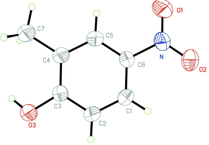
In the molecule of the title compound (Fig 1), the bond lengths (Allen et al., 1987) and angles are within normal ranges.
Ring A (C1-C6) is, of course, planar. Atoms O1, O2, O3, N and C7 are 0.112 (3), 0.023 (3), 0.049 (3), 0.026 (4) and
-0.042 (3) Å away from the ring plane, respectively. So, the molecule is nearly planar.
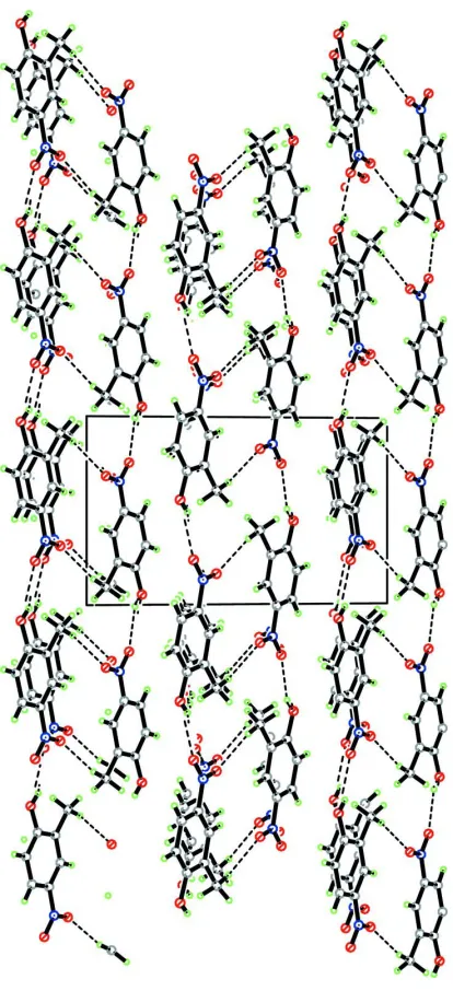
In the crystal structure, intermolecular O-H···O and C-H···O interactions (Table 1) link the molecules into a network, in
which they may be effective in the stabilization of the structure.
S2. Experimental
For the preparation of the title compound, ethyl acetate (150 ml), 2-methyl-phenol (5.9 g) and zinc chloride (7.4 g) are
placed in an ultrasonic cleaning bath equipped with a round botton flask, and then nitric acid (5.9 g) was added dropwise
in 3 min. After the reaction was completed, water (200 ml) was added. After evaporation of the organic layer, the
obtained product (Ahmed & Ashwini, 2004) was crystallized by slow evaporation of a methanol solution.
S3. Refinement
H atoms were positioned geometrically, with O-H = 0.82 Å (for OH) and C-H = 0.93 and 0.96 Å for aromatic and methyl
H, respectively, and constrained to ride on their parent atoms, with Uiso(H) = xUeq(C,O), where x = 1.2 for aromatic H and
Figure 1
The molecular structure of the title molecule, with the atom-numbering scheme. Displacement ellipsoids are drawn at the
Figure 2
A partial packing diagram of the title compound. Hydrogen bonds are shown as dashed lines.
2-Methyl-4-nitrophenol
Crystal data
C7H7NO3 Mr = 153.14 Monoclinic, P21/n
Hall symbol: -P 2yn a = 5.6210 (11) Å b = 8.7420 (17) Å c = 14.300 (3) Å β = 100.71 (3)° V = 690.4 (2) Å3 Z = 4
F(000) = 320 Dx = 1.473 Mg m−3
Mo Kα radiation, λ = 0.71073 Å Cell parameters from 25 reflections θ = 9–13°
Enraf–Nonius CAD-4 diffractometer
Radiation source: fine-focus sealed tube Graphite monochromator
ω/2θ scans
Absorption correction: ψ scan (North et al., 1968)
Tmin = 0.966, Tmax = 0.988
1378 measured reflections
1245 independent reflections 870 reflections with I > 2σ(I) Rint = 0.027
θmax = 25.3°, θmin = 2.7° h = 0→6
k = 0→10 l = −17→16
3 standard reflections every 120 min intensity decay: 1%
Refinement
Refinement on F2
Least-squares matrix: full R[F2 > 2σ(F2)] = 0.054 wR(F2) = 0.181 S = 1.01 1245 reflections 102 parameters 0 restraints
Primary atom site location: structure-invariant direct methods
Secondary atom site location: difference Fourier map
Hydrogen site location: inferred from neighbouring sites
H-atom parameters constrained w = 1/[σ2(F
o2) + (0.08P)2 + 0.74P]
where P = (Fo2 + 2Fc2)/3
(Δ/σ)max < 0.001
Δρmax = 0.25 e Å−3
Δρmin = −0.24 e Å−3
Extinction correction: SHELXL97 (Sheldrick, 2008), Fc*=kFc[1+0.001xFc2λ3/sin(2θ)]-1/4
Extinction coefficient: 0.017 (4)
Special details
Geometry. All e.s.d.'s (except the e.s.d. in the dihedral angle between two l.s. planes) are estimated using the full covariance matrix. The cell e.s.d.'s are taken into account individually in the estimation of e.s.d.'s in distances, angles and torsion angles; correlations between e.s.d.'s in cell parameters are only used when they are defined by crystal symmetry. An approximate (isotropic) treatment of cell e.s.d.'s is used for estimating e.s.d.'s involving l.s. planes.
Refinement. Refinement of F2 against ALL reflections. The weighted R-factor wR and goodness of fit S are based on F2,
conventional R-factors R are based on F, with F set to zero for negative F2. The threshold expression of F2 > σ(F2) is used
only for calculating R-factors(gt) etc. and is not relevant to the choice of reflections for refinement. R-factors based on F2
are statistically about twice as large as those based on F, and R- factors based on ALL data will be even larger.
Fractional atomic coordinates and isotropic or equivalent isotropic displacement parameters (Å2)
x y z Uiso*/Ueq
C7 −0.3278 (6) 0.1019 (4) 0.9313 (3) 0.0531 (9) H7A −0.2143 0.0360 0.9707 0.080* H7B −0.4172 0.0441 0.8793 0.080* H7C −0.4375 0.1443 0.9684 0.080*
Atomic displacement parameters (Å2)
U11 U22 U33 U12 U13 U23
O1 0.093 (2) 0.0485 (16) 0.104 (2) 0.0137 (14) 0.0576 (18) −0.0051 (15) O2 0.0861 (19) 0.0303 (13) 0.097 (2) −0.0030 (12) 0.0392 (16) 0.0063 (13) O3 0.0661 (15) 0.0341 (13) 0.0802 (17) 0.0000 (11) 0.0405 (13) −0.0053 (11) N 0.0594 (18) 0.0337 (15) 0.0567 (17) 0.0039 (13) 0.0180 (14) 0.0005 (13) C1 0.0539 (19) 0.0371 (17) 0.059 (2) −0.0036 (14) 0.0281 (17) 0.0044 (15) C2 0.0496 (19) 0.0413 (18) 0.063 (2) −0.0036 (15) 0.0302 (17) −0.0024 (16) C3 0.0438 (17) 0.0339 (15) 0.0503 (18) −0.0019 (14) 0.0168 (14) −0.0029 (14) C4 0.0389 (16) 0.0397 (17) 0.0473 (18) −0.0032 (13) 0.0156 (14) −0.0012 (14) C5 0.0403 (16) 0.0407 (17) 0.0469 (17) 0.0021 (14) 0.0171 (14) 0.0005 (14) C6 0.0461 (17) 0.0312 (16) 0.0500 (18) 0.0009 (13) 0.0177 (14) −0.0006 (13) C7 0.054 (2) 0.045 (2) 0.068 (2) −0.0034 (15) 0.0285 (17) 0.0007 (16)
Geometric parameters (Å, º)
O3—C3 1.357 (4) C2—H2A 0.9300 O3—H3A 0.8200 C3—C4 1.393 (4) N—O1 1.217 (3) C4—C5 1.379 (4) N—O2 1.225 (4) C4—C7 1.496 (4) N—C6 1.451 (4) C5—C6 1.379 (4) C1—C2 1.370 (5) C5—H5A 0.9300 C1—C6 1.382 (4) C7—H7A 0.9600 C1—H1A 0.9300 C7—H7B 0.9600 C2—C3 1.382 (4) C7—H7C 0.9600
C3—O3—H3A 109.5 C5—C4—C7 121.1 (3) O1—N—O2 122.9 (3) C3—C4—C7 121.2 (3) O1—N—C6 118.4 (3) C6—C5—C4 120.5 (3) O2—N—C6 118.7 (3) C6—C5—H5A 119.8 C2—C1—C6 118.4 (3) C4—C5—H5A 119.8 C2—C1—H1A 120.8 C5—C6—C1 121.6 (3) C6—C1—H1A 120.8 C5—C6—N 119.9 (3) C1—C2—C3 120.4 (3) C1—C6—N 118.5 (3) C1—C2—H2A 119.8 C4—C7—H7A 109.5 C3—C2—H2A 119.8 C4—C7—H7B 109.5 O3—C3—C2 115.9 (3) H7A—C7—H7B 109.5 O3—C3—C4 122.7 (3) C4—C7—H7C 109.5 C2—C3—C4 121.5 (3) H7A—C7—H7C 109.5 C5—C4—C3 117.7 (3) H7B—C7—H7C 109.5
O1—N—C6—C1 177.3 (3) O3—C3—C4—C7 1.4 (5) O2—N—C6—C1 −2.6 (5) C2—C3—C4—C7 −178.3 (3) C6—C1—C2—C3 −1.0 (5) C3—C4—C5—C6 −1.5 (5) C2—C1—C6—C5 1.2 (5) C7—C4—C5—C6 178.6 (3) C2—C1—C6—N −177.5 (3) C4—C5—C6—C1 0.0 (5) C1—C2—C3—O3 179.7 (3) C4—C5—C6—N 178.8 (3) C1—C2—C3—C4 −0.5 (5)
Hydrogen-bond geometry (Å, º)
D—H···A D—H H···A D···A D—H···A
O3—H3A···O2i 0.82 2.10 2.770 (4) 138
C7—H7C···O1ii 0.96 2.57 3.505 (5) 165