Bostrycin
Shao-Yuan Chen,a* Chao Huang,aChu-Long Zhang,a Yu-Zhe Chenband Fu-Cheng Lina
a
State Key Laboratory for Rice Biology, Institute of Biotechnology, Zhejiang University, People’s Republic of China, andbCollege of Pharmaceutical Sicence, Zhejiang University, People’s Republic of China
Correspondence e-mail: [email protected]
Received 6 September 2008; accepted 6 October 2008
Key indicators: single-crystal X-ray study;T= 293 K; mean(C–C) = 0.003 A˚; Rfactor = 0.035;wRfactor = 0.105; data-to-parameter ratio = 6.9.
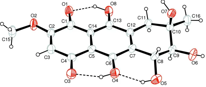
The title compound, C16H16O8, is a potent nonspecific phyto-toxin. The crystal structure is the average of two tautomers, 5,6,7,9,10-pentahydroxy-2-methoxy-7-methyl-1,4,5,6,7,8-hexa-hydroanthracene-1,4-dione and 1,4,5,6,7-pentahydroxy-2- methoxy-7-methyl-5,6,7,8,9,10-hexahydroanthracene-9,10-di-one. The cyclohexene rings in both tautomers display a half-chair conformation. An extensive O—H O hydrogen-bonding network is present in the crystal structure.
Related literature
For general background, see: Charudattan & Rao (1982); van Eijk (1975). For a related structure, see: Kelly & Saha (1985).
Experimental
Crystal data
C16H16O8
Mr= 336.29 Monoclinic,P21
a= 8.280 (2) A˚ b= 6.644 (2) A˚ c= 13.1535 (12) A˚ = 102.12 (2)
V= 707.5 (3) A˚3 Z= 2
MoKradiation = 0.13 mm1
T= 293 (2) K 0.300.200.08 mm
Data collection
Rigaku R-AXIS RAPID IP diffractometer
Absorption correction: multi-scan (ABSCOR; Higashi, 1995) Tmin= 0.953,Tmax= 0.990
6151 measured reflections 1517 independent reflections 1282 reflections withI> 2(I) Rint= 0.025
Refinement
R[F2> 2(F2)] = 0.035
wR(F2) = 0.105 S= 1.08 1517 reflections 220 parameters
1 restraint
H-atom parameters constrained max= 0.24 e A˚
3
min=0.19 e A˚
3
Table 1
Hydrogen-bond geometry (A˚ ,).
D—H A D—H H A D A D—H A
O1—H1A O3i
0.85 2.55 3.229 (3) 137 O3—H3A O8ii 0.84 2.40 2.808 (3) 111 O4—H4A O8ii
0.90 2.54 3.223 (3) 132 O5—H5A O4 0.92 1.85 2.687 (3) 152 O6—H6A O7iii
0.97 1.92 2.821 (3) 154 O7—H7A O1iv
0.90 2.11 2.966 (3) 159 O7—H7A O2iv
0.90 2.40 3.066 (3) 131
Symmetry codes: (i) xþ1;y;z; (ii) x1;y;z; (iii) xþ1;y1
2;zþ2; (iv) xþ1;yþ1
2;zþ1.
Data collection: PROCESS-AUTO (Rigaku, 1998); cell refine-ment:PROCESS-AUTO; data reduction:CrystalStructure(Rigaku/ MSC, 2002); program(s) used to solve structure:SHELXS97 (Shel-drick, 2008); program(s) used to refine structure: SHELXL97 (Sheldrick, 2008); molecular graphics: ORTEP-3 (Farrugia, 1997); software used to prepare material for publication:WinGX(Farrugia, 1999).
The work was supported by the Science and Technology Project of Zhejiang Province (grant Nos. 2004 C22008 and 2006 C12088) and the National Natural Science Foundation of China (grant No. 30600002).
Supplementary data and figures for this paper are available from the IUCr electronic archives (Reference: XU2453).
References
Charudattan, R. & Rao, K. V. (1982).Appl. Environ. Microbiol.43, 846–849. Eijk, G. W. van (1975).Cell. Mol. Life Sci.31, 783–784.
Farrugia, L. J. (1997).J. Appl. Cryst.30, 565. Farrugia, L. J. (1999).J. Appl. Cryst.32, 837–838.
Higashi, T. (1995).ABSCOR. Rigaku Corporation, Tokyo, Japan. Kelly, T. & Saha, J. K. (1985).J. Org. Chem.50, 3679–3685.
Rigaku (1998).PROCESS-AUTO. Rigaku Corporation, Tokyo, Japan. Rigaku/MSC (2002).CrystalStructure. Rigaku/MSC, The Woodlands, Texas,
USA.
Sheldrick, G. M. (2008).Acta Cryst.A64, 112–122. Acta Crystallographica Section E
Structure Reports Online
supporting information
Acta Cryst. (2008). E64, o2226 [doi:10.1107/S1600536808032030]
Bostrycin
Shao-Yuan Chen, Chao Huang, Chu-Long Zhang, Yu-Zhe Chen and Fu-Cheng Lin
S1. Comment
Bostrycin, a nonspecific phytotoxin, was identified as a metabolite of the fungus Arthrinium phaeospermum in 1975 (van
Eijk, 1975; Charudattan & Rao, 1982). It is active against Bacillus subtilis but inactive against the fungus Geotrichum
candidum (Charudattan & Rao, 1982). We report here the crystal structure of the title compound.
The crystal structure of the title compound is the average structure of two tautomers,
5,6,7,9,10-pentahydroxy-2-meth-oxy-7-methyl-1,4,5,6,7,8-hexahydroanthracene- 1,4-dione (I) and
1,4,5,6,7-pentahydroxy-2-methoxy-7-methyl-5,6,7,8,9,10-hexahydroanthracene- 9,10-dione (II). The molecular structures of the two tautomers are shown in
Fig. 1 and Fig. 2, respectively. Both of tautomer molecules contains three six-membered rings, among which the
C9-containing ring displays a half-chair conformation. Within the molecule the carbonyl group is hydrogen bonded to the
neighboring hydroxyl group(s). The bond distances and angles agree with those found in a derivative of bostrycin,
bostrycin acetonide (Kelly & Saha, 1985). The extensive O—H···O hydrogen bonding network helps to stabilize the
crystal structure (Table 1).
S2. Experimental
For morphological identification (Arthrinium sp. (CGMCC 2082), a fungi from Polygonum hydropiper L.) cultures were
grown on OA, PDA, and SNA media for 7–14 days at room temperature (293 K) under ambient daylight. Microscopic
observations and measurements were made from slides mounted in water. For metabolite production, the strains were
inoculated onto PDA media and incubated for 10 days at 298 K in the dark. Selected strains were also cultivated in liquid
media placed in a rotary shaker at 120 rpm for 7 days at 298 K in the dark. After cultivation, the bottles were stored at
253 K until extraction.
Liquid cultures were extracted with trichloromethane. The trichloromethane phase was filtered and evaporated in vacuo.
Samples were then redissolved in trichloromethane, then filtered to remove solid. The trichloromethane solution was
evaporated in vacuo. The single crystals were obtained from an ethanol solution.
S3. Refinement
Hydroxyl H atoms were located in a difference Fourier map and refined as riding in as-found relative positions with
Uiso(H) = 1.5Ueq(O). Methyl H atoms were placed in calculated positions with C—H = 0.96 Å and torsion angles were
refined to fit the electron density, Uiso(H) = 1.5Ueq(C). Other H atoms were placed in calculated positions with C—H =
0.93 (aromatic), 0.97 (methylene) or 0.98 Å (methine), and refined in riding mode with Uiso(H) = 1.2Ueq(C). In the
absence of significant anomalous scattering effects, Friedel pairs were merged; the absolute configuration was not
Figure 1
The molecular structure of (I) with 50% probability displacement ellipsoids (arbitrary spheres for H atoms), dashed line
indicates hydrogen bonding.
Figure 2
The molecular structure of (II) with 50% probability displacement ellipsoids (arbitrary spheres for H atoms), dashed line
indicates hydrogen bonding.
5,6,7,9,10-pentahydroxy-2-methoxy-7-methyl-1,4,5,6,7,8-hexahydroanthracene-
1,4-dione–1,4,5,6,7-pentahydroxy-2-methoxy-7-methyl-5,6,7,8,9,10- hexahydroanthracene-9,10-dione
Crystal data
C16H16O8 Mr = 336.29 Monoclinic, P21
Hall symbol: P 2yb a = 8.280 (2) Å b = 6.644 (2) Å c = 13.1535 (12) Å β = 102.12 (2)° V = 707.5 (3) Å3 Z = 2
F(000) = 352 Dx = 1.579 Mg m−3
Mo Kα radiation, λ = 0.71069 Å Cell parameters from 5814 reflections θ = 6.1–54.9°
µ = 0.13 mm−1 T = 293 K Chunk, red
[image:3.610.129.484.282.432.2]Data collection
Rigaku R-AXIS RAPID IP diffractometer
Radiation source: fine-focus sealed tube Graphite monochromator
Detector resolution: 10 pixels mm-1 ω scans
Absorption correction: multi-scan (ABSCOR; Higashi, 1995) Tmin = 0.953, Tmax = 0.990
6151 measured reflections 1517 independent reflections 1282 reflections with I > 2σ(I) Rint = 0.025
θmax = 26.0°, θmin = 3.2° h = −10→10
k = −8→8 l = −16→16
Refinement
Refinement on F2
Least-squares matrix: full R[F2 > 2σ(F2)] = 0.035 wR(F2) = 0.105 S = 1.08 1517 reflections 220 parameters 1 restraint
Primary atom site location: structure-invariant direct methods
Secondary atom site location: difference Fourier map
Hydrogen site location: inferred from neighbouring sites
H-atom parameters constrained w = 1/[σ2(F
o2) + (0.0667P)2 + 0.0817P]
where P = (Fo2 + 2Fc2)/3
(Δ/σ)max = 0.001
Δρmax = 0.24 e Å−3
Δρmin = −0.19 e Å−3
Extinction correction: SHELXL97 (Sheldrick, 2008), Fc*=kFc[1+0.001xFc2λ3/sin(2θ)]-1/4
Extinction coefficient: 0.009 (3)
Special details
Geometry. All e.s.d.'s (except the e.s.d. in the dihedral angle between two l.s. planes) are estimated using the full covariance matrix. The cell e.s.d.'s are taken into account individually in the estimation of e.s.d.'s in distances, angles and torsion angles; correlations between e.s.d.'s in cell parameters are only used when they are defined by crystal symmetry. An approximate (isotropic) treatment of cell e.s.d.'s is used for estimating e.s.d.'s involving l.s. planes.
Refinement. Refinement of F2 against ALL reflections. The weighted R-factor wR and goodness of fit S are based on F2,
conventional R-factors R are based on F, with F set to zero for negative F2. The threshold expression of F2 > σ(F2) is used
only for calculating R-factors(gt) etc. and is not relevant to the choice of reflections for refinement. R-factors based on F2
are statistically about twice as large as those based on F, and R- factors based on ALL data will be even larger.
Fractional atomic coordinates and isotropic or equivalent isotropic displacement parameters (Å2)
x y z Uiso*/Ueq Occ. (<1)
O1 0.4354 (2) 0.3424 (4) 0.34450 (13) 0.0399 (5)
H1A 0.5153 0.3341 0.3968 0.060* 0.50
O2 0.1814 (2) 0.3555 (4) 0.19361 (12) 0.0414 (5)
O3 −0.1725 (2) 0.3863 (4) 0.43528 (13) 0.0384 (5)
H3A −0.1659 0.3844 0.4995 0.058* 0.50
O4 −0.0420 (2) 0.3933 (4) 0.63061 (13) 0.0370 (5)
H4A −0.1226 0.4012 0.5725 0.055* 0.50
O5 0.1000 (3) 0.2810 (5) 0.82507 (17) 0.0571 (7)
H5A 0.0233 0.2963 0.7644 0.086*
O6 0.3619 (2) 0.4004 (4) 0.97960 (12) 0.0417 (5)
H6A 0.4364 0.3205 1.0303 0.063*
O7 0.5068 (2) 0.6526 (3) 0.84879 (13) 0.0336 (5)
H7A 0.5511 0.7105 0.7990 0.050*
H8A 0.5559 0.3636 0.4666 0.060* 0.50
C1 0.2943 (3) 0.3552 (4) 0.37142 (18) 0.0302 (6)
C2 0.1464 (3) 0.3632 (4) 0.28875 (18) 0.0303 (6)
C3 −0.0063 (3) 0.3742 (5) 0.31216 (18) 0.0314 (6)
H3 −0.0996 0.3781 0.2587 0.038*
C4 −0.0247 (3) 0.3798 (4) 0.41735 (18) 0.0284 (5)
C5 0.1177 (3) 0.3742 (4) 0.50050 (17) 0.0263 (5)
C6 0.1023 (3) 0.3820 (4) 0.60602 (17) 0.0268 (5)
C7 0.2492 (3) 0.3761 (5) 0.68900 (17) 0.0291 (6)
C8 0.2301 (3) 0.3980 (5) 0.80142 (17) 0.0311 (6)
H8 0.2063 0.5396 0.8130 0.037*
C9 0.3863 (3) 0.3403 (4) 0.87974 (18) 0.0323 (6)
H9 0.3996 0.1938 0.8790 0.039*
C10 0.5382 (3) 0.4387 (4) 0.85300 (17) 0.0309 (6)
C11 0.5585 (3) 0.3595 (5) 0.74721 (17) 0.0319 (6)
H11A 0.6425 0.4383 0.7240 0.038*
H11B 0.5970 0.2213 0.7552 0.038*
C12 0.4022 (3) 0.3671 (4) 0.66537 (17) 0.0294 (6)
C13 0.4195 (3) 0.3563 (4) 0.55826 (18) 0.0296 (6)
C14 0.2770 (3) 0.3626 (4) 0.47627 (17) 0.0262 (5)
C15 0.0456 (3) 0.3549 (6) 0.10420 (18) 0.0435 (8)
H15A −0.0220 0.2384 0.1068 0.065*
H15B 0.0881 0.3517 0.0417 0.065*
H15C −0.0196 0.4743 0.1048 0.065*
C16 0.6936 (3) 0.3933 (6) 0.93488 (19) 0.0408 (7)
H16A 0.7885 0.4434 0.9117 0.061*
H16B 0.7039 0.2505 0.9453 0.061*
H16C 0.6861 0.4575 0.9991 0.061*
Atomic displacement parameters (Å2)
U11 U22 U33 U12 U13 U23
C9 0.0411 (15) 0.0348 (14) 0.0232 (10) 0.0016 (13) 0.0117 (10) 0.0021 (11) C10 0.0363 (15) 0.0338 (15) 0.0224 (12) 0.0012 (12) 0.0055 (10) 0.0004 (11) C11 0.0283 (13) 0.0429 (16) 0.0251 (11) 0.0016 (13) 0.0067 (9) −0.0028 (12) C12 0.0304 (13) 0.0340 (14) 0.0248 (11) 0.0024 (13) 0.0079 (9) −0.0028 (12) C13 0.0270 (13) 0.0370 (15) 0.0262 (11) 0.0028 (12) 0.0087 (9) −0.0019 (11) C14 0.0250 (12) 0.0309 (13) 0.0230 (10) 0.0014 (12) 0.0059 (9) −0.0019 (12) C15 0.0381 (15) 0.068 (2) 0.0225 (11) 0.0052 (17) 0.0028 (10) −0.0018 (15) C16 0.0368 (15) 0.0555 (17) 0.0279 (12) 0.0023 (15) 0.0019 (10) −0.0011 (14)
Geometric parameters (Å, º)
O1—C1 1.293 (3) C5—C6 1.421 (3)
O1—H1A 0.8501 C5—C14 1.423 (3)
O2—C2 1.344 (3) C6—C7 1.454 (3)
O2—C15 1.447 (3) C7—C12 1.368 (4)
O3—C4 1.294 (3) C7—C8 1.527 (3)
O3—H3A 0.8350 C8—C9 1.524 (4)
O4—C6 1.304 (3) C8—H8 0.9800
O4—H4A 0.9046 C9—C10 1.523 (4)
O5—C8 1.415 (4) C9—H9 0.9800
O5—H5A 0.9154 C10—C16 1.525 (3)
O6—C9 1.427 (3) C10—C11 1.530 (3)
O6—H6A 0.9667 C11—C12 1.501 (3)
O7—C10 1.444 (3) C11—H11A 0.9700
O7—H7A 0.9012 C11—H11B 0.9700
O8—C13 1.296 (3) C12—C13 1.448 (3)
O8—H8A 0.9624 C13—C14 1.422 (3)
C1—C14 1.417 (3) C15—H15A 0.9600
C1—C2 1.458 (3) C15—H15B 0.9600
C2—C3 1.364 (4) C15—H15C 0.9600
C3—C4 1.424 (3) C16—H16A 0.9600
C3—H3 0.9300 C16—H16B 0.9600
C4—C5 1.431 (3) C16—H16C 0.9600
C1—O1—H1A 112.1 C10—C9—C8 111.2 (2)
C2—O2—C15 118.3 (2) O6—C9—H9 108.9
C4—O3—H3A 108.7 C10—C9—H9 108.9
C6—O4—H4A 110.2 C8—C9—H9 108.9
C8—O5—H5A 99.7 O7—C10—C9 106.3 (2)
C9—O6—H6A 106.8 O7—C10—C16 109.9 (2)
C10—O7—H7A 110.5 C9—C10—C16 111.6 (2)
C13—O8—H8A 106.3 O7—C10—C11 110.9 (2)
O1—C1—C14 123.4 (2) C9—C10—C11 108.4 (2)
O1—C1—C2 117.6 (2) C16—C10—C11 109.7 (2)
C14—C1—C2 119.0 (2) C12—C11—C10 113.5 (2)
O2—C2—C3 127.1 (2) C12—C11—H11A 108.9
O2—C2—C1 112.4 (2) C10—C11—H11A 108.9
C2—C3—C4 120.9 (2) C10—C11—H11B 108.9
C2—C3—H3 119.5 H11A—C11—H11B 107.7
C4—C3—H3 119.5 C7—C12—C13 120.6 (2)
O3—C4—C3 118.4 (2) C7—C12—C11 122.6 (2)
O3—C4—C5 121.4 (2) C13—C12—C11 116.7 (2)
C3—C4—C5 120.2 (2) O8—C13—C14 121.8 (2)
C6—C5—C14 119.9 (2) O8—C13—C12 118.2 (2)
C6—C5—C4 121.1 (2) C14—C13—C12 120.1 (2)
C14—C5—C4 119.0 (2) C1—C14—C13 120.0 (2)
O4—C6—C5 121.3 (2) C1—C14—C5 120.5 (2)
O4—C6—C7 118.7 (2) C13—C14—C5 119.5 (2)
C5—C6—C7 120.0 (2) O2—C15—H15A 109.5
C12—C7—C6 119.9 (2) O2—C15—H15B 109.5
C12—C7—C8 120.9 (2) H15A—C15—H15B 109.5
C6—C7—C8 119.0 (2) O2—C15—H15C 109.5
O5—C8—C9 106.9 (2) H15A—C15—H15C 109.5
O5—C8—C7 113.5 (2) H15B—C15—H15C 109.5
C9—C8—C7 112.6 (2) C10—C16—H16A 109.5
O5—C8—H8 107.8 C10—C16—H16B 109.5
C9—C8—H8 107.8 H16A—C16—H16B 109.5
C7—C8—H8 107.8 C10—C16—H16C 109.5
O6—C9—C10 112.1 (2) H16A—C16—H16C 109.5
O6—C9—C8 106.7 (2) H16B—C16—H16C 109.5
Hydrogen-bond geometry (Å, º)
D—H···A D—H H···A D···A D—H···A
O1—H1A···O3i 0.85 2.55 3.229 (3) 137
O3—H3A···O8ii 0.84 2.40 2.808 (3) 111
O4—H4A···O8ii 0.90 2.54 3.223 (3) 132
O5—H5A···O4 0.92 1.85 2.687 (3) 152
O6—H6A···O7iii 0.97 1.92 2.821 (3) 154
O7—H7A···O1iv 0.90 2.11 2.966 (3) 159
O7—H7A···O2iv 0.90 2.40 3.066 (3) 131