University of New Orleans University of New Orleans
ScholarWorks@UNO
ScholarWorks@UNO
University of New Orleans Theses and
Dissertations Dissertations and Theses
1-20-2006
Synthesis of Molecular Baskets and Introduction of Inward Facing
Synthesis of Molecular Baskets and Introduction of Inward Facing
Functionality
Functionality
Zachary Laughery
University of New Orleans
Follow this and additional works at: https://scholarworks.uno.edu/td
Recommended Citation Recommended Citation
Laughery, Zachary, "Synthesis of Molecular Baskets and Introduction of Inward Facing Functionality" (2006). University of New Orleans Theses and Dissertations. 328.
https://scholarworks.uno.edu/td/328
SYNTHESIS OF MOLECULAR BASKETS AND INTRODUCTION OF
INWARD FACING FUNCTIONALITY
A Dissertation
Submitted to the Graduate Faculty of the
University of New Orleans
in partial fulfillment of the
requirement for the degree of
Doctor of Philosophy
in
Chemistry
by
Zachary R. Laughrey
A.D., Louisiana Tech University
B.A., University of New Orleans
ACKNOWLEDGEMENTS
I would like to express my undying gratitude to my advisor, Professor Bruce C. Gibb for
his patience, encouragement and open discussions during the winding course of my research.
My gratitude is also expressed to the members of my research committee, Dr. Mark Trudell, Dr.
Branko Jursic, Dr. Guijun Wang, and Dr. Steven Rick for their assistance and guidance.
I would also like to thank all the past and present members of the Gibb research group
especially Corinne Gibb for her assistance, knowledge and guidance. I would also like to thank
Dr. John Wiley for his assistance and Dr. Richard Cole for mass spectroscopy analysis.
Finally, I would like to thank my family. My greatest gratitude goes out to my daughters,
Maggie, Cassie and Audrey for their support and love, and to my ever-patient wife, Stacey, who
TABLE OF CONTENTS
List of Tables...v
List of Figures ... vii
List of Schemes ... xii
Abbreviations ...xiv
Abstract ...xv
I. Introduction...1
1.1 Host-Guest interactions...1
1.2 Enzymes and enzyme mimics ...5
1.3 Molecular hosts...6
1.3.1 Cyclodextrins...7
1.3.2 Calixarenes ...10
1.3.3 Cavitands ...22
1.3.4 Cyclotriveratrylene...28
1.3.5 Cucurbituril...33
1.3.6 Inward facing functionality ...37
II. Synthesis and Characterization of m-baskets...44
2.1 Synthesis and conformation of molecular hosts ...44
2.2 NMR binding studies of molecular hosts...48
2.3 Synthesis and hosting properties of a deuterated molecular host...59
III. Functionalization of molecular concavity...66
3.2 Base strength and product yield...77
3.3 Synthesis of carboxylic acids m-baskets ...81
3.4 Carboxylic acid m-baskets as acid catalysts...86
3.5 Binding properties of carboxylic acid m-baskets...87
IV. Synthesis of a Carbonic anhydrase mimic...101
4.1 Synthesis of the enzyme mimic ...106
4.2 Zinc binding and physical studies of the enzyme mimic ...117
V. Conclusion...120
VI. Experimental Section...121
6.1 General ...121
6.2 Synthesized Compounds ...121
6.3 Binding Studies of 5-Methyl-m-basket, 2-Methyl-m-basket, and d4-m-basket ...147
6.4 1D- and 2D-NMRs of aldehyde m-baskets ...160
6.5 NMR and UV-Vis titrations of 157...163
6.6 Crystal structure of 145...164
6.7 Crystal structure of 149...191
VII.References...204
LIST OF TABLES
Table 1.1 Dimensions of cyclodextrins...8
Table 1.2 Binding Constants of Host 12...13
Table 1.3 Binding Constants of Calix[4]arene with Increasing Alkyl Groups ...14
Table 1.4 Dimensions of Cucurbit[n]uril ...35
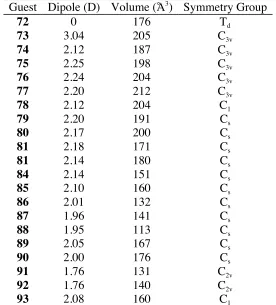
Table 2.1 Physical Properties of Guests...51
Table 2.2 Association Constants of Hosts 36 and 70...52
Table 2.3 Thermodynamic Characteristics of Host 70...54
Table 2.4 Association Constants of Host 71...56
Table 2.5 Thermodynamic Characteristics of Host 71...56
Table 2.6 Association Constants of Hosts 36 and 99 with Bromide Guests ...63
Table 2.7 Association Constants of Hosts 36 and 99 with Iodide Guests...64
Table 3.1 Product distribution and yields of lithiation/formylation reaction ...78
Table 3.2 Product Yield in the Presence of Guest ...80
Table 3.3 Yield of methyl ester m-basket utilizing three different bases...84
Table 4.1 Yields of Partially/Fully Weaved 2-Me m-baskets Products ...108
Table 4.2 Yields of Partially/Fully Weaved m-baskets Products ...111
Table 4.3 Yields of 153...114
Table 6.1 Crystal data and structure refinement for 145...165
Table 6.2 Atomic coordinates (x104) and equivalent isotropic displacement parameters (Å2 x10³) for 145...166
Table 6.3 Bond lengths [Å] and angles [deg] for 145...170
Table 6.5 Hydrogen coordinates ( x104) and isotropic displacement parameters
(Å2 x10³) for 145...183
Table 6.6 Torsion angles [deg] for 145...185
Table 6.7 Crystal data and structure refinement for 149...192
Table 6.8 Atomic coordinates ( x104) and equivalent isotropic displacement
parameters (Å2 x10³) for 149...193
Table 6.9 Bond lengths [Å] and angles [deg] for 149...195
Table 6.10 Anisotropic displacement parameters (Å2 x10³) for 149...198
Table 6.11 Hydrogen coordinates ( x104) and isotropic displacement parameter
(Å2 x10³) for 149...200
LIST OF FIGURES
Figure 1.1 Potassium cation bound by benzo[18]crown-6...1
Figure 1.2 The two possible orientations of dipole-dipole interactions of carbonyl groups ...2
Figure 1.3 Watson-Crick base pairing ...2
Figure 1.4 Quadrapole moments of aromatic rings ...3
Figure 1.5 Orientations of aromatic rings ...3
Figure 1.6 Peptidocalixarenes used to bind amino acids in CDCl3 and D2O ...5
Figure 1.7 Cyclodextrin ...7
Figure 1.8 Space filling model of β-cyclodextrin...7
Figure 1.9 Modified cyclodextrin and rocuronium bromide...9
Figure 1.10 Conformations of calix[4]arene ...11
Figure 1.11 Conformations of calix[6]arene found in the solid state ...16
Figure 1.12 Bridged calixarene regioisomers...18
Figure 1.13 Ribose in solution...23
Figure 1.14 Crystal structure of 36 and iodoadamantane ...28
Figure 1.15 Requirements of functionalized benzyl alcohols ...29
Figure 1.16 Space filling model of CB[6]...34
Figure 1.17 Stabilization of tetrahedral intermediate by pyridone...42
Figure 2.1 Open and closed conformations of 70...46
Figure 2.2 Optimized geometry of 70 with restrained dihedral angles ...46
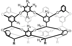
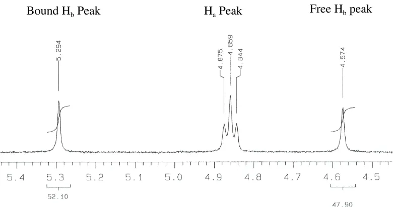
Figure 2.3 Proton designation for hosts 36, 70 and 71...47
Figure 2.4 Potential density maps of 36, 70 and 71...47
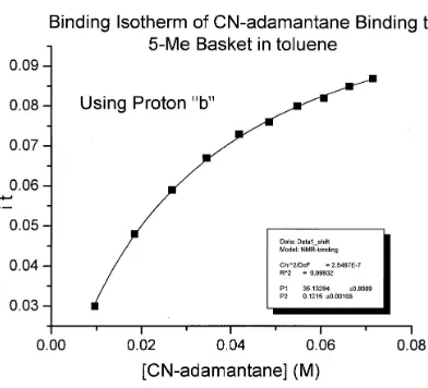
Figure 2.6 Binding isotherm of host 70 and 73...49
Figure 2.7 Guests used in binding studies...50
Figure 2.8 von Hoft plot of host 70 and 77 in CDCl3...54
Figure 2.9 Upfield portion of EXSY NMR of 70 and 77...57
Figure 2.10 Upfield portion of EXSY NMR of 71 and 86...58
Figure 2.11 Model of 86 binding to 71...59
Figure 2.12 Hydrogen bond preference of protium and deuterium ...60
Figure 2.13 Percent change of association constants as a function of cycle size...64
Figure 3.1endo- and exo- position and nomenclature...68
Figure 3.2 Possible substitution patterns from per-lithiation ...70
Figure 3.3 ChemDraw and cartoon structures of aldehydes produced under reaction conditions...71
Figure 3.41 H-NMR of 101...72
Figure 3.51 H-NMR of 100...73
Figure 3.6 Indicator protons used to identify aldehyde m-baskets ...73
Figure 3.7 d1 array of 101...76
Figure 3.8 Product distribution using a) 5.5 equivalent or b) 10 equivalent of base...78
Figure 3.9 Substitution patterns seldom or never seen in the lithiation/formylation of 36...79
Figure 3.10 Methyl esters formed from lithio-m-basket and methyl chloroformate ...83
Figure 3.11 Products formed from the saponification of the methyl esters m-baskets...85
Figure 3.12 Cyclic and bicyclic acetals ...87
Figure 3.15 Conformations of guest inside hosts ...90
Figure 3.16 Upfield NMR region of 123/137 showing bound 137...91
Figure 3.17 Upfield NMR region of 124/137 showing bound 137...92
Figure 3.18 Upfield 2D-EXSY showing bound 137 by 124...92
Figure 3.19 Model of 124 bound to 137...93
Figure 3.20 Upfield NMR region of 129/137 showing bound 137...94
Figure 3.21 Upfield NMR region of 131/137 showing bound 137...95
Figure 3.22 2D-EXSY of 124/137...95
Figure 3.23 Model of 131 bound to 137...96
Figure 3.24 Two possible orientations of a guest within a host with both endo- and exo-carboxylic acids ...96
Figure 3.25 Upfield NMR region of 128/137 showing bound 137...97
Figure 3.26 Two binding orientations of 137 within 128...97
Figure 3.27 Upfield NMR region of 130/137 showing bound 137...98
Figure 3.28 2D-EXSY of 130/137...99
Figure 4.1 Common tripodal ligands used to mimic carbonic anhydrase...102
Figure 4.2 Tris(pyridyl) ligands with amino acid blocking groups ...104
Figure 4.3 Funnel complex using imidizole as the nitrogen donor with zinc ...105
Figure 4.4 Products formed attempting to make 2-Me-tris-m-basket...107
Figure 4.5 Space filling model of mimic synthesized from 2-Me tris-m-basket...109
Figure 4.6 Products formed attempting to make tris-m-basket ...109
Figure 4.7 Crystal structure of 145...110
Figure 4.9 NMR zinc titration of 156 a) in the absence of zinc b) with one
equivalent of zinc...118
Figure 4.10 Binding isotherm of 156 titration with zinc ...119
Figure 6.1 Binding isotherm of host 70 and adamantane in toluene-d8...147
Figure 6.2 Binding isotherm of host 70 and cyanoadamantane in toluene-d8...148
Figure 6.3 Binding isotherm of host 70 and bromocyclohexane in DMSO-d6...148
Figure 6.4 Binding isotherm of host 70 and iodocyclohexane in DMSO-d6...149
Figure 6.5 Binding isotherm of host 70 and bromocyclopentane in DMSO-d6...149
Figure 6.6 Binding isotherm of host 70 and iodocyclopentane in DMSO-d6...150
Figure 6.7 Binding isotherm of host 70 and iodopentane in DMSO-d6...150
Figure 6.8 Binding isotherm of host 70 and bromobenzene in DMSO-d6...151
Figure 6.9 Binding isotherm of host 70 and iodobenzene in DMSO-d6...151
Figure 6.10 Binding isotherm of host 71 and bromocycloheptane in DMSO-d6...152
Figure 6.11 Binding isotherm of host 71 and cyclohexane in DMSO-d6...152
Figure 6.12 Binding isotherm of host 71 and bromocyclohexane in DMSO-d6...153
Figure 6.13 Binding isotherm of host 71 and iodocyclohexane in DMSO-d6...153
Figure 6.14 Binding isotherm of host 71 and bromocyclobutane in DMSO-d6...154
Figure 6.15 Binding isotherm of host 71 and iodopentane in DMSO-d6...154
Figure 6.16 Binding isotherm of host 99 and bromocyclooctane in DMSO-d6...155
Figure 6.17 Binding isotherm of host 99 and iodocyclooctane in DMSO-d6...155
Figure 6.18 Binding isotherm of host 99 and bromocycloheptane in DMSO-d6...156
Figure 6.19 Binding isotherm of host 99 and iodocycloheptane in DMSO-d6...156
Figure 6.22 Binding isotherm of host 99 and bromocyclopentane in DMSO-d6...158
Figure 6.23 Binding isotherm of host 99 and iodocyclopentane in DMSO-d6...158
Figure 6.24 Binding isotherm of host 99 and exo-2-bromonorborane in DMSO-d6...159
Figure 6.25 1 H-NMR of (+/-)-105 in CDCl3...160
Figure 6.26 2D-ROESY of (+/-)-105 in DMSO-d6...160
Figure 6.27 1H-NMR of 107 in CDCl 3...161
Figure 6.28 2D-NOESY of 107 in DMSO-d6...161
Figure 6.29 1 H-NMR of (+/-)-109 in CD2Cl2...162
Figure 6.30 2D-ROESY of (+/-)-109 in DMSO-d6...162
Figure 6.31 Crystal structure of 145...164
LIST OF SCHEMES
Scheme 1.1 Synthesis of calixarenes...11
Scheme 1.2 Synthesis of resorcinarenes...21
Scheme 1.3 Synthesis of ditopic host 29...24
Scheme 1.4 Synthesis of 30...25
Scheme 1.5 Synthesis of deep cavity cavitands...27
Scheme 1.6 Synthesis of m-basket ...27
Scheme 1.7 Synthesis of cyclotriveratrylene (37) ...29
Scheme 1.8 Mock’s pseudorotaxane switch...34
Scheme 1.9 Cyclization of cyclopropanes...40
Scheme 2.1 Synthesis of 5-Me m-basket (70) ...45
Scheme 2.2 Synthesis of 2-Me m-basket (71) ...45
Scheme 2.3 Synthesis d4-m-basket (99) ...61
Scheme 2.4 Synthesis of 82...62
Scheme 2.5 Synthesis of 80...62
Scheme 3.1 Reaction mechanism of Directed Ortho Metallation ...67
Scheme 3.2 Synthesis of benzyl alcohol m-baskets ...75
Scheme 3.3 General synthesis of ester m-baskets...82
Scheme 3.4 General synthesis of carboxylic acid m-baskets ...84
Scheme 3.5 Synthesis of acetals ...86
Scheme 4.1 Mechanism of reversible hydrolysis of carbon dioxide by Carbonic Anhydrase ...102
Scheme 4.4 Proposed scheme for the synthesis of m-basket CA mimic...106
Scheme 4.5 Synthesis of 2-Me-m-tris-basket ...106
Scheme 4.6 Synthesis of tris-m-basket...110
Scheme 4.7 Synthesis of tripodal ligand ...112
Scheme 4.8 Model reaction to test the Pd source ...113
Scheme 4.9 Model Suzuki reaction using 35...113
Scheme 4.10 Model Suzuki reaction using 149...114
Scheme 4.11 Synthesis of tris-m-basket diol...115
ABBREVIATIONS
t
BuOH tert-butanol n-BuLi n-butyllithium sec-BuLi sec-butyllithium tert-BuLi tert-butyllithium
CB Cuccurbituril
CD Cyclodextrin
CDCl3 Chloroform-d
COSY Correlated spectroscopy
CTC Cyclotricatechylene
CTV Cyclotriveratrylenes
DMA N,N-dimethylacetamide DME Ethylene glycol dimethyl ether
DMF N,N-dimethylforamide DMSO Dimethyl sulfoxide
Eq Equivalents
EXSY Exchange spectroscopy
ITC Isothermal titration calorimetry
i
PrOH Isopropyl alcohol
Ka Association constant
NOESY Nuclear Overhauser Effect spectroscopy
PNPCC para-nitrophenyl choline carbonate
ABSTRACT
As a first step to producing a shape selective catalysts or enzyme mimic, two
preorganized host molecules were synthesized. Binding studies of the two hosts with a variety of
guests in three solvents demonstrated that an important driving force in the association was the
formation of C-H⋅⋅⋅X-R hydrogen bonds (X = halogen). A deuterated host was utilized to further
examine the formation of the C-H⋅⋅⋅X-R hydrogen bonds.
In an effort to place functionality in the hydrophobic pocket of these hosts, two methods
were developed. The first utilized directed ortho metallation to place electrophiles above and/or directed into the cavity. Perlithiation of the host could lead to sixty-nine products but reaction
conditions and host rigidity limited product formation. This reaction technique led to the
placement of carboxylic acid groups onto the host and the isolation of twelve products. Two
different positions of the carboxylic acids (endo- and exo-) direct the orientation of the guest. 1D- and 2D-NMR were utilized to examine how the was orientated inside the host.
The second method employed to place functionality on the host, sited a tripodal zinc
binding ligand on the side of the hydrophobic pocket of the host. The synthesized host was able
I.
INTRODUCTION
1.1 Host-Guest Interactions
Host-Guest chemistry has evolved from cation binding crown ethers and macrocyclic
Schiff bases, to larger molecules that have the ability to encapsulate biologically relevant and
complex molecules. Hosts have been designed to bind cations, anions, both (ditopic), or neutral
molecules.[1]
Hosts can utilize many different, subtle interactions to increase selectivity and
affinity for a particular guest. These noncovalent interactions are:
1) Ion-ion interactions are particularly strong intermolecular interactions with energies
ranging from 100-350 kJ/mol.[1]
An example of an ion-ion interaction is a salt bridge found in
proteins (-COO- +
NH3-). [1]
2) Ion-dipole interactions have been extremely important in supramolecular chemistry.
These interactions are often used to build extended molecular architectures.[2, 3]
These
interactions range in energy from 50-200 kJ/mol and are the driving force in the association
between cations and crown ethers (Figure 1.1).[1]
O
O O
O
O O K+
Figure 1.1 Potassium cation bound by benzo[18]crown-6
3) Dipole-dipole interactions arise from the alignment of two different dipoles. One
example is the interaction between two carbonyl groups (Figure 1.2)[1]
of which there are two
possible alignments. The orthogonal alignment (a) allows for one point of interaction with the
The anti-parallel alignment (b) has the dipoles arranged so that there are two points of
interaction.
O R
R O R
R O R R O R R
a b
Figure 1.2 The two possible orientations of dipole-dipole interactions of carbonyl groups
4) Hydrogen bonding can be considered a type of dipole-dipole interaction.[1]
There are
however additional components to hydrogen bonds, such as electrostatic, charge transfer,
dispersion, and polarization forces.[4, 5] Strong hydrogen bonds, such as F-H
⋅⋅⋅F- and N-H⋅⋅⋅O, are
primarily electrostatic in nature. In contrast, weaker hydrogen bonds, such as C-H⋅⋅⋅O, are more
dispersive in nature.[6, 7] Hydrogen bonding, along with !-! stacking and hydrophobic forces
(see later), are among the most important in biological systems. They participate in DNA double
helix in addition to protein secondary and higher order structure formation, along with countless
other recognition events. An example of biologically important hydrogen bonds is the
Watson-Crick base-pairing model (Figure 1.3).[8]
N N N H N N N H N O O H H H N N N H N O N N NH N O H H H H H
Figure 1.3 Watson-Crick Base Pairing
5) Cation-! interactions have been widely utilized in calixarene and cavitand chemistry
as recognition elements.[9-13] Their energy can range from 5-80 kJ/mol.[1] Researchers have also
6) !-! interactions, as mentioned above, play a key role in biological systems as a driving
force for DNA and protein assembly.[16] The interaction is governed by the quadrapole moments
of the aromatic rings (Figure 1.4).[1]
Figure 1.4 Quadrapole moments of aromatic rings
There are two orientations that can be adopted by interacting aromatic systems (Figure 1.5). The
edge to face orientation (a) can be considered a C-H⋅⋅⋅! interaction[17] and is primarily
electrostatic. The offset stacked interaction (b) is the orientation found in DNA duplex. This
orientation allows for greater van der Waals interaction and thereby reduces the surface area
exposed to solvent.[18, 19]
a
b
H H H
Figure 1.5 Orientations of aromatic rings
7) The weakest of all intermolecular forces are van der Waals interactions.[1] When the
nucleus of one atom approaches a second atom, the electron cloud polarizes and gives rise to
these attractive forces. The individual interaction energy is minimal, and the molecules must be
in very close proximity for this interaction to have any effect. However, many of these,
interacting in concert, can give rise to relatively large association constants. Several of the
previously mentioned forces partially involve van der Waal interactions.
The “hydrophobic effect” can promote association, although it is not a force of
interaction.[20]
entropy. There are two means by which an increase in the enthalpic gain can be achieved.
Solvent molecules present in the host are higher in energy than bulk solvent due to the reduced
interaction between the host and solvent as compared with solvent-solvent interactions. When
these solvent molecules are released through guest binding, an enthalpic gain is seen.
Additionally, the solvent molecules around the guests are higher in energy than in bulk solvent.
When these are released through association, there is a decrease in enthalpy. Entropy is
increased by the release of these high-energy solvent molecules. For example, there are twelve
water molecules that solvate the interior of β-cyclodextrin. When a guest is bound, there is an
increase in entropy. However, the ordering of the host and guest often “hides” this increase in
entropy.
A host-guest complex arises from many of these forces and it is often difficult to
determine which force is dominant. Additionally, the interaction that dominates depends on
external conditions such as solvent and temperature. One example is the complexation studies of
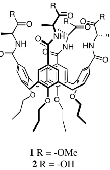
hosts 1 and 2 (Figure 1.6).[21]
When 1 binds amino acids in CDCl3 the major driving force is the
formation of hydrogen bonds between the ammonium and the host’s amide carbonyl.
Alternately, when using 2 as a host in water, the main driving force is the hydrophobic effect.
R
O
O O O
O O NH
R O
O NH
O R
NH O
O HN
O R
1 R = -OMe 2 R = -OH
Figure 1.6 Peptidocalixarene used to bind amino acids in CDCl3 and D2O
1.2 Enzymes and Enzyme Mimics
Enzymes are extremely active and selective catalysts and serve as an inspiration to
host-guest chemists.[1]
They derive their catalytic power from several phenomena.[22, 23]
They increase
the effective molarity of substrate and reactive groups by binding the substrate in an
active-site.[24]
Additionally, enzymes can stabilize a transition state and/or destabilize a ground state.
The selectivity of an enzyme arises from the active site. The active site is generally a concave
part of the enzyme that contains all the functionality required to affect a transformation. The
amino acid residues in the active site are arranged in such a way that only the desired substrate
will be catalyzed, while the residues in or around the active site can direct the substrate into the
active site. For example, the opening into the active site of acetylcholine esterase has a negative
charge that attracts the positively charged acetylcholine.[25]
While enzymes are exceptional catalysts, studying them can prove challenging. Proteins
are very large, conformationally complex molecules and are usually available in very small
quantities. They are, therefore, difficult to study using traditional spectroscopic techniques.
Accordingly, it is advantageous to use small molecule mimics of the active site to determine
Nature has not required that all possible reactions and substrates of interest to synthetic
organic chemistry be catalyzed. As a result, the majority of reactions and substrates of interest to
organic chemistry are not catalyzed by enzymes. Additionally, life has evolved in relatively
narrow limits. Enzymes are only active at certain temperatures, salt concentrations, solvents, and
pH. When not within these windows, enzymes lose much of their catalytic power. Another
consideration is that not all organic molecules are soluble in water. It would therefore be
beneficial to develop enzyme mimics that would catalyze reactions and substrates not essential to
natural selection.
1.3 Molecular Hosts
There have been hundreds of hosts described in the literature. To limit the number of
hosts described here, several requirements were put into place, with the primary restriction being
that the host molecule must be concave. The host must also be organic in nature. This
requirement removes zeolites and other inorganic host molecules from consideration. Thirdly,
the host must be monomeric, which eliminates “molecular capsules” and other self-assembling
systems.[26]
The host must be large enough to encapsulate greater than 50% of the guest. Many
rotaxanes and catenanes are ineligible for consideration due to this restraint.[27]
Finally, the host
must have a permanent entrance/exit, thereby excluding carceplexes and hemicarceplexes.[28]
The
host molecules that fulfill all these requirements are: cyclodextrins (CD); calixarenes; cavitands;
cyclotriveratrylenes (CTV) and cucurbiturils (CB).
1.3.1 Cyclodextrins
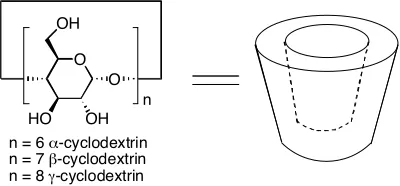
Cyclodextrins (CD) are a group of macrocyclic host molecules synthesized from α
host-guest chemistry as well as enzyme mimicry. Industrially, CDs are made from the
degradation of starch by cyclodextrin glucosyl transferase.[29]
Depending on the desired CD size
(see later) several templates may be added to form complexes with the selected cyclodextrin that
remove it from solution. All CDs are cone-shaped molecules with a hydrophobic interior made
structurally rigid from a ring of hydrogen bonds. There are two portals to the hydrophobic
interior. The largest portal is the “secondary face”, so called because it is ringed by secondary
alcohols. The smaller portal is the primary face. The secondary alcohols hydrogen bond to each
other in an interglucose fashion, which gives the host rigidity. The hydrophobic interior is lined
with C-H groups and glycosidic oxygens. This interior is less hydrophobic as compared with
calixarenes or cavitands, but it does allow for favorable van der Waals interactions and shields
the hydrophobic guest from bulk water (Figure 1.8).
O
HO OH
O OH
n
n = 6 α-cyclodextrin n = 7 β-cyclodextrin n = 8 γ-cyclodextrin
Figure 1.7 Cyclodextrin
Figure 1.8 Space filling model of β-cyclodextrin
volume is 174 Å3.[29-31]
β-CD has seven α-(+)-glycopyranose units, a cavity diameter of 6.6 Å and
a cavity volume of 262 Å3
. Finally the largest cyclodextrin, γ-CD, is made from eight α
-(+)-glycopyranose units. Its cavity diameter and volume are 8.4 Å and 427 Å3
respectively.
Table 1.1 Dimensions of cyclodextrins
A
C B
CD A (Å) B (Å) C (Å)
α 13.7 5.2 4.7
β 15.3 6.6 6.0
γ 16.9 9.5 7.5
Due to the relative ease of their synthesis, CDs have been widely studied. They have
been found to form stabile complexes with a wide range of guests including aliphatic and
aromatic hydrocarbons, aliphatic and aromatic alcohols, amino acids, sugars and a variety of
drug molecules.[30, 31]
CDs prefer binding to guests that will interact simultaneously with the
hydrophobic interior as well as the alcohols at the rim of the hosts. Therefore, α-CD prefers to
bind amino acids, β-CD prefers to bind adamantane carboxylic acid, and γ-CD prefers to bind
larger guests such as anionic methyl orange (3).[31]
NN S
O
O O -N
3
Several noncovalent forces drive these complexations. The most important being the
hydrophobic effect in which the organic guest as well as the CD is desolvated (β-CD has been
provides a large increase in entropy upon guest binding. Other forces include the formation of
hydrogen bonds and beneficial van der Waals interactions between host and guest.[31]
One recent example of using a modified CD is the binding of rocuronium bromide, 4
(Figure 1.9).[32]
Rocuronium bromide is a neuromuscular blocker used in anesthesia to relax
muscle tissue and facilitate access to internal cavities and organs. The physiological action of
rocuronium bromide involves stopping the action of acetylcholine and nicotine acetylcholine.
To reverse this action, drugs are given that inhibit the action of acetylcholine esterase, thereby
increasing the concentration of acetylcholine. These “recovery” drugs can have deleterious side
effects such as nausea, vomiting and hypotension. If however, the neuromuscular blockers could
be encapsulated and removed from the body, the reversal drugs would not be necessary.
Initially, α, β, and γ-CD were tested but the only γ-CD demonstrated any binding ability.
O O O O O O O O O O O O O O O O HO OH HO OH HO HO HO HO OH HO OH OH OH OH OH OH S NaO2C
S CO2Na
S CO2Na
S
CO2Na
S
CO2Na
S
CO2Na
S NaO2C
S NaO2C
N OAc N O HO H
4 5
Figure 1.9 Modified cyclodextrin and rocuronium bromide
To increase the association of 4, a γ-cyclodextrin was modified to produce 5. The modifications
increase the lipophilicity of CD, and the anionic groups near the cavity increase the association
both enthalpically and entropically favored. Researchers have found, using Rhesus monkeys,
that when administered 0.5 µmol/kg of 5, the action of 4 is reduced to 90% within three minutes
and there were no changes in vital signs even when 5 was used up to 10 µmol/kg. This is an
improvement of half the recovery time without any of the adverse side effects.
While CDs are excellent host molecules, they still possess several drawbacks. There is
no position to place functionality so that it would continually face into the hydrophobic pocket.
Additionally, there are three different types of hydroxyl groups on the two faces of cyclodextrin
(Figure 1.7). D’Souza has developed methods to place functionality where it is desired on
cyclodextrins.[33] The 6-position is the most nucleophilic and the most basic. The 2-position is
the most acidic and the 3-position is most inaccessible. Using this knowledge it is possible to
promote one product over another. However, it is not possible to eliminate the formation of the
unwanted regioisomers.
The secondary alcohols rigidify the structure of CD through a ring of hydrogen bonds. If
substitution takes place on these alcohols, then the rigidity is subsequently reduced. Researchers
must therefore be aware of the increased flexibility of the host CD. This becomes important
when attempting to functionalize CD to allow for solubility in solvents other than water and
polar protic solvents.
1.3.2 Calixarenes
Calixarenes are macrocyclic compounds made from the condensation of p-alkyl phenols with formaldehyde (Scheme 1.1).[34]
Scheme 1.1 Synthesis of calixarenes
R
OH
CH2O OH
-R
OH n
n = 4-8
The products formed are determined by reaction conditions (amount of base, temperature,
etc). The largest yields are with n = 4, 6, and 8 (50, 85 and 63%) and are therefore the most
studied. Calix[4]arenes (6) and calix[6]arenes (7) have four and six aromatic units
respectively.[35] R R R R R R OH OH OH OH HO HO OH OH OH HO R R R R
6 7
Calix[4]arene have a cup shaped structure. Four different conformations arise from the
rotation about the σ-bonds of the Ar-CH2-Ar: the cone; partial cone; 1,3 alternate; and the
1,2-alternate conformation (Figure 1.10). The unmodified calix[4]arene is in equilibrium between
these four conformations. The cone conformation is stabilized by the formation of
intramolecular hydrogen bonds between the phenols.
OH R R OH R OH OH R OH R R R OH OH OH R R OH R R OH OH OH R OH R OH R OH OH R R
In the cone conformation there are two sides to the calixarene. Traditionally the rim with
the alkyl groups is known as the upper rim and the side with the phenols is known as the lower
rim.
Solid-state structures have shown that calix[4]arenes can bind neutral organic molecules.
They do not however, act as host in solution due to rapid conformational changes.
Rigidification of the cone conformation is required to allow the calix[4]arene to act as a
host in solution. There are two places to rigidify the cone conformation: the upper and lower
rim. One method is to position bulky groups on the phenolitic oxygens of the lower rim. This
mechanically prevents the rotation around the Ar-CH2-Ar bonds. An example is 8.
[36] While the
bulky amide is able to maintain the calixarene in the cone conformation, there is still residual
movement around the Ar-CH2-Ar bonds. The calixarene adopts two conformations, a C4v
conformation (cone) and a C2v conformation (“pinched cone”). If sodium is added to a solution
of 8, the carbonyl oxygens bind the sodium (9) that in turn increases the rigidity of the
calixarene. This rigidity allows it to act as a host for nitromethane in CDCl3 (Ka = 34 M -1
).
O Na+
O O
O O
O N O N
N N O O But tBu
tBu
tBu
O O
O O
O N O N
N N O But tBu
tBu
tBu
8 9
The oxygens of the calixarene may also be linked covalently. Various spacers have been
used.[36]
230 and 50 M-1
respectively. The higher association constants of 10 with nitromethane
demonstrate the importance of preorganization and rigidity of hosts.
O O O O But t
Bu
tBu
tBu
O O
O O O O But t
Bu tBu tBu O O O O
10 11
The upper rim may also be covalently joined to reduce the residual motions. Ungaro et al. used a variety of aromatic spacers to bridge the upper rim (12).[37]
This technique also allowed
for the introduction of a pyridine nitrogen to act as an additional recognition point. These hosts
were able to bind solvent sized molecules with acidic protons (Table 1.2).
O O O
O
O O O O
O X O
O O
Z
12
Table 1.2 Binding Constants of host 12a
Guest (Solvent)
X = CH Z = H
X = N Z = H
X = N Z = OMe
X = N Z = N(Me)2
CH3CN (CCl4) - 36 13
-CH3NO2 (CCl4) - 57 124
-CH2(CN)2
(CDCl3)
- 79 50 37
Overall, modified calix[4]arenes can act as hosts for small, neutral, solvent-sized
molecules. The driving force for complexation is the formation of favorable van der Walls
interactions and C-H-π interactions. To further develop the chemistry of these molecules,
researchers have used electrophilic substitution at the upper rim to both enlarge the cavity and
add recognition points to increase selectivity and binding strength.
Additional depth has been used to increase a molecule’s ability to act as a host. In the
case of calixarenes, this has been achieved by increasing the cavity walls.[38]
A range of
calixarenes (6) was synthesized and their Kas were determined (Table 1.3). It does appear that
deepening the cavity will increase the affinity of the guest.
Table 1.3 Binding Constants of calix[4]arene with increasing alkyl groupsa
R = Ka(M
-1
) CDCl3
H 5
Tert-Bu 27
Cyclohexyl 36
Phenyl
-Key: aThe symbol “-“ indicates that binding was not noted
An example of adding a recognition point is the calixarene 13.[36]
The researchers use two
points of binding to allow the modified calixarene to act as a host. The carbonyl group of the
amide guest forms hydrogen bonds with the urea group of the calixarene. Additionally, the NH
group of the guest forms an NH-π interaction with the arenes of the host. The guest species
O O O O O O N N O H H Me O N H CH3 H N O HN
O O O
O
O O
13 14 15
To further enhance the binding ability of calixarenes, the Ungaro group has placed
peptides at the upper rim. This substitution has the advantage of stabilizing the cone
conformation by formation of intramolecular hydrogen bonds.[21, 39]
The calixarenes 16 and 17 both bind carboxylic acids and ammonium cations in CDCl3.
However, 16 had larger association constants. The greater flexibility of 16 allows it to form
more effective hydrogen bonds.[21]
It is worthwhile to note that the researchers do not believe that
the hydrophobic pocket is involved in the reception of these guests. The calixarene acts as a
scaffold for the hydrogen bond donors/acceptors to be arranged.
MeO O O O O O O NH MeO O O NH O MeO NH O O HN
O OMe MeO
O
O O O O O NH MeO O O NH O MeO NH O O HN O OMe O O
16 17
Hydrolysis of the ester makes the calixarenes (18 and 19) water-soluble. When these
driving force of association has changed from the formation of hydrogen bonds to hydrophobic
effects and the esters bind into the cavity of the calixarene.
HO
O
O O O
O O NH HO O O NH O HO NH O O HN O OH HO O O O O O O NH HO O O NH O HO NH O O HN O OH O O
18 19
Calix[6]arenes (7) are significantly more flexible. While calix[4]arene has always been
found in the cone conformation in the solid state, two conformations of calix[6]arene have been
described. One conformation has all of its –OH groups on the same side of the molecule (cone
conformation, Figure 1.11a). The second conformation has two sets of three neighboring phenol
–OH groups on opposite sides of the molecule (1,2,3 alternate conformation, Figure 1.11b).
OH R OH OH R OH R R OH R OH R OH R OH OH R R R OH R OH R OH
a b
Figure 1.11 Conformations of calix[6]arene found in the solid state
One might consider rigidifying the calix[6]arene by alkylating each phenol individually.
This proves to be unsatisfactory for two reasons. Large alkyl groups are required to slow the
rotation around the Ar-CH2-Ar bonds. For example, the calixarene 20 has a great amount of
Cs+
, it can then act as a host for C60. [40]
Secondly, incomplete alkylation gives rise to a large
number of regioisomeric products.[41]
These reasons have led researchers to bridge the phenols
with a variety of aromatic spacers.[42]
tBu
But
But
tBu
tBu tBu OR
OR
OR OR
RO RO
R = -CH2COOEt
20
Gutsche’s group reported A/D linkage by reacting tert-butyl calix[6]arene with α,α ’-dibromo-p-xylene. When the remaining phenols are methylated a “self-anchored rotaxane” 21
was formed, a clear indication that the cone conformation was not rigid.[43]
R
R
R
R
R O O
O
O O
O R
R = tBu
21
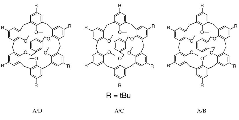
Several other groups have bridged the calix[6]arene A/D positions.[44, 45]
The Shinkai
regioisomers arising from bridging with aromatic spacers at different positions (A/B v. A/C v.
A/D). They found that all bridges stabilized the calixarene in the cone conformation relative to
the unmodified calixarene. Under these circumstances, the most stable was the A/C bridged
calix[6]arene (Figure 1.12).
R
R
R
R
R R O
O
O O
O O
R
R
R
R
R R O
O
O O
O O
R
R
R
R
R R O
O
O O
O O
R = tBu
A/D A/C A/B
Figure 1.12 Bridged Calixarene regioisomers
Another way to rigidify the calix[6]arene is to bridge three phenol positions, the A/C/E
positions. These compounds are also known as “capped” calixarenes. The capping can be
accomplished in several ways.
Shinkai group used covalent bonds to link the phenolitic oxygens on the lower rim.[44]
They bridged 1,3,5-tri-O-methylated tert-butyl calixarene with 1,3,5-tris(bromomethyl)benzene and obtained a 91% yield of 22. The cone conformation of this compound was found to be
R
R
R
R
R R O
O
O O
O O
R = tBu
22
An unfortunate result of this method is the effective sealing of the calix[6]arene. The
aromatic cap seals the lower rim while alternating tert-butyl groups effectively seals the upper
rim. The capped calix[6]arenes do act as hosts toward primary ammonium cations, but are
selective for Cs+
.[46]
Quadruple bridging of calix[6]arene has been accomplished at the A/B/D/E positions of
the lower rim using 1,2,4,5-tetrakis(bromomethyl)benzene with a surprisingly high yield
(65-80%)(23). NMR studies have demonstrated that these capped calixarenes are also stable in the
cone conformation.[47]
These molecules have been used to encapsulate Cs+
.[48, 49]
R
R
R
R
R R OH
O
O OH
O O
Reinaud et al. have synthesized a variety of functionalized capped calix[6]arenes.[50-52]
One example is 24 which has a triphenylphosphine cap on the lower rim of calix[6]arene.[52]
This
stabilizes the cone conformation on the NMR time scale and allows the calixarene to act as a
host for several primary amines. The primary driving force appears to be the formation of
hydrogen bonds with the amines of the cap and the guest.
OMe HN
O OOMeO MeO NH
P
HN
24
Another way to stabilize the calix[6]arene is to bridge sequential aromatic rings. One
example is 25. Shinkai bridged a benzyl chloride calixarene with N,N-dihexyl-1,3 m -aminobenzene.[53] Shinkai have called these hosts “stapled calixarenes”. Such hosts can bind C
60
in toluene due to charge transfer interactions between the m-phenyl diamine bridges and C60. [40]
N
N C6H13
C6H13
3
25
The upper rim of calix[6]arenes can also be capped.[54]
1,3,5-tris(mercaptomethyl)benzene reacted with tetrachloromethyl calixarene to give the capped
product 26 in 28% yield. The association constant of 26 with PhNMe3I is >5 times greater than
OMe
MeO
MeO
OMe
OMe OMe
tBu
tBu But
S S
S
26
Calixarenes act as molecular hosts. However, they suffer from several drawbacks. All
calixarenes are conformationally flexible and care must be taken to ensure that the conformation
is sufficiently rigid. Calix[4]arenes’ hydrophobic pocket is only large enough to encapsulate
solvent size molecules. Extension of the cavity is required to encapsulate larger guests. Finally,
the lack of any inward facing functional groups usually requires that functionality be added
above or below the cavity, which might react with substrates not bound by the cavity.
1.3.3 Cavitands
Cavitands have been defined by Cram as “synthetic organic compounds that contain
enforced cavities of dimensions at least equal to those of the smaller ions, atoms or
molecules”.[55] This broad definition has been narrowed by other authors to include only
molecules that arise from functionalization of resorcinarenes.
Resorcinarenes are synthesized by the acid catalyzed[56]
condensation of resorcinol with
aldehydes (Scheme 1.2).
HO OH
X
RCHO H+
HO OH
X
R n
The yield of this reaction is very high when n = 4. The macrocycle precipitates out of solution
and therefore is a thermodynamic sink. There have been recent attempts to synthesize
resorcinarenes with n > 4.[57-59]
So far, they have only been obtained in low yield (n = 5, 4%; n =
6, 14%; and n = 8, 1%). A large range of resorcinarenes can be produced and the properties
changed by altering the aldehyde.
Resorcinarenes have a bowl shaped structure that is held in place by four hydrogen bonds
at the rim. These bowls are slightly flexible but not nearly as flexible as the corresponding
calixarenes.
Resorcinarenes themselves have been used as hosts for polar organic molecules. Aoyama
et al. have bound sugars, diols, and amino acids with resorcinarenes.[60-65]
One example is the
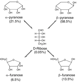
binding of ribose with 27. There are five different constitutional isomers of ribose in solution
(Figure 1.13).
HO
HO OH HO OH
R
R R R
H H H H
OH OH
HO
R = -(CH2)10CH3
CHO OH H OH H OH H
CH2OH
D-Ribose O OH OH OH OH O OH OH OH OH
α−pyranose β−pyranose
O HOH2C
OH OH OH
α−furanose
O HOH2C
OH OH β−furanose OH (21.5%) (58.5%) (6.5%) (13.5%) (0.05%)
Figure 1.13 Ribose in Solution
The major isomer is β-pyranose in solution. However, when ribose is extracted into CCl4 with
27, pyranose is the only isomer found and the α- is preferred to the β- in a 10:1 ratio. The
selectivity can be accounted for by the fact that the α-pyranose can form two hydrogen bonds
with the resorcinarene host and the β-pyranose can form only one.
Resorcinarenes have some residual flexibility. To further rigidify the structures and
enlarge the cavities, Cram et al. used different groups to link neighboring phenolitic oxygens (28) to synthesize the first cavitands.[55, 66]
Initially, Cram used simple alkyl groups to link the oxygens.[55, 66]
When X = CH2, the
cavity is rigid enough to bind small molecules in the solution and the solid state.[56, 67, 68]
The
host-guest systems were, unfortunately, fast on the NMR time scale and further elaboration of
the cavity was required.
The Cram group also used silyl bridges (X = R2Si). The inward facing alkyl groups
reduced the volume of the cavity and only linear guests could bind (carbon disulfide, acetylene
and molecular oxygen).[67-69]
Thermodynamic evaluation demonstrates that the binding is
enthalpically driven with an entropic penalty.
To entrap larger molecules and to increase the association constant, larger cavitands were
synthesized. One way is to build up over the resorcinol rings.[70, 71]
An example is the cavitand
29.[72]
Atwood et al. used the Mannich reaction to form several of these examples.[72]
These
cavitands were used as ditopic receptors for tetrabutylammonium chloride.
Scheme 1.3 Synthesis of ditopic hosts 29
Cram continued to link neighboring phenolitic oxygens to make larger cavitands. He
synthesized 30 in 34% yield by reacting resorcinarene and 2,3-dichloro-1,4-diazanapthalene.[66]
Scheme 1.4 Synthesis of 30
HO
HO
HO OH
OH OH OH HO
R
R R
R
N
N Cl
Cl
O
O O O O
R
R R R
H H H H
O
O O
N N
N N N N N N
30
The host-guest chemistry of cavitand 30 was studied by Dalcanale et al.[73, 74]
They found that 30
acted as a good host for substituted aromatic rings.
Cavitands such as 30 have high conformational freedom. The two extreme
conformations are known as the “vase” and the “kite”: the “vase” conformation with the
aromatic walls of the cavity up and the “kite” conformation with the aromatic walls extended.
The extent to which conformation is dominant depends on several factors including substitution,
electrostatic factors, temperature and solvent.[66, 75, 76] The extended cavitands form dimmers in
solution and are known as “velcrands”. The considerable conformational freedom enjoyed by
these cavitands reduces their ability to act as hosts.
In order to stabilize the vase conformation of these hosts, Rebek synthesized a group of
cavitands known as “self-folding cavitands”.[77, 78]
They began with resorcinarenes that were
bridged with 1,2-diflouro-4,5-dinitobenzene to give the octa nitro compound 31. This was
reduced to the octa amine compound 32. 32 was in turn acylated with various acid chlorides to
R R R R O O O O O O O O H2N
H2N
H2N
H2N NH2
NH2 NH2 NH2 R R R R O O O O O O O O O2N
O2N
O2N
O2N NO2
NO2 NO2 NO2 R R R R O O O O O O O O HN H N O R O R N H HN O R O R NH N H O R O R NH H N R O O R
31 32 33
The upper rim of 33 allows for the formation of a seam of hydrogen bonds that stabilize
the cavitand in a vase formation to the point where guest exchange is slow on the NMR
timescale. This cavitand has also been used as a chiroselective receptor by placing strereocenters
on the R groups of the amide.[79]
The intermediate octa amine 32 is also a host for choline and other tetramethyl
ammonium salts.[9]
It has been used by the Rebek group to make a range of deep-cavity
cavitands. Other groups can be condensed with the amines to build even deeper cavitands. 34
was synthesized from the condensation of acenaphthenequinone and 32.[80]
The cavity of 34 is
~14Å deep and can act as a host for C60 with a Ka of 900 M -1
.
O
O O O O
R
R R R
H H H H
O
O O
N
N N N N N N N
34
Deep cavity cavitands have also been synthesized using substituted benzal bromides as
bridging groups (Scheme 1.5). This reaction was largely tolerant of functionality (X = H, Br,
-I, -Me, -Phenyl, -CO2Et, -OCH2OEt, -NO2, -CN). Unfortunately, these concave molecules were
Scheme 1.5 Synthesis of Deep cavity cavitands
O
O O O O O
R
R R R
H H H H
O O
H H H H
HO
HO OH HO OH
R
R R R
H H H H
OH OH
HO
polar aprotic solvent base
X X X X
R = CH2CH2Ph
R = CH2CH2Ph
X
Br Br
In an attempt to eliminate this rotation, a new host (36) was synthesized from the deep
cavity cavitand 35 and resorcinol utilizing the Ullmann ether synthesis (Scheme 1.6).[84, 85]
This
new host (36) was called the meta-basket (m-basket) because of the relationship between the phenols on resorcinol.
Scheme 1.6 Synthesis of m-basket
O
O O O O O
R
R R R
H H H H
OH HO H H
BrBr Br Br Br
Br Br Br
O
O O O O O
R1
R1 R1 R1
H H H H
OH HO H
O O O O O O H O O
35 36
The host 36 was found to selectively bind halogenated guests, especially halogenated
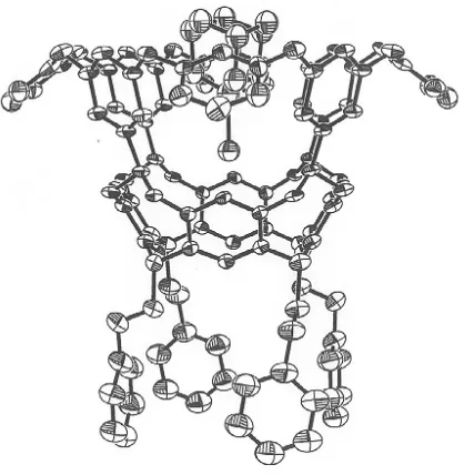
substituted adamantanes. A crystal structure of 36 binding iodoadamantane was obtained.
Interestingly, the adamatane guest does not completely fill the concave host but instead hovers
over its base. The reason for this inefficient packing is the van der Waal overlap of the iodine of
the guest, and the ring of four interior acetal hydrogens of the host (Figure 1.14).[84] The x-ray
crystal structure as well as 1D- and 2D-NMR indicated the formation of C-H⋅⋅⋅X-R hydrogen
Figure 1.14 Crystal Structure of 36 and Iodoadamantane
C-H⋅⋅⋅X-R interactions had previously been noted in several other systems. This was
however, exclusively in the solid state, and it was unknown if these interactions were
attractive.[86-88]
Other researchers have since noted C-H⋅⋅⋅X-R hydrogen bonds in inter- and
intramolecular interactions in solution.[89, 90]
Cavitand and resorcinarene hosts have been a fruitful area of research and many new
hosts have been developed from the original Cram models. This evolution has allowed the
accommodation of larger guests as well as placement of possible reaction centers along the rim.
The thermodynamic driving force for complexation is enthalpic gain while there is usually an
accompanying entropic loss. The enthalpic gain arises primarily from C-H-π interactions, as
well as van der Waals contacts.
1.3.4 Cyclotriveratrylene
Cyclotriveratrylene (CTV, 37) is a shallow bowl shaped molecule obtained by the
condensation of veratryl alcohols under strong acidic conditions (Scheme 1.7).[91-93]
resorcinarenes and calixarenes, it is unclear whether CTV is a thermodynamic or kinetic
product.[92]
Scheme 1.7 Synthesis of cyclotriveratrylene (37)
MeO OMe OMe
OMe MeO
OMe MeO
MeO
OH Acid
37
Many CTV analogs have been prepared. The requirements of the 3, 4-substituted benzyl
alcohols to react are very few (Figure 1.15). The 3-position must be electron donating, which
will activate the 6-position toward electrophilic attack. The 4-position’s only role is to block this
position from undergoing reaction leading to unwanted polymer products.
X
Y
OH 1 2 3
4 5 6
Figure 1.15 Requirements offunctionalized benzyl alcohols
If the benzyl alcohol is suitably substituted (X ≠ Y) the resulting CTV will be chiral. An
example is 38.[92]
The rasemization rate of this compound was obtained and from that the
crown-crown interconversion barrier was found to be 26.5 kcal mol-1
. Examination of other substituted
CTVs demonstrates similar energy barriers. However the rates of interconversion can be very
different.
D3CO OCH3
OCH3
OCD3
H3CO
OCD3
38
neither has been shown to act as a host in solution. This inability is due to the shallowness of the
cavity and the lack of functionality. To expand the range of host-guest chemistry of this class of
molecule, researchers have attempted to deepen the cavity and place recognition points onto the
cavity.
HO OH OH
OH HO
OH
39
Some of the first attempts to increase the binding ability of CTV were made by Hyatt
with “octopus” molecules 40.[92]
These long chain polyethers (n = 1-4) are able to non-selectively
complex monocations but not dications in aprotic solvent.
O O
O O O
O
O
R O
R O R
O
R O
R O R
n n
n n n n
40
Menger et al. placed six long chain carboxylic acids on CTV which makes the molecule soluble in basic (pH = 8.6) water.[99]
These molecules form micelles (nine molecules) at low
concentrations. These micelles will complex a variety of organic, aromatic molecules. For
example, p-Nitrophenyl butylate is bound and protected from base catalyzed hydrolysis.
In order to expand the concavity and stop the crown-to-crown conversion at higher
temperature, Cram bridged neighboring phenols on different aromatic rings with a variety of
aromatic molecules 41.[100]
The bridging did rigidify the CTV but the molecule has not been used
O O O
O O
41
To arrange metal coordination sites, as well as to extend the hydrophobic pocket of
CTVs, Weiss has bridged the phenolitic oxygens with a variety of pyridine groups (for example,
42).[101, 102]
This locks the CTV in one crown conformation. They are currently exploring the
ligand possibilities and host-guest chemistry of this type of CTV.
O O O
O O
N
N N
42
Another way to increase the size of the cavity is to build up directly above the aromatic
walls of the CTV. The Bohle group has employed metal coordination to increase the size of the
cavity.[103, 104] The group has used Schiff base chemistry to place a coordinated nickel on the
N O
N
O N
O
O
H
H
O
H
O
Ni L
Ni
L Ni
L
43
Aromatic nucleophilic substitution has been used to increase the volume of the CTV
cavity by Pochin.[105]
Various substituted aryl fluorides have been utilized to increase the size of
the walls of the hydrophobic pocket. The CTV 44 has been shown to bind tetramethyl
ammonium tosylate in CDCl3.
N
O HO OH
O O OH
N
O O
N
O O O O
44
CTVs can also act as molecular scaffolds[106, 107]
to allow the folding of collagen peptides
into triple helixes. This ability to form a helix model using native collagen peptides sequence
will allow researchers to evaluate how collagen interacts with peptides and small molecules.
Modified CTVs have also been used as scaffolds in both solution and solid phase
combinatorial chemistry.[108-110]
Solution studies show that the triple amino acids derivatives (45)
2197 molecules to identify small peptide trios that were selective for D-Ala-D-Ala or D-Ala-D
-Lac dipeptides, which are important in the study of vancomycin resistant bacteria.
OMe O
O
MeO OMe O
NH HN
NHBoc
NH HN
NHBoc
R O
O R
O O
HN NH BocHN
O O R R
R R
45
CTVs and modified CTVs are beginning to become viable host molecules. Much
research has been done to rigidify and increase the size of the cavity while adding recognition
points to the cavity. These molecules are unique in that they are chiral when appropriately
substituted which may be of use in future research.
1.3.5 Cucurbituril
Cucurbiturils (CB) (46) are barrel-shaped molecules which are the product of acid
condensation between glycouril and formaldehyde.[111-114]
This method gives primarily the cyclic
hexamer (n = 6) (Figure 1.15) in 40-70% yield.[115]
N N
N N O
O
H H
CH2
CH2
n
46
Like cyclodextrin, CB has two portals. However, unlike cyclodextrin, which has two
portal has a ~4Å diameter. It opens into a hydrophobic interior which is ~5.5Å in diameter. The
combination of the carbonyl oxygens and the hydrophobic interior allow CB to act as hosts.
Figure 1.16 Space filling model of CB[6]
The best guests for 46 are linear alkyl chains with multiple amine groups to interact with
both the portals’ carbonyls simultaneously through ion-dipole and hydrogen bond
interactions.[111, 116]
These interactions are pH dependent. Therefore, pH has been used to control
the motions of molecular machines synthesized with CB.[117, 118]
For example, Mock synthesized a
pseudorotaxane using CB[6] as a “bead” and a triamine “string” (Scheme 1.8).[119]
At acidic pH,
CB[6] resides at the doubly protonated diaminohexane “station” because CB[6] forms a more
stable complex with diaminohexane. However, when the pH is raised (pH > 6.7), deprotonation
of the aniline nitrogen causes the CB[6] to migrate to the fully protonated diaminobutane station
to allow the carbonyl oxygens to interact with the protonated amines.
Scheme 1.8 Mock’s pseudorotaxane switch
H2 N
N H2
NH3
-H+
+H+ H
N
N H2
NH3
CB[6]
There have been three limitations to using CB as molecular hosts, several of which have
macrocyclic rings of the CB. Secondly, there was no general way to functionalize the exterior of
the CB to make it soluble in common solvents. CB[6] is only soluble in formic acid/water
solutions. Lastly, there was no general way to functionalize the interior of the hydrophobic
pocket to allow for enzyme mimicry or supramolecular catalysis.
In an effort to increase the host-guest chemistry of CB, the Kim group carried out a group
of experiments to change the number of glycourils incorporated into the macrocycle.[112, 113, 120]
They found that by reducing the temperature of the reaction, CB[5], CB[6], CB[7], and CB[8]
could be produced in 15%, 50%, 20% and 15% respectively. The different sizes of the cavities
are shown in Table 1.4
Table 1.4 Dimensions of Cucurbit[n]uril
CB[5] CB[6] CB[7] CB[8]
Outer diameter (Å)
13.1 14.4 16.0 17.5
Portal Diameter (Å)
2.4 3.9 5.4 6.9
Cavity Diameter (Å)
4.4 5.8 7.3 8.8
The change in the size of the host also modifies which guests will be preferred. CB[5]
binds Pb2+
and ammonium cation. CB[7] prefers to bind 4,4’-dimethyl bipyridinium and
adamantaneamine.[121]
CB[8] is large enough to encapsulate several guests in its interior.[122]
It is also able to
encapsulate cyclen (47) or cyclam (48). The encapsulated macrocycle can then be metallated
with copper(II). The redox chemistry of the copper is significantly different when encapsulated
HN
HN NH NH
NH NH
HN HN
47 48
Larger CBs have also been observed by MS.[112]
CB[10] has been characterized by NMR,
MS, and x-ray crystallography with CB[5] as a guest. The two CBs rotate independently of each
other and are called “gyroscane”.[123]
CB[10] is never found without encapsulated CB[5] and
therefore it has been postulated that the relative high yield of CB[10] compared to CB[9] and
CB[11] is due to temptation by CB[5].
Having increased the number of possible cucurbiturils available for study, researchers
then turned to the problem of external functionalization to increase solubility in common
solvents. Initially, groups attempted to use different glycourils. The first successful attempt was
reported by several groups using dimethylglycouril (49) .[116, 124]
This gives the
decamethylcucurbit[5]uril (50) in 36% yield with the portals capped by ammonium chloride
coordinated to the portal oxygens. When these caps are removed 50 can be used as a molecular
sieve in the solid state. However, these homologues are still insoluble in common solvents.
N N
N N
O
O
Me Me
CH2
CH2
5 NH
HN
HN NH
O
O
Me Me
49 50
Kim’s research group used cyclohexanoglycouril and formed 51 with n = 5, 6 in 16% and 2% respectively.[112, 125]
Both of these modified CB are soluble in water, DMF, DMSO and
N N
N N O
O CH2
CH2
n
51
Modifying the glycouril does introduce groups on the outside of the CBs. They are
however, limited to the few glycourils that will undergo reaction and the sizes of the CBs that
are produced. Kim’s group developed a more direct synthetic method. Reacting CB[5-8] with
K2S2O8 in water results in perhydroxylation around the equator of the CB (52). [126]
These alcohols
can be further modified for solvation in a particular solvent or whatever purposes the researcher
may have.
N N
N N O
O
HO OH
CH2
CH2
n
52
These recent advances in cucurbituril chemistry have helped to solve many of the
problems presented by the original cucurbituril as a host. Further work is required to
functionalize the interior and allow development of an enzyme mimic or catalyst.
1.3.6 Inward Facing Functionality
As demonstrated by the previous examples, there are relatively few concave molecules
that have inward facing functionality. Cyclodextrins, the most studied hosts, have few examples
of functionality pointed into the cavity, although they have been used successfully as enzyme
mimics.[127, 128]