_________________________________________________________ Journal of Experimental Biology and Agricultural Sciences http://www.jebas.org
KEYWORDS
Cytotoxicity
Antitumor activity
Apoptosis
Sulphorhodamine assay
ABSTRACT
Microemulsions, isotropic mixture of oil, water, surfactant and most frequently cosurfactant, have been involved recently for solubilizing many anticancer drugs in order to improve their efficacy and reduce their side effects. The antitumor effects of three microemulsion formulas were evaluated against MCF-7 breast cancer cells, blank microemulsion (ME1), freshly prepared docetaxelloadedmicroemulsion (ME2) and stored docetaxel-loaded-microemulsion for around two weeks (ME3). It has been found that their cytotoxicity, determined by the sulphorhodamine B (SRB) assay, were more than Taxotere at 1 and 10µM. However, among the microemulsion formulations, there were no significant differences at the same concentrations. At 5µM, there were significant differences between Taxotere and both of ME2 and ME3 but there were no significant differences between ME1 and Taxotere. According to the light microscopy images and FITC apoptosis detection kit, microemulsion formulations induced apoptosis in MCF-7 cells. Results of study revealed that the microemulsion formula improved the efficiency of docetaxel against MCF-7 cells.
Mayson H Alkhatib* and Wafaa A Al-qaidi
Department of Biochemistry, College of Science, King Abdulaziz University, P.O. Box 42801, Jeddah 21551, Saudi Arabia
Received – January 17, 2014; Revision – February 10, 2014, Accepted – February 24, 2014 Available Online - February 28, 2014
ANTICANCER EFFECT OF DOCETAXEL ENCAPSULATED IN A CAPRYOL 90/
CREMOPHOR EL/TRANSCUTOL MICROEMULSION AGAINST MCF-7 BREAST
CANCER CELLS
E-mail: [email protected] (Mayson H Alkhatib)
Peer review under responsibility of Journal of Experimental Biology and Agricultural Sciences.
* Corresponding author
Journal of Experimental Biology and Agricultural Sciences, February - 2014; Volume – 2(1)
Journal of Experimental Biology and Agricultural Sciences
http://www.jebas.org
ISSN No. 2320 – 8694
1 Introduction
The physical properties of microemulsions grabbed the attention of the pharmaceutical industry (Gan et al., 2009). They can be applied as drug delivery systems for many hydrophobic drugs because of their great solubilization and diffusion capacity, thermodynamic and kinetic stability, transparency and ease of preparation (Jadhav et al., 2006; Yin et al., 2009; Tsai et al., 2010).Additionally, the small sizes of microemulsion droplets allow them to penetrate the cancer cells without undergoing enzymatic degradation (Zhang et al., 2012).
Docetaxel, a member of taxane family, has antitumor activity against breast, ovarian, non-small cell lung and prostate cancers. Its current formulation contains Tween 80 that have severe side effects, including hypersensitivity reactions, cumulative fluid retention, nausea, mouth sores, fatigue, hair loss, peripheral neuropathy, and anemia. Many alternative delivery formulations with free Tween-80 or with addition of a low concentration of Tween 80 were invented. Docetaxel was encapsulated in several delivery systems that improved its efficiency(Yin et al., 2009; Zhao et al., 2010; Mu et al., 2010; Ma et al., 2011).
In this study, the microemulsion formula, consisted of weight percentages of 37 of cremophor/transcutol mixture (2:1, w/w), 29 of capryol 90 and 34 of water, was produced according to a method described by Yin et al. (2009). It was used to encapsulate the antimitotic and anticancer drug, docetaxel which has side effects due to solubilizing it in Tween 80.
2 Materials and Methods
2.1 Materials
Cremophor EL, transcutol, capryol 90 and docetaxel were purchased from Jassomah Establishment (Jeddah, Saudi Arabia). Taxotere was generously gifted from King Abdulaziz University Hospital. Modified eagle medium (MEM).Along this vitamins solution, fetal calf serum (FCS), non-essential amino acid, penicillin streptomycin, phenol red, phosphate buffered saline, 4-(2-hydroxyethyl)-1-piperazineethanesulfonic acid buffer solution (HEPES), trypsin, sulforhodamine B (SRB) and trichloroacetic acid (TCA) were purchased from Sigma-Aldrich Chemical Co, St Louis, MO, USA. ApopNexin FITC Apoptosis Detection Kit was purchased from Millipore, MA, USA. All other reagents were of analytical grades.
The human cell line of MCF-7 breast cancer was obtained from the Tissue Culture Bank at King Fahd Medical Research Center, Jeddah, KSA.
2.2 Methods
2.2.1Preparation of the docetaxel -loaded -microemulsion
The Capryol 90/ Cremophor EL/Transcutol microemulsion formulation was prepared as described elsewhere (Yin et al., 2009). The examined microemulsion formulations involved in this study were blank microemulsion (ME1), freshly prepared loaded-microemulsion (ME2) and stored docetaxel-loaded-microemulsion for around two weeks (ME3).
2.2.2 In Vitro evaluation of antitumor activity
2.2.2.1 Cell culture
MCF-7 breast cancer cells were cultured in a tissue culture flask (75 cm2)containing 10 ml of MEM media supplemented with 10% (v/v) heat inactivated fetal calf serum at 37°C in a 95% air and 5% humidified CO2 incubator. Medium was discarded from the tissue culture flask and changed at each 48 h intervals. Cells were fed on cultured media until confluence. Confluent cells were collected by trypsinization, washed and passaged every 3 days. Cells, used for experiments, were between passages 7 and 11 days. They were dissociated with 2 ml of trypsin (0.15M) added to the tissue culture flask, left for few seconds and then discarded two times with expanding the second time to three minutes. The experimental cells were incubated on a culture medium MEM media (10% FBS) for 24 h in a 95% air and 5% humidified CO2 incubator at 37°C.
2.2.2.2 Cytotoxicity screening using sulphorhodamine B (SRB) assay
The anti-proliferative assay (SRB assay) was performed according to method of Skehanet al. (1990). Cultured MCF-7cells were counted using hemocytometer and seeded at a density of 1 x10⁵ cells per well into 96-well in 100 µl culture media, flat-bottomed tissue culture plates containing 0.1 ml of growth medium per well. Cells were incubated with 0.1 ml of media containing (1, 5 and 10) μM of ME1, ME2 and ME3 and Taxotere solubilized in the media (triplicate wells were prepared for each individual concentration) and re-incubated for additional 48 h at 37°C in a humidified 5% CO2. Untreated cells were used as control.
After incubation, cells were fixed by gentle layering with 50 µl of cold 50% TCA on the top of growth media in each well. The cultures were incubated at 4ºC for one hour and then washed five times with tap water to remove TCA and left for drying at room temperature. TCA-fixed cells were stained for 30 min with 0.4% (wt/vol) of SRB dissolved in 1% acetic acid. At the end of the staining period, SRB removed and culture were quickly rinsed four times with 1% acetic acid to remove unbound dye. Bound dye was solubilized with 10 mMunbufferedTris-EDTA (100 µl/well) for 5 min on a gyratory shaker. The numbers of living cells were assayed by measuring the color intensity using enzyme-linked immunosorbent assay (ELISA) reader at wave length of 490 nm.
_________________________________________________________ Journal of Experimental Biology and Agricultural Sciences
The ratios of vital cell to dead cells were determined to evaluate the cytotoxicity of ME1, ME2, ME3 and Taxotere against MCF-7 cells.The cytotoxicity effect was determined by measuring the percentages of cell viability using the following equation :
Cell viability (%) = (Absorbance of the sample)/(Absorbance of the control)×100 ,
where the absorbance of the sample and the absorbance of the control were defined as the absorbance of the treated and untreated cells, respectively, measured at 490 nm.
2.2.2.3 Characterization of cell morphology using light microscope
MCF-7 Cultured cells were counted and plated at a density of 1 x10⁵
cells per well into 96-well flat-bottomed tissue culture plates containing 0.1 ml of growth medium per well. A 200 μl of media containing (1, 5, 10) μM of ME1, ME2 and ME3 were added to the cells which were incubated for 48 h. Then, they were washed with 100 µl of PBs for 5 min and after that, 4% of formaldehyde was added for 5 min and then, discarded and stained with 10% Coomassie blue dye(100 µl) for 10 min. Finally, the stain was discarded and cells were washed with tap water five times, and left to dry overnight at room temperature. Morphological changes were observed by phase contrast inverted microscope (1X17 Olympus, Japan).
2.2.2.4 Apoptosis detection using ApopNexin FITC assay
The signs of apoptosis induced by 5 µM of ME1, ME2, ME3 and Taxotere were inspected by ApopNexin FITC Apoptosis Detection kit (Millipore, Lot. No. 2053919, Billerica, MA, USA). This kit used a staining protocol in which the apoptotic cells were stained with annexin V conjugated with fluorescein isothiocyanate (FITC) (green fluorescence) which stains phosphatidylserine (PS). MCF-7 cultured cells were plated in 24-well plates (2×10⁴ cells per well) and incubated for 24 h. The formulations of 5 µM of ME1, ME2, ME3 and Taxotere were introduced to the cells and incubated for another 48 h. The positive of Annexin V-FITC indicates the out-releasing of phospholipid phosphatidylserine (PS), which happens in the early stage of apoptosis. Therefore, the apoptotic cells were identified as Annexin V-FITC+ and PI−. The nonviable cells were identified as Annexin V-FITC+ and PI+ and viable cells as Annexin V-FITC− and PI−.
2.2.3 Statistical analysis
All values were expressed as mean ± standard deviation X ± SD of the obtained data from the experiments (each experiment was performed in triplicate). Statistical analyses were performed using one- way analysis of variance (ANOVA) test, two- way ANOVA test and independent sample t-test using the MegaStat. The statistical significance differences were considered if P< 0.05.
3 Results and Discussions
3.1 Cytotoxicity screening using SRB assay
Different micromolar concentrations (1, 5 and 10) of ME1, ME2, ME3 and Taxotere were applied into MCF-7 cells. As illustrated in Table 1, it has been found that all of the microemulsion formulations were more cytotoxic than Taxotere. According to the statistical analyses using one – factor ANOVA and Fisher’s LSD post –hoc test, there were significant differences between all of the microemulsion formulations and Taxotere at 1 and 10µM (P< 0.05). However, among the microemulsion formulations, there were no significant differences at the same concentrations (P> 0.05). At 5µM, there were significant differences between Taxotere and both of ME2 and ME3(P< 0.05) but there were no significant differences between ME1 and Taxotere.
Cell toxicity or cell death might occur due to necrosis (cell membrane lysis), apoptosis (Programmed cell death) or change on the topoisomerase activity (Leteurtre et al., 1994; Riss & Moravec, 2004). There are several assays that detect the toxicity of the cells when they are subjected into different kinds of compounds, such as pharmaceuticals, cosmetics or food additives. Many of them depend on testing the alteration on cell membrane by staining the intracellular components with either trypan blue (Tolnai 1975; Dougherty et al., 2006),the red PI (Deitch et al., 1982; Duchler and & Stepnik, 2008) or SRB which is a fluorescent kiton red (C27H30N2O7S2) (Skehan et al., 1990; Coppeta et al., 1998). In this work, SRB assay, as recommended by National Cancer Institute for cytotoxicity screening of drugs (Monks et al., 1991), was involved in detecting the viability of the cells when subjected into the drug formulations. In particular, SRB penetrates the non-viable cells with altered cell membrane and hence binds into the basic amino acids of the cells that are fixed with TCA.
Table 1 The percentages of cell viability of MCF-7 breast cancer cells subjected for 48 h into different micromolar concentrations (1, 5 and 10) of blank microemulsion (ME1), freshly prepared microemulsion (ME2), stored
docetaxel-loaded-microemulsion for around two weeks (ME3) and Taxotere. The percentages of cell Viability were expressed as 𝑋 ± 𝑆𝐷 .
Formula % Cell Viability at certain micromolar concentration
1 5 10
ME1 25.35 ± 4.02 26.21 ± 7.52 24.71 ± 3.39
ME2 19.54 ± 5.42 16.39 ± 3.74 27.61 ± 2.88
ME3 24.69 ± 3.76 16.63 ± 1.28 23.74 ± 1.20
Taxotere 38.12 ± 11.09 32.73 ± 10.98 45.33 ± 26.10
Figure 1 Light microscopic images of MCF-7 breast cancer cells treated with blank microemulsion (ME1) at different concentrations of (a) 1 µM, (b) 5 µM, (c) 10 µM
and (d) 0 µM (control). Images were magnified at 400 µm.
Figure 2 Light microscopic images of MCF-7 breast cancer cells treated with fresh docetaxel-loaded- microemulsion (ME2) at different concentrations of (a) 1 µM, (b) 5
µM, (c) 10 µM and (d) 0 µM (control). Images were magnified at 400 µm.
Figure 3 Light microscopic images of MCF-7 breast cancer cells treated with old docetaxel- loaded- microemulsion (ME3) at different concentrations of (a) 1 µM, (b)
5 µM, (c) 10 µM and (d) 0 µM (control). Images were magnified at 400 µm.
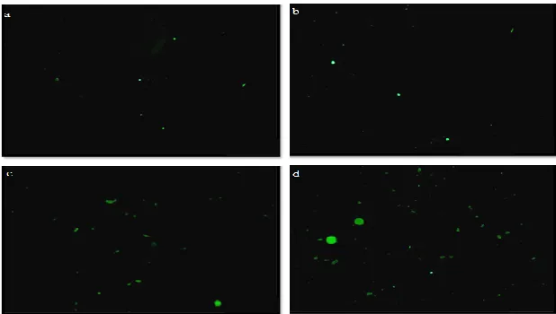
Figure 4 Fluorescent microscopic images of MCF-7 breast cancer cells labelled with Annexin-V-FITC and propidium iodide. Images were magnified at 200 µm. (a) Untreated cells and cells treated with 5 µM of (b) Taxotere, (c) blank microemulsion
_________________________________________________________ Journal of Experimental Biology and Agricultural Sciences
3.2 Characterization of cell morphology using light microscope
The MCF-7 cells were incubated with different micromolar concentrations (1, 5 and 10) of ME1, ME2 and ME3 for 48 h. The cell morphologies were analyzed by using light microscope. The apoptotic effect of the 5 µM of ME1, ME2 and ME3 and 10 µM of ME2 was very noticeable, as more condensed chromatids, intracellular spaces, membrane blebbing and formation of apoptotic bodies were exposed in Figures 1-3. In contrast, it was only the shape of MCF-7 cells that got changed, when administered with 1 µM of ME1, ME2 and ME3 and 10 µM of ME1 and ME3.
Our study was in agreement with other studies that formulated docetaxel in emulsion systems. A 0.8 mg/ml of docetaxel was encapsulated in emulsion that composed of 10% oil phase (Soya oil and Miglyol 812), 1.2% soybean lecithin and 0.3% Pluoronic F68 (Gaoet al. 2008). The submicron lipid emulsion showed promising intravenous carrier in place of the available commercial ones with more efficiency. Furthermore, Zhao et al. (2010) have encapsulated docetaxel in lipid emulsion that exhibited higher plasma concentrations in rats than Taxotere but bioequivalent to it in beagle dogs. The docetaxel loaded-lipid emulsion displayed safe effect and exhibited antitumor activities againstthe A549, BEL7402 and BCAP-37 cell lines in nude mice, similar to Taxotere.
3.3 Apoptosis detection using ApopNexin FITC assay
In order to clarify the mechanism of cell death whether it occurred due to apoptosis or necrosis, ApopNexin FITC apoptosis detection kit was employed. After the treatment of MCF-7 cells with 5µM of ME1, ME2 and Taxotere for 48 h, the cells were labeled with annexin V conjugated with FITC and PI, then viewed by fluorescence microscope. As shown in Figure 4, the untreated cells didn’t stain positively with neither dyes which indicates the viability of cells, while the treated MCF-7 cells with ME1, ME2 and Taxoterewere stained positively green fluorescent with annexin-V-FITC but not with PIwhich implies signs of apoptosis with no detectable necrotic effect due to the externalization of PS caused by the cell surface outbreak (Casciola-Rosen et al., 1996).It should be noted that ME2 was having the best apoptotic induction effect.
The microemulsion delivery system improved the docetaxel efficiency against MCF-7 breast cancer cells. It is recommended to apply this formula in vivo to give a comprehensive study on the docetaxel-loaded-microemulsion formula.
Acknowledgements
The authors wish to express a sincere thanks and appreciation to King Abdulaziz City for Science and Technology for its financial support to the research project designated by number (P-S-11-0613), King Abdulaziz University Hospital for providing cell cultures and King Fahd Medical Research Center, Jeddah, KSA, for technical support.
References
Casciola-Rosen La, Rosen A, Petri M, Schlissel M (1996) surface blebs on apoptotic cells are sites of enhanced procoagulant activity: Implications for coagulant events and antigenic spread in systemic Iupusery thermatosus. Proceedings of the National Academy of Sciences.93: 1624-1629.
Coppeta J, Rogers C (1998) Dual Emission Laser Induced Fluorescence for Direct Planar Scalar Behavior Measurements. Experiments in Fluids. l25: 1–15.
Deitch AD, Law H, White RDV (1982) A stable propidium iodide staining procedure for flow cytometry .Journal of Histochemistry&Cytochemistry.30: 967-972.
Dougherty EM, Narang N, Leob M, Lynn DE, Shapiro M (2006) Fluorescent brightener inhibits apoptosis in baculovirus-infected gypsy moth larval midgut cells in vitro. Biocontrol Science and Technology.16: 157-168.
Duchler M, Stepnik M (2008) Cytotoxic effects of a combination of three natural compounds to leukemia cells in vitro. Cancer Therapy.6: 733-740.
Gan L, Gan Y, Zhu C, Zhang X, Zhu J (2009) Novel microemulsion in situ electrolyte-triggered gelling system for ophthalmic delivery of lipophilic cyclosporine A: in vitro and in vivo results. International Journal of Pharmaceutics. 365: 143-149.
Gao K., Sun J, Liu K, Liu X, He Z (2008) Preparation and characterization of a submicron lipid emulsion of docetaxel: submicron lipid emulsion of docetaxel. Drug Development and Industrial Pharmacy.34:1227–1237.
Jadhav KR, Shaikh IM, Ambade KW, Kadam VJ (2006) Applications of microemulsion based drug delivery system. Current Drug Delivery.3: 267-273.
Leteurtre F, Fujimori A, Tanizawa A, Chhabra A, Mazumder A, Kohlhagen G, Nakano H, Saintopin P (1994) A dual inhibitor of DNA topoisomerases I and II, as a probe for drug-enzyme interactions. The Journal of Biological Chemistry.269:28702–28707.
Ma Y, Zheng Y, Zeng X, Jiang L, Chen H, Liu R, Huang L, Mei L (2011) Novel docetaxel loaded nanoparticles based on PCL-Tween 80 copolymer for cancer treatment. International Journal of Nanomedicine. 6: 2679–2688.
Monks A, Scudiero D, Skehan P, Shoemaker R, Paull K, Vistica D, Hose C, Langley J, Cronise P, Vaigro-Wolff A, Gray-Goodrich M, Campbell H, Mayo J, Boyd M (1991) Feasibility of a high-flux anticancer drug screen using a diverse panel of cultured human tumor cell lines. Journal of the National Cancer Institute. 83: 757-766.
Mu CF, Balakrishnan P, Cui FD, Yin YM, Lee YB, Choi HG, Yong CS, Chung SJ, Shim CK, Kim DD (2010) The effects of mixed MPEG-PLA/Pluronic copolymer micelles on the bioavailability and multidrug resistance of docetaxel. Biomaterials. 31:2371-2379.
Riss TL, Moravec RA (2004) Use of multiple assay endpoints to investigate the effects of incubation time, dose of toxin and plating density in cell-based cytotoxicity assays. Assay and Drug Development Technologies.2: 51-62.
Skehan P, Storeng R, Scudiero D, Monks A, McMahon J, Vistica D, Warren JT, Bokesch H, Kenney S, Boyd MR (1990) New colorimetric cytotoxicity assay for anticancer-drug screening. Journal of the National Cancer Institute.82:1107– 1112.
Tolnai SA (1975) Method for viable cell counting, Tissue Culture Association Manual Methods. 1: 37-38.
Tsai YH, Hsieh YH, Huang YB, Chang JS, Huang CT, Wu PC (2010) Microemulsions for intravesical delivery of gemcitabine. Chemical and Pharmaceutical Bulletin.58: 1461-1465.
Yin YM, Cui FD, Mu CF, Choi MK, Kim JS, Chung SJ, Shim CK, Kim DD (2009) Docetaxel microemulsion for enhanced oral bioavailability: preparation and in vitro and in vivo evaluation. Journal of Controlled Release.140: 86-94.
Zhang YT, Huang ZB, Zhang SJ, Zhao JH, Wang Z (2012) In vitro cellular uptake of evodiamine and rutaecarpine using a microemulsion. International Journal of Nanomedicine. 7: 2465-2472.
Zhao M, Su M, Lin X, Luo Y, He H, Cai C, Tang X (2010) Evaluation of docetaxel-loaded intravenous lipid emulsion: pharmacokinetics, tissue distribution, antitumor activity, safety and toxicity. Pharmaceutical Research.27:1687-1702.
6 Mayson and Wafaa