183
Copyright © 2018. IJEMR. All Rights Reserved.
Volume-8, Issue-1 February 2018
International Journal of Engineering and Management Research
Page Number: 183-188
Toxicity Effects of Aspartame on Embryonic Development of Zebrafish
(Danio Rerio)
M.S.Weerasooriyagedara
Lecturer (Temporary), Department of Animal Science, Uva Wellassa Univeresity, SRI LANKA
Corresponding Author:[email protected]
ABSTRACT
Zebrafish (Danio rerio) is a widely used biological model to investigate different chemicals since it has certain similarities with human biology. Present study investigated the impact of Aspartame (APM); an excessively used food additive all over the world, on the embryonic development of zebrafish. Acute toxicity experiments were performed for a 4-day period using zebrafish eggs. Ten different test concentrations of aspartame 100, 250, 300, 500, 1000, 2000, 5000, 10000, 15000 and 20000 mg/L were used as treatment concentrations. The results clearly indicated that with the
increase of the aspartame concentrations, different
observable deformities are formed in zebrafish embryo. At the low concentrations of aspartame such as 100, 200, 300 mg/L there were no observable malformations in zebrafish embryonic development. However, at high concentrations such as 10,000, 15,000, 20,000mg/L there were distinguishable negative alterations such as growth retardation, shrinkage of chorion, yolk sac edema, lack of pigmentation, tail deformities and scoliosis in developing embryos. Zebrafish embryo can be successfully used to investigate food additives such as aspartame. However, the impacts of these concentrations on internal anatomical and physiological changes in zebrafish embryo should be comprehensively investigated.
Keywords— Zebrafish, Danio rerio, Aspartame, Embryonic development
I.
INTRODUCTION
Zebrafish (Danio rerio), the cyprinid schooling teleost, who is an ideal model for studying genetics. They are extensively used as a renowned vertebrate model over the past twenty years (Jusuf and Harris, 2009). Due to their several specialties Zebra fish have been considered as amicable research models in biomedical and health research and well apposite as a model system to perform
chemical biology experiments efficiently in educational and research settings (Grunwald and Eisen, 2002). They are easy to maintain in large numbers, readily reproducing under laboratory conditions (Brand etal, 2002), high in fecundity, possess transparent embryos, have rapid development and ease in culture (Lammer, 2009; Oliveira
et al., 2009). Their embryos are strong enough for
manipulations in scientific experiments such as cell transplanting and microinjection experiments. Moreover the zebrafish shares a high degree of homology with the human genome (Howe, 2013). Thus, the zebrafish is becoming an influential model organism for studying genetics (Kawahara etal,2009), environmental toxicology (Yang etal,2009), DNA damage repair (Pei, 2011), cancers (Konantz etal, 2012), and other diseases (Sullivan and Kim, 2008).
Non-sugar sweeteners have been used for centuries, mostly in natural forms derived from the Stevia plant family (Misra et al., 2011).Commercial production of chemically synthesized artificial sweeteners began with saccharine in the 1890s (De la Peña, 2010). Artificial sweeteners were primarily used to make pharmaceuticals than more palatable, and as a sugar substitute in foods designed for patients with diabetes until the 1970s (Talbo and Fisher, 1978). With that, huge industry has developed that focuses on low calorie foods and drinks for the general public. With the demand, number of new artificial sweeteners have been introduced with increasing use in a wide variety of food products.
184
Copyright © 2018. IJEMR. All Rights Reserved.
in 1981 (FDA, 1981), in soft drinks in 1983 (FDA, 1983), and in all foods in 1996 (FDA, 1996). In 1994, the use of APM was approved in Europe (European Parliament and Council, 1994).
The additives are subjected to a variety of toxicological tests, including long term rodent bioassays to test for potential of carcinogenic effects. Melnick et al. (2008) noted that in order to minimize dissension over the results of different carcinogenic bioassays and to extrapolate the results from bioassays to humans. According to him test protocols must at least employ animal models who are sensitive to the study endpoint, have to thoroughly characterize both the test chemical and the dose administered to the animals, use challenging doses and exposure durations, use sufficient numbers of animals per dose group, use multiple dose groups to detect dosage effects, employ complete and peer-reviewed histological evaluations and evaluate data using pairwise comparisons and analyses of trends that rely on survival-adjusted tumor incidence.
Speculation about the relationship between aspartame and brain cancer risk was raised in an article which temporally linked increasing incidence rates in the U.S. during the period from 1975 to 1992, especially an upward shift of more aggressive glioblastomas during the 1980s, to the entry of aspartame in the food supply in 1981. From an earlier positive animal study reviewed by the Food and Drug Administration the authors cited data and an in vitro nitrosation experiment (Shephard etal., 1993) to support their hypothesis. More recent and extensive animal trials have failed to show the carcinogenic activity of aspartame (Butchko etal., 2002). Additionally, a population-based case-control study found a null association between childhood brain tumors and aspartame intake among both children and their mothers during pregnancy and lactation (Gurney etal., 1997). In contrast, however, a recent study found that female rats fed aspartame developed more lymphomas and leukemias than controls, in a dose-dependent manner, starting from a dose that may be relevant to human intake (as low as 20 mg per kg body weight), which is lower than the acceptable daily intake established by the Food and Drug Administration at 50 mg per kg body weight (Butchko etal., 2002). But very few studies have conducted regarding the effect of food additive like aspartame for the embryonic development in humans.
More recently, use of renowned or novel small molecules (i.e. chemicals) to study phenotypic changes and identify the corresponding cellular target(s) has gained popularity in the scientific community. Thus, zebrafish can be used as an effective tool for studying such type of chemical biology approaches. Zebrafish embryo is a model with high potential to investigate the effects of a wide range of chemical compounds including flavour enhancers, food additives and different controversial compounds (Abdelkader et al, 2012). Therefore this study was
conduted to investigate the toxic effect of aspartame embryonic development of zebrafish as they can use as a human model.
II.
METHODOLOGY
Zebrafish maintenance and egg collection
Domestic aquarium starin of adult male and female zebrafish were maintained in a separate glass tanks with aerators, filters in the Aquaculture Laboratory, Uva Wellassa University of Badulla, Sri Lanka. All the physico-chemical water quality parameters inside the tanks were maintained at optimum levels (Temperature at 26 ± 3 C and pH 5 to 6) and fish were fed with Artemia nauplii and commercial feed twice a day. Initially, matured male and female brooders were selected for the breeding. Breeding tank was prepared with artificial plants and breeding traps because of their predatory activity on laid eggs. The bottom of the tank was covered with a stainless steel grid which helps to collect all the eggs on the bottom. In addition, artificial plants were used to stimuli spawning. Selected males and females were placed in the spawning tank day before the test in the ratio of 2:1, male to female. Mating and spawning were taken place 30 minutes after the onset of sunlight in the following morning. About 30- 60 minutes after spawning, spawn traps were removed and eggs were carefully collected using a pipette. Fertilized viable eggs were selected under the binocular microscope for further investigations
Toxicity assessment
Acute Aspartame toxicity experiment was performed for a four days period using Zebrafish (Danio rerio) eggs. Ten test concentration of aspartame (100, 250, 300, 500, 1000, 2000, 5000, 10000, 15000 and 20000 mg/L) were prepared by diluting the stock solution in distilled water. A control of distilled water was used for each concentration of pesticide. The test was started as soon as possible after fertilization of the eggs. Fertilized eggs were separated from non-fertilized eggs under a stereo microscope. Distilled water was used as negative control and as internal plate control. Ethanol was used as a positive control (Reimers, Flockton and Tanguay, 2004). As a positive control concentration of 2% of ethanol (100% embryo mortality) was performed with each egg batch used for testing.
185
Copyright © 2018. IJEMR. All Rights Reserved.
control on each above plates, 24 eggs in distil water as negative control on one set plate, twenty eggs as positive control on one plate.
The eggs in standard 24- well plate were covered by shelf adhesive foil and maintain at a temperature of 26 ± 1 °C. Dead eggs were removed immediately since they can adversely affect on the other viable eggs. Three replicates were performed individually. In this study, the acute toxic effect of aspartame on the zebrafish embryo was observed and identified using binocular stereomicroscope.
III.
RESULTS AND DISCUSSION
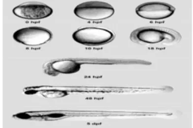
Embryonic development of the zebrafish (Danio
rerio) has been described in several studies. Zebrafish has
seven development stage at their embryonic stage such as zygote, cleavage, blastula, gastrula, segmentation, pharyngula and hatching period. Figure 1 demonstrated main developing stages of zebrafish. That stages are very helpful in for conducting and identifying toxicological test (Kimmel et al., 1995).
Figure 1: Embryonic development of the zebrafish
In zebrafish embryo toxicity test, several lethal, sub- lethal and teratogenic endpoints were observed. According to OECD guideline for testing chemicals -236, 2013, Fish Embryo Toxicity Assay, fish embryos are considered as dead when it is coagulated, lack of somite formation, non-detachment of tail and lack of heart beat.
Figure 2: (a) Coagulated embryo, (b) Lack of somite formation, (c) Non-detachment of tail
By conducting Zebrafish (Danio rerio) embryo toxicity test developmental morphological deformities were observed in many concentration during the exposure
time of embryonic development of the zebrafish from 24 hours onwards. Several sub lethal and teratogenic endpoints on zebrafish embryos which are treated with the aspartame were observed over the four major lethal endpoints. Sub lethal or teratogenic end point were not observed in embryos of zebrafish which are treated with negative control. Lethal endpoints were observed in embryos of zebrafish which are treated with positive control.
The digital photograph of developmental deformities in zebrafish embryos exposed to different concentrations of aspartame after 24 hours were observed under binocular stereomicroscope. Fish embryo stage is highly sensitive to chemicals and pollutants. Since embryos are covered by semi permeable protective membrane chorion, it does not fully protect the embryo against all chemical penetration. In aspartame low concentrations of 100mg/L and 200mg/L. there were no observable deformities and they showed normal embryonic development. Also in control, no dead embryos and deformities were observed and it was fully developed without malformations.
Figure 3: Normal embryonic development
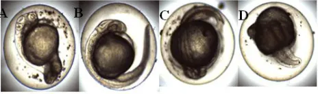
Embryos exposed in concentrations 300 and 500mg/L displayed edema formation in yolk sac region. The concentration of 500, 1,000 and 2,000mg/L aspartame treated embryos were observed with growth retard during embryonic development of zebrafish. Pericardial edema was found 15,000 and 20,000 concentration. Malformation of head region were observed at 1,000, 5,000, 10,000 and 20,000mg/L concentrations.
Figure 4: A: edema formation in yolk sac region, B: growth retard, C: Pericardial edema, D: Malformation
of head region
A
B
C
A
186
Copyright © 2018. IJEMR. All Rights Reserved.
Some embryo demonstrated more than one deformities in single embryo. Growth retard, malformation of head and edema formation were observed at 20,000mg/L concentration. Yolk sac edema, lack of eye and malformation of tail were observed at 15,000 and 20,000mg/L concentrations. Growth retard, lack of tail, eye, head and modification of cord identified at 10,000, 15,000 and 20,000 mg/L concentrations.
Figure 5: A: Growth retard, malformation of head and edema formation, B: Yolk sac edema, lack of eye and
malformation of tail, C: Growth retard, lack of tail, eye, head and modification of cord
Zebrafish embryos exposed to different concentrations of aspartame after 48 hours demonstrated some abnormalities. In negative controller no any malformation was indicated. Embryos exposed to 5,000, 10,000, and 15,000mg/L concentrations showed degeneration of tail. Pericardial edema was observed at 500, 1,000, 10,000 and 20,000mg/L concentrations. The concentration of 300, 500, 5,000mg/L aspartame treated embryos were observed with fluid accumulation of pericardial region during embryonic development of zebrafish. Embryo exposed to 1,000, 5,000, and 15,000 mg/L concentration showed growth retard. In 20,000mg/L concentration level lack of head formation and deformities in cord region were observed. Lack of eye, head, cord and somite formation were observed at 15,000 and 20,000 mg/L concentration. Growth retard, pericardial edema and degeneration of tail region were observed at 20,000 mg/L concentration level.
Figure 6: Degeneration of tail, B: Fluid accumulation of pericardial region, C: lack of head formation and deformities in cord region, D: Lack of eye, head, cord
and somite formation
Zebrafish embryos exposed to the treatment solution observed some deformities at 96 hours also. In negative controller there was no any malformation recorded. Embryos exposed to 5,000, 15,000, and 20,000mg/L concentrations showed growth retard.
Scoliosis was observed at 10,000, 15,000, and 20,000mg/L concentrations. Scoliosis might have resulted from impaired ionic regulation (Khayatzaden and Abbasi, 2010) or damage of the vertebral column (Stouthart et al., 1994; Cheng et al., 2000; Nguyen and Janssen, 2002; Hallare et al., 2005) or fractured vertebrae through tetanic muscular contraction (Holcombe et al., 1976). In 20,000mg/L concentration level no eye formation was observed. Some embryo showed more than one deformities in single embryo. Lesion, lack of tail, head and eye formation were observed at 10,000 and 15,000mg/L concentration level. Growth retard, lack of tail formation were observed at 20,000mg/L concentration level.
Figure 7: A: Scoliosis B: No eye formation
As results indicate in this study, there is a negative impact on zebrafish embryonic development by aspartame, especially when the concentrations are increased. Certain neurological disorders, abnormal neural development, certain endocrine disorders and neuropsychiatric disorders can be derived by the excessive intake of aspartame to the zebrefish embryos.
Different animal models have been used so far to demonstrate the toxicity of aspartame such as Swiss mice. The studies investigated possible health risks relating to the consumption of low calorie sweeteners including aspartame and it was included an epidemiological study on the association between intakes of low calorie sweetened soft drinks and increased incidence of preterm delivery (Halldorsson et al. 2010) and a carcinogenicity study in mice exposed to aspartame through feed (Soffritti et al.
187
Copyright © 2018. IJEMR. All Rights Reserved.
There is an emerging trend in the world with the sophisticated lifestyles of human towards the fast food which contain lot of food additives (Qudsi and Al-Jahdali). The high concentrations of food additives can be adversely affect on human health, especially the early developmental stages. Hence, this has to be comprehensively investigated using animal models. Because of exposure to nutritional and environmental challenges during critical stages of early development of life, the metabolism in later life can be remarkably affected (Jackson et al., 2010). Zebrafish embryonic development can provide an ideal model for these types of studies due to its easiness as well as more similarities with human genome.
IV.
CONCLUSION
The results of this study clearly indicate that with the increase of the aspartame concentrations, different observable deformities are formed in zebrafish embryo. Although at the low concentrations of aspartame (100, 250, 300mg/L) there were no observable malformations in zebrafish embryonic development, at high concentrations (i.e. 10,000, 15,000 and 20,000mg/L) there were distinguishable negative alterations such as growth retardation, shrinkage of chorion, yolk sac edema, lack of pigmentation, tail deformities and scoliosis. Zebrafish embryo can be successfully used to investigate excititoxins such as food additives. However, further studies on internal anatomical and physiological changes can be recommended to carryout for more comprehensive understanding on this regards.
REFERENCES
[1] Al-Qudsi, F. & Al-Jahdali, A. (2012). Effect of monosodium glutamate on chick embryo development.
Journal of American Science, 8(10), 499-509.
[2] Hoffman, E.J., Turner, K.J., Fernandez, J.M., Cifuentes, D., Ghosh, M., Ijaz, S., Jain, R.A., Kubo, F., Bill, B.R., Baier, H., Granato, M., Barresi, M.J., Wilson, S.W., Rihel, J., State, M.W., & Giraldez, A.J. (2016). Estrogens suppress a behavioral phenotype in zebrafish mutants of the autism risk gene. CNTNAP2 Neuron, 89(4), 725-33.
[3] Butchko HH, Stargel WW, Comer CP, et al. (2002). Aspartame: Review of safety. Intake of aspartame vs acceptable daily intake. Regul Toxicol Pharmacol, 35(2 Pt 2), S1-93.
[4] Cheng, S. H., Wai, A. W. K., So, C. H. & Wu, R. S. S. (2000). Cellular molecular basis of cadmium‐induced deformities in zebrafish embryos. Environmental
toxicology chemistry, 19(12), 3024-3031.
[5] FDA. (1981). Aspartame: commissioner’s final decision. Fed Reg (Food and Drug Administration), 46,
38285–38308.
[6] FDA. (1983). Food additives permitted for direct addition to food for human consumption: Aspartame. Fed
Reg (Food and Drug Administration),48, 31376–31382.
[7] FDA. (1996). Food additives permitted for direct addition to food for human consumption: Aspartame. Fed
Reg (Food and Drug Administration),61, 33654–33656.
[8] Soffritti M, Belpoggi F, Manservigi M, Tibaldi E, Lauriola M, Falcioni L, & Bua L. (2010). Aspartame administered in feed, beginning prenatally through life span, induces cancers of the liver and lung in male Swiss mice. American Journal of Industrial Medicine, 53(12), 1197-206.
[9] Gilmour DT, Jessen JR, & Lin S. (2002). Manipulating gene expression in the zebrafish. In: Zebrafish: A Practical Approach, Nusslein- Volhard C, Dahm R, (eds.). Oxford:
Oxford University Press, 121-43.
[10] Grunwald, D. J. & Eisen, J. S. (2002). Headwaters of the zebrafish—emergence of a new model vertebrate.
Nature Reviews Genetics, 3(9), 717-724.
[11] Hallare, A. V., Schirling, M., Luckenbach, T., Köhler, H. R. & Triebskorn, R. (2005). Combined effects of temperature cadmium on developmental parameters biomarker responses in zebrafish (Danio rerio) embryos.
Journal of Thermal Biology, 30(1), 7-17.
[12] Holcombe, G. W., Benoit, D. A., Leonard, E. N., & McKim, J. M. (1976). Long-term effects of lead exposure on three generations of brook trout (Salvelinus fontinalis).
Journal of the Fisheries Board of Canada, 33(8),
1731-1741.
188
Copyright © 2018. IJEMR. All Rights Reserved.
Gehring I, Berger A, Dooley CM, Ersan-Urun Z, Eser C, Geiger H, Geisler M, Karotki L, Kirn A, Konantz J, Konantz M, Oberlander M, Rudolph-Geiger S, Teucke M, Osoegawa K, Zhu B, Rapp A, Widaa S, Langford C, Yang F, Carter NP, Harrow J, Ning Z, Herrero J, Searle SM, Enright A, Geisler R, Plasterk RH, Lee C, Westerfield M, de Jong PJ, Zon LI, Postlethwait JH, Nusslein-Volhard C, Hubbard TJ, Roest Crollius H, Rogers J, & Stemple DL. (2013). The zebrafish reference genome sequence and its relationship to the human genome. Nature, 496(7746), 498–503.
[14] Khayatzaden, J. & Abbasi, E. (2010). The effects of heavy metals on aquatic ecosystem. The 1st International
Applied Geological Congress, 688-694.
[15] Kimmel, C. B., Ballard, W. W., Kimmel, S. R., Ullmann, B. & Schilling, T. F. (1995). Stages of embryonic development of the zebrafish. Developmental
Dynamics, 203(3), 253-310.
[16] Konantz M, Balci TB, Hartwig UF, Dellaire G, Andre MC, Berman JN, & Lengerke C. (2012). Zebrafish xenografts as a tool for in vivo studies on human cancer.
Annals of the New York Academy of Sciences, 1266, 124–
137.
[17] Nguyen, L. T. H. & Janssen, C. R. (2002). Embryo-larval toxicity tests with the African catfish (Clarias gariepinus): comparative sensitivity of endpoints. Archives
of Environmental Contamination Toxicology, 42(2),
256-262.
[18] Oliveira, R., Domingues, I., Grisolia, C. K. & Soares, A. M. (2009). Effects of triclosan on zebrafish early-life stages adults. Environmental Science Pollution Research, 16(6), 679-688.
[19] Olney JW, Farber NB, Spitznagel E, et al. (1996). Increasing brain tumor rates: is there a link to aspartame?
Journal of Neuropathol Exp Neurol, 55, 1115 – 23.
[20] Pei DS, Yang XJ, Liu W, Guikema J, Schrader C, & Strauss PR. (2011). A novel regulatory circuit in base excision repair involving AP endonuclease 1, Creb1 and DNA polymerase beta. Nuclear Acids Research, 39(8), 3156–3165.
[21] Shephard SE, Wakabayashi K, & Nagao M. (1993). Mutagenic activity of peptides and the artificial sweetener aspartame after nitrosation. International Journal of
Genomics, 31(5), 323–9.
[22] Stouthart, A. J. H. X., Spanings, F. A. T., Lock, R. A. C. & Bonga, S. W. (1994). Effects of low water pH on lead toxicity to early life stages of the common carp (Cyprinus carpio). Aquatic toxicology, 30(2), 137-151.
[23] Sullivan C & Kim CH. (2008). Zebrafish as a model for infectious disease and immune function. Fish Shellfish
Immunol, 25(4), 341–350.