DEVICES BY USING THREE-DIMENSIONAL
KNITTED STRUCTURES FOR HEALTHCARE AND
WELLBEING
BY
CAROL DIANE HEPBURN
CText. ATI (Textile Technology); PhD (Textile Technology)
A thesis submitted in partial fulfilment of the requirements of the
University of Bolton for the degree of Doctor of Philosophy
This dissertation has been submitted in partial fulfilment for the award of Doctor of
Philosophy in Technical Textiles at the University of Bolton, United Kingdom. I hereby
confirm that the work contained in this dissertation is my own work and that the work or
contribution of others have been fully acknowledged.
___________________ __________________________
i
A critical and recurrent problem for wheel chair users are the development of
decubitus ulcers, also known as pressure ulcers, pressure sores or bed sores. For
some wheelchair users, this can be life threatening, whereas for the majority, it is a
huge disruption and major complication in their personal life.
During this research a series of pressure relieving cushions were created.
Three innovative contoured prototype wheelchair cushions were developed and fully
characterised, utilising 3D warp knitted spacer fabrics. A uniquely constructed
contoured recess was developed for each prototype wheelchair cushion, which
successfully protected the most vulnerable part of the human buttocks, the Ischial
Tuberosities. Using a methodology which utilised a pressure mapping system; a
simulated human buttocks template, the Rigid Loading Cushion Indenter and a
computerised universal tester, the prototype cushions were characterised for their
pressure distribution properties. This allowed the measuring of pressure distribution
while under simulated loading conditions. These prototype wheelchair cushions were
able to demonstrate low peak pressures in the area of the vulnerable Ischial
Tuberosities, which ranged from 17.7mmHg – 32.9mmHg. This measuring system was
also used to compare, the prototype 3D spacer wheelchair cushions against a small
selection of commercially available foam wheelchair cushions. Pressure results for the
commercial foam cushions ranged from 50.4mmHg – 79.0mmHg peak pressure in
Ischial Tuberosities area. The flammability behaviour of 3D warp knitted spacer fabrics,
in the context of pressure relieving cushions, were also investigated by using a
modified ‘Mydrin’ test method, adapted to test the multiple layers found within the
prototype cushions. The resulting outcome was a better understanding of the
flammability behaviour of the warp knitted spacer fabrics within multiple layers, as well
as enhancing the flame retardancy properties of the prototype wheelchair cushions.
These prototype cushions satisfied the requirements of BS 5852 Ignition Source 5 (Crib
ii
cushions, ASD4, ASD4-S and ASD450-S.
The prototype wheelchair cushions have been designed and developed with the
following characteristics:
1. A shaped/contoured surface which can re-distribute high pressure points
normally located in vulnerable areas of an immobile and seated person, to
under 50.0mmHg in the Ischial Tuberosities.
2. Peak pressures could be reduced and distributed evenly over a much larger
area of these cushions than basic PU foam.
This series of prototype 3D warp knitted spacer fabric wheelchair cushions will assist in
iii
S.C.Anand MBE for his encouragement, enthusiasm and support throughout the
experimental phase of this research, also for his guidance and academic supervision
during the completion of this thesis.
I would also like to express my sincere thanks to my industrial supervisor Mr Charles
Wood, Managing Director of Baltex Ltd, UK, for his generous supervision, support and
encouragement during this research programme. I would also like to express my
thanks to Baltex Ltd, Ilkeston, UK, for providing financial sponsorship and for providing
the materials for undertaking this project.
I would also like to thank Mr Jason Bolton for his assistance during the testing and
characterisation of the prototypes.
I would also like to thank Mr Akbar Zarei, Mr Tony Ratcliffe and Mrs Donna Zarei, for
their support and assistance.
Finally, I would like to express my warm and sincere thanks to my parents, brothers
and my friends for their support and encouragement throughout this project.
iv
TABLE OF CONTENTS
ABSTRACT i
ACKNOWLEDGEMENTS iii
TABLE OF CONTENTS iv
LIST OF TABLES viii
LIST OF FIGURES xi
CHAPTER 1 GENERAL INTRODUCTION 1
1.0 Introduction 2
1.1 Background 2
1.2 Aims of the Research 3
1.3 Structure of the thesis 4
CHAPTER 2 LITERATURE REVIEW 6
2.0 Introduction 7
2.1 Human Anatomy 8
2.2 Pressure Ulcers and their causes 11
2.2.1 Decubitus Ulcers 11
2.2.2 Types of pressure ulcers 12
2.2.3 Complications of pressure ulcers 16
2.2.4 Predicting the risk of pressure ulcers – Assessment
Methods. 16
2.2.5 Aetiology or causes. 19
2.2.5.1 Interface pressure 21
2.2.5.2 Shear 23
2.2.5.3 Friction 24
2.2.5.4 Additional causes of pressure ulcers 24
v
2.5.1.1 Cushion material characteristics 29
2.5.2 Cushion surface characteristics 32
2.5.3 Additional feature characteristics 32
2.5.4 Secondary characteristics 33
2.6 Materials 36
2.6.1 Three-dimensional Spacer fabric 37
2.6.2 Knitted spacer fabrics 37
2.6.3 Weft knitted spacer fabrics 37
2.6.4 Warp knitted spacer fabrics 38
2.6.4.1 Construction 38
2.6.4.2 Characterisation 39
2.6.4.2.1 Thermophysiological characteristics 41
2.6.4.2.2 Positive mechanisms 42
CHAPTER 3 EXPERIMENTAL WORK 44
3.0 Experimental Work 45
3.1 Materials 45
3.1.1 Warp knitted spacer fabrics 45
3.2 Equipment Development 47
3.2.1 Rigid Cushion Loading Indenter (RCLI) 49
3.3 Experimental pressure distribution methodology 51
3.4 Flammability behaviour 53
3.4.1 Equipment 54
3.4.2 Experimental methodology 55
vi
4.1 Requirements 59
4.2 Cushion development 61
4.2.1 Contouring/shaping 61
4.2.1.1 Moulding 62
4.3 Partially contoured cushion development 63
4.3.1 Development of a pressure relieving recess 64
4.3.2 Construction of the partially contoured pressure relieving
cushion. 65
4.4 Fully contoured Wheelchair cushion development 70
4.4.1 Abductors and Adductors 70
4.4.2 Bariatric 74
4.5 Comparative products 76
CHAPTER 5 RESULTS AND DISCUSSION – PRESSURE MAPPING 81
5.0 Experimental Work 82
5.1 Preliminary experimental work 82
5.1.1 Phase One 82
5.1.2 Phase Two 85
5.1.2.1 Analysis of the basic flat cushions 97
5.2 Experimental pressure mapping - shaped cushions 100
5.2.1 Phase Three contouring development 100
5.2.1.1 Analysis 102
5.2.2 Phase Four full contouring development 107
vii
6.2 Preliminary experimental results 142
6.3 Experimental flammability behaviour 147
6.4 Experimental flammability results 148
6.4.1 Samples with the PU cover 149
6.4.2 Samples with the Knitted spacer fabric cover 162
6.5 Analysis of the experimental results 178
CHAPTER 7 CONCLUSIONS AND SUGGESTIONS FOR FURTHER
WORK 181
7.0 Conclusion 182
7.1 Introduction 182
7.2 Conclusions 182
7.2.1 Pressure distribution properties 183
7.2.2 Flammability properties 185
7.3 Suggestions for future work 187
REFERENCES 189
APPENDICES 199
viii
Table 2.1 The Braden Scale: a score of 16 or below indicates that the
patient is AT RISK of pressure sore development. The lower
the score the higher the risk. 17
Table 2.2 The Waterlow Scale. 18
Table 2.3 Norton Scale: a total score of 16 or below indicates a patient is
AT RISK and preventative measures should be taken. The lower
the total, the greater the risk. 19
Table 2.4 Comparison of pressure ulcer risk assessment methods. 19
Table 2.5a A summary of cushion material characteristics– Cellular materials – A 31
Table 2.5b A summary of cushion material characteristics – Fluid materials – B 32
Table 2.5c A summary of cushion material characteristics–Other constructions–C 32
Table 2.6 Properties of 100% polyester warp knitted spacer fabric. 40
Table 2.7 Thermal properties of polyurethane foam and warp knitted
spacer fabrics. 42
Table 2.8 Air permeability of samples. 42
Table 3.1 Warp knitted spacer fabric characteristics 45
Table 3.2 Additional fabric characteristics (cover fabrics) 45
Table 4.1 Specification: AS100 – overlay cushion 57
Table 4.2 Specification: AS200 – flat cushion 58
Table 4.3 Specification: AS200PRO – flat cushion 59
Table 4.4 Specification: Prototype ASD4(Fully recessed cushion) 68
Table 4.5 Specification: Prototype ASD5(Partially recessed cushion) 69
Table 4.6 Specification: Prototype cushion ASD4-S 74
Table 4.7 Specification: Prototype cushion ASD450-S(bariatric specification) 76
ix
Table 5.1 Average cycle results for AS100 Overlay cushion at initial loading
of 0.500kN. 86
Table 5.2 Average cycle results for AS200 wheelchair cushion at initial loading
of 0.500kN. 86
Table 5.3 Compression test loads (conversion) 87
Table 5.4 Pressure mapping data for Airospring® AS100 overlay cushion 87
Table 5.5 Pressure mapping data for Airospring® AS100 overlay cushion
on a foam base. 88
Table 5.6 Pressure mapping data for Airospring® AS200 cushion 88
Table 5.7 Pressure mapping data for ASD3 prototype cushion 88
Table 5.8 Pressure mapping data for 66Fit® viscoelastic foam cushion 89
Table 5.9 Pressure mapping data for basic PU foam cushion 89
Table 5.10 Comparative pressure mapping data on the flat cushions –
time ‘0 mins’. 95
Table 5.11 Comparative pressure mapping data on the flat cushions –
time ’15 mins’. 96
Table 5.12 Comparative summary of pressure mapping results at initial
loading. 98
Table 5.13 Comparative summary of pressure mapping results after 15mins
loading. 99
Table 5.14 Pressure mapping data for ASD4-S fully contoured prototype cushion
at 0.500kN to 1.000kN loads 107
Table 5.15 Pressure mapping data for ASD4-S fully contoured prototype cushion
at 1.500kN to 2.000kN loads 107
Table 5.16 Pressure mapping data for ASD450-S fully contoured bariatric
prototype cushion at 0.750kN to 1.500kN loads. 107
Table 5.17 Pressure mapping data for ASD450-S fully contoured bariatric
x
prototype cushion at 2.500kN and 3.000kN loads 108
Table 5.19 Pressure mapping data for Invacare-Flo-tech contoured foam
cushion at 0.500kN to 1.000kN loads 108
Table 5.20 Pressure mapping data for Invacare-Flo-tech contoured foam
cushion at 1.500kN to 2.000kN loads 109
Table 5.21 Pressure mapping data for the Emerald profiled foam cushion at
0.500kN to 1.000kN loads. 109
Table 5.22 Pressure mapping data for the Emerald profiled foam cushion at
1.500kN to 2.000kN loads. 109
Table 5.23 Comparison of average pressure after 15 mins at the load 1.500kN 126
Table 5.24 Peak pressures in the vulnerable IT area and whole cushion area
compared across all pressure relieving cushions 127
Table 6.1 Initial experimental sample descriptions 137
Table 6.2 PU cover samples – (prototype representation) 138
Table 6.3 Spacer cover samples – (prototype representation) 138
Table 6.4 Experimental composite samples 138
Table 6.5 Preliminary experimental samples 138
Table 6.6 Comparative average burn times of the experimental assemblies
(PU cover). 147
Table 6.7 Comparative average burn times of the experimental assemblies
(Spacer cover). 147
Table 6.8 Summary of flame test results for the cushion assembly samples
Table 6.9 Flammability test results tested under BS 5852:2006 Ignition Source 5.148
Table 6.10 Basic composite construction: Prototype ASD4-S with PU cover 180
Table 6.11 Basic composite construction: Prototype ASD4-S with spacer fabric
Cover 180
Table 7.1. Pressure relieving cushions ranked by the mean pressure results in
xi
Figure 2.2 Potential areas for pressure ulcers in lateral, seated and supine
Positions. 8
Figure 2.3 Anatomy of the female and male pelvis. 9
Figure 2.4 Drawing showing the position of the ischial tuberosities in
relationship to the wheelchair back panel with pelvis in a sitting
position. 9
Figure 2.5 Drawing showing the increased distance of the ischial tuberosities
away from the wheelchair back panel with pelvis in a slumped
position. 9
Figure 2.6 Drawing of the pelvis tilted significantly backward making the
weight-bearing surface the sacrum and coccyx. 10
Figure 2.7 Category I Pressure ulcer 12
Figure 2.8 Category II Pressure ulcer 12
Figure 2.9 Category III Pressure ulcer, with curled under wound edges (with
Epibole). 13
Figure 2.10 Category IV Pressure ulcer 14
Figure 2.11 Unstageable/Unclassified Pressure ulcer 15
Figure 2.12 Deep tissue pressure injury. 16
Figure 2.13 Diagrammatical definition of pressure. 21
Figure 2.14 Tissue distortion due to pressure. 22
Figure 2.15 MRI showing the effect on the tissues during the application &
non-application of load during sitting. 22
Figure 2.16 Effect of shear stress on body tissue layers. 23
Figure 2.17 Immersion and Envelopment 28
Figure 2.18 Antithrust cushion shape. 34
Figure 2.19 Medial and Lateral thigh support features. 34
Figure 2.20 A selection of current pressure relieving cushions 35
Figure 2.21 Technit D3 weft knit spacer fabric. 37
Figure 2.22 Warp knitted spacer fabric construction 39
xii
Figure 2.25 Compression / Recovery curves of different materials. 41
Figure 2.26 Uniaxial and hydrostatic loading mechanisms (a) No load.
(b) Loading in one direction. (c) Hydrostatic loading. 42
Figure 2.27 Hydrostatic Loading 42
Figure 2.28 Uniaxial Loading 43
Figure 3.1 Front and bottom view of Staarink’s test buttocks with positions
of pressure sensors. 46
Figure 3.2 RCLI template based on ISO Buttock model. 47
Figure 3.3 Photographs of the template – Rigid Cushion Loading
Indenter (RCLI). 47
Figure 3.4 Denison Universal Tester– computer controlled 48
Figure 3.5 Diagram of the universal tester – set up. 48
Figure 3.6 ‘BodiTrak’ pressure mat and testing screen. 49
Figure 3.7 Cycle sequence showing areas of data capture. 50
Figure 3.8 Example of a pressure profile. 51
Figure 3.9 Vertical flame test apparatus – ‘Mydrin’ test. 52
Figure 3.10 Side view diagram of the modified ‘Mydrin’ test set up. 53
Figure 3.11 Adjusted vertical flame height, in millimetres. 54
Figure 3.12 Ignition surface set up. 54
Figure 3.13 Example Assembly for flammability testing. 55
Figure 4.1 Airospring® AS100 – overlay cushion 58
Figure 4.2 Airospring® AS200 – flat wheelchair cushion. 59
Figure 4.3 Deeper cushioning by itself increases the area over which
force is applied, thus lowering peak pressures. 60
Figure 4.4 Area of high pressure – comparison of high and low
vulnerabilities. 62
Figure 4.5 Pressure profile at initial load of 0.750kN (75.5kg/12st). 63
Figure 4.6 Cross-sectional view of the development of the recess. 64
Figure 4.7 Construction of castellated layer. 64
Figure 4.8 Core layers of the partially contoured wheelchair cushion
prototype ASD4 – construction of the recess. 65
Figure 4.9 Core layers of the partially contoured wheelchair cushion
xiii
Figure 4.14 Adductor construction techniques. 73
Figure 4.15 Top layer cut for abductor and recess. 74
Figure 4.16. Top layer assembled for recess and abductor. 74
Figure 4.17 Side profile of the Emerald® foam wheelchair cushion 78
Figure 4.18 Front profile of Invacare® Matrx® Flotech contour wheelchair
cushion. 78
Figure 4.19 Construction sequence for prototype ASD4-S wheelchair cushion. 79
Figure 4.20 Front and back cross-sectional views of prototype ASD4-S 80
Figure 4.21 Front and side profiles of the fully contoured prototype cushions. 81
Figure 4.22 Photograph showing the soft spacer layer moulding over the top
of the contours. 81
Figure 4.23 Diagram showing positioning. 81
Figure 5.1 AS100 Overlay Cushion: Preliminary pressure profile – Cycle 1
results. 84
Figure 5.2 AS100 Overlay Cushion: Preliminary pressure profile – Cycle 2
results. 85
Figure 5.3 AS100 Overlay Cushion: Preliminary pressure profile – Cycle 3
results. 85
Figure 5.4 Pressure mapping profiles for Airospring© AS100 overlay
cushion using the new methodology. 90
Figure 5.5 Pressure mapping profiles for Airospring© AS200 flat wheelchair
cushion using the new methodology. 91
Figure 5.6 Pressure mapping profiles for ASD3 (comfort improved AS200)
wheelchair cushion. 92
Figure 5.7 Pressure mapping profiles for Basic PU foam cushion. 93
Figure 5.8 Comparative pressure mapping profiles for flat cushions – at initial
loading of 0.500kN. 94
Figure 5.9 Pressure profile of IT area on foam cushion – 66Fit® 96
Figure 5.10 Pressure profile of the IT area on foam cushion – Basic PU
xiv
Figure 5.12 Pressure mapping profile images of ASD4 prototype wheelchair
cushion by using the new methodology 101
Figure 5.13 Pressure mapping profile images of ASD5 prototype wheelchair
cushion by using the new methodology 102
Figure 5.14. Comparative peak pressures found in the IT area of flat and
contoured cushions – PU foam; Prototype ASD4 and 66Fit
viscoelastic foam. 103
Figure 5.15 Comparative peak pressures found in the IT area of flat and
contoured cushions – Prototypes ASD4, ASD5; 66Fit viscoelastic
foam and PU foam - Load = 0.750kN after 15 mins. 104
Figure 5.16 Pressure mapping profile images of ASD4-S prototype fully
contoured wheelchair cushion by using the new methodology –
Load 0.750kN and 1.000kN 110
Figure 5.17 Pressure mapping profile images of ASD4-S prototype fully
contoured wheelchair cushion by using the new methodology
- Load 1.500kN and 1.7500Kn. 111
Figure 5.18 Pressure mapping profile images of ASD4-S prototype fully
contoured wheelchair cushion by using the new methodology -
Load 1.750kN and 2.000kN. 112
Figure 5.19 Pressure mapping profile images of ASD450-S Bariatric prototype
fully contoured wheelchair cushion by using the new methodology
- Load 0.750kN and 1.000kN 113
Figure 5.20 Pressure mapping profile images of ASD450-S Bariatric prototype
fully contoured wheelchair cushion by using the new methodology
- Load 1.500kN and 1.750kN. 114
Figure 5.21 Pressure mapping profile images of ASD450-S Bariatric prototype
fully contoured wheelchair cushion by using the new methodology
xv
contoured foam wheelchair cushion by using the new methodology
- Load 0.500kN and 0.750kN. 117
Figure 5.24 Pressure mapping profile images of INVACARE® Flo-Tech®
contoured foam wheelchair cushion by using the new methodology
- Load 1.000kN and 1.500kN 118
Figure 5.25 Pressure mapping profile images of INVACARE® Flo-Tech®
contoured foam wheelchair cushion by using the new methodology
- Load 1.750kN and 2.000kN 119
Figure 5.26 Pressure mapping profile images of EMERALD® profiled foam
wheelchair cushion by using the new methodology - Load
0.500kN and 0.750kN. 120
Figure 5.27 Pressure mapping profile images of EMERALD® profiled foam
wheelchair cushion by using the new methodology - Load
1.00kN and 1.500kN. 121
Figure 5.28 Pressure mapping profile images of EMERALD® profiled foam
wheelchair cushion by using the new methodology - Load
1.750kN and 2.000kN. 122
Figure 5.29 Comparison of pressure in the IT area – load 0.750kN after
15mins 128
Figure 5.30 Comparative commercially available foam cushions – Recess
areas 128
Figure 5.31 Pressure profile images of ASD4-S showing the effect of the
‘recess area’ under 3 different common weights. 129
Figure 5.32 Pressure profiles indicating the location of the ‘Recess’ at the
xvi
Overall): Load = 0.750kN). 130
Figure 5.34 Comparison of peak pressure in different sections of the prototype
cushion ASD4-S – (Section areas – IT; Thigh and Overall):
Load = 1.0kN). 131
Figure 5.35 Area of high pressure exceeding 200mmHg at a load of 2.0kN –
(i) Pressure profile. (ii) Areas of high pressure exceeding
200mmHg. 131
Figure 5.36 Comparison of peak pressure in different sections of the prototype
cushion ASD450-S – (Section areas – IT; Thigh and Overall):
Load = 1.0kN). 132
Figure 5.37 Comparison of peak pressure in different sections of the prototype
cushion ASD450-S – (Section areas – IT; Thigh and Overall):
Load = 1.5kN). 132
Figure 5.38 Comparison of peak pressure in different sections of the prototype
cushion ASD450-S – (Section areas – IT; Thigh and Overall):
Load = 2.0kN. 133
Figure 5.39 Comparison of peak pressure in different sections of the prototype
cushion ASD450-S – (Section areas – IT; Thigh and Overall):
Load = 2.5kN. 133
Figure 5.40 Comparative pressure mapping profiles of the contoured cushions
– at 15 mins after loading of 1.0kN. 134
Figure 5.41 Comparative pressure mapping profiles of the contoured cushions
– at 15 mins after loading of 1.500kN. 135
Figure 6.1. Sample N – Single layer assembly (after flame test) 140
Figure 6.2. Sample P – Single layer assembly (after flame test) 140
Figure 6.3. Sample N – Experimental assembly (after flame test) 141
Figure 6.4 Sample P – Experimental assembly (after flame test) 141
Figure 6.5 Sample G – Experimental assembly (after flame test) 142
Figure 6.6 Sample G - Scorching seen at each layer after exposure to flame
for 10 seconds. 144
Figure 6.7 Close-up of the scorched area of M3250 in sample G. 145
Figure 6.8 Sample F - Scorching as seen on each layer after exposure to
a flame for 10 seconds. 146
xvii
Figure 6.13. Sample A - Scorching as seen on each layer after exposure to
a flame for 20 seconds 154
Figure 6.14. Sample A - Scorching as seen on each layer after exposure to
a flame for 30 seconds 155
Figure 6.15. Sample B - Scorching as seen on each layer after exposure to
a flame for 10 seconds 156
Figure 6.16. Sample B - Scorching as seen on each layer after exposure to
a flame for 30 seconds. 157
Figure 6.17. Sample B - Scorching as seen on each layer after exposure to
a flame for 40 seconds 158
Figure 6.18. Sample L - Scorching as seen on each layer after exposure to
a flame for 30 seconds. 159
Figure 6.19. Sample L - Scorching as seen on each layer after exposure to
a flame for 40 seconds 160
Figure 6.20. Graph showing average burning times compared between samples
A, B and L which are using a PU cover. 161
Figure 6.21. Sample K – Experimental assembly (after flame test). 164
Figure 6.22. Sample J – Experimental assembly (after flame test). 164
Figure 6.23. Sample K - Scorching as seen on each layer after exposure to
a flame for 10 seconds. 165
Figure 6.24. Sample K - Scorching as seen on each layer after exposure to
a flame for 20 seconds 166
Figure 6.25. Sample K - Scorching as seen on each layer after exposure to
a flame for 30 seconds 167
Figure 6.26. Sample K - Scorching as seen on each layer after exposure to
a flame for 40 seconds. 168
Figure 6.27. Close-up of Sample K, the burn pattern of layer 4 (Spacer fabric
A1301-235) after 30 secs flame application. 169
Figure 6.28. Close-ups of sample K at 40 secs flame application: (A) – whole
xviii
Figure 6.30. Sample J - Scorching as seen on each layer after exposure to
a flame for 20 seconds. 172
Figure 6.31. Close-up photograph of Layer 3 (A1301-235) in sample J at
20 second flame exposure 173
Figure 6.32. Comparison of the burning patterns of the experimental cushion
composites – representing ASD4-S at 10 secs exposure. 174
Figure 6.33. Comparison of the burning patterns of the experimental cushion
composites – representing ASD4-S at 30 secs exposure. 175
Figure 6.34. Comparison of the burning patterns of the experimental
composites at 10 secs flame exposure. 176
Figure 6.35. A comparison of ‘After burn’ time for the PU fabric cover (A) and
CHAPTER 1
Carol Diane Hepburn 2 | P a g e
Chapter 1
Introduction
1.1
Background
Decubitus ulcers are a worldwide healthcare concern, affecting tens of
thousands of patients and individuals. Susceptibility to Decubitus ulcers comes from a
combination of external factors (e.g. pressure, friction, shear force, heat and moisture),
and internal factors (e.g. fever, malnutrition, anaemia, and endothelial dysfunction) [1]
and is unfortunately a common manifestation in an immobile patient.
“A review of epidemiological studies in Europe, Canada and the USA described the
reported prevalence of pressure ulcers in European hospitals as ranging from 8.3% to
23%. In the UK, the overall prevalence of pressure ulcers within care settings was
10.2%, with 59% of these being hospital-acquired. In the USA and Canada, prevalence
ranged from 12.3% in US health care facilities, to 33% in patients in the community
with spinal cord injury, and the overall estimate of pressure ulcer incidence in Canadian
healthcare settings has been reported as 26%. The presence of pressure ulcers has
been associated with a two- to four-fold increase in risk of death in older people in
intensive care units, however, these findings were not adjusted for other prognostic
factors” [2, 3, 4].
The development of decubitus ulcers in community and hospital environments
represent a significant cost burden in the UK, both to patients and to the healthcare
providers. For the average wheelchair user, the development of pressure ulcers can be
a major complication in that person’s life, and for some it can be life changing or even
life threatening. This cost is likely to increase in the future as the population ages. A
final and poignant aspect is that globally decubitus ulcers have resulted in over 28,000
deaths in 2013, increasing from 14,000 deaths in 1990 [5], given the continued aging of
the population this figure can only increase [6, 7, 8, 9, 10].
Part of this research will review some of the current commercial products on the
market today and how they contribute to the intervention put in place by various health
organisations in the fight to prevent and treat pressure ulcers.
This research will look at how a pressure ulcer develops and the causes, the risk
Carol Diane Hepburn 3 | P a g e relieving cushions as an intervention. Research into the various types of materials and
the necessary construction features of many types of pressure relieving cushions on
the markets at present, have shown that very little has been researched in the use of
knitted 3D spacer fabrics being utilised more fully in the core design of pressure
relieving cushions. It is this area which will be used to tackle the problem of creating an
effective composite of materials and construction to create an effective pressure
relieving wheelchair cushion by using a smart material.
1.2 Aims of the research
U
sing an understanding of the causes of ‘decubitus ulcers or pressure ulcers’, theaims of this research were to develop novel pressure relieving devices utilising 3D
warp knitted spacer fabrics.
This research will review the current commercially available pressure relieving cushion
products, as well as focusing on the development of appropriate test methods or
methodologies, as part of the design and development of novel pressure relieving
devices by using 3D warp knitted spacer fabrics, i.e. pressure relieving wheelchair
cushions, for wheelchair-bound or immobile patients or individuals.
This research would look at the development of a pressure relieving cushion, that can
successfully reduce peak pressure found in the Ischial Tuberosities, the most
vulnerable part of the human buttocks, in the seated position. This would help decrease
the overall peak pressure exhibited in the whole area of a wheelchair cushion, when a
person is seated. This research would also investigate incorporating flame retardant
properties within these prototypes. This would ensure the prototypes could meet the
requirements of BS 5852: 2006 Ignition Source 5 (Crib 5) and BS 7175: 1989 Section 3
Ignition source 5 test, which are a requirement of the healthcare sector for these
cushions.
In summary, the main aim of this research is to develop a fully contoured pressure
relieving wheelchair cushion, utilising 3D warp knitted spacer fabric, using a unique
contouring method. This should be a flame-retardant product that can meet the
regulatory requirement for the healthcare sector, which are the BS 5852: 2006 Ignition
Carol Diane Hepburn 4 | P a g e actively reduce pressure in the vulnerable Ischial Tuberosities area at a range of
applied loads, simulating the average weight of an average person, ie. 8st – 10st.
1.3 Structure of the thesis
In order to develop an understanding of the problems caused by pressure ulcers
and the solutions currently in use, Chapter 2 discusses the published literature on how
a pressure ulcer develops and its causes, as well as the risk factors affecting the
development of different types of pressure ulcers. What intervention is put in place in
relation to the support surface(s), including the effect of pressure reducing and
pressure re-distributing devices, i.e. pressure relieving cushions as an intervention.
Chapter 2 also reviews the advantages and disadvantages of the various types of
wheelchair cushions, with particular interest in the latest smart materials and how they
are being used in these devices.
Chapter 3 describes the methodology created for the characterisation of the pressure
distribution properties that were measured in the 3D knitted spacer fabric wheelchair
cushions as well as a small selection of commercially available foam wheelchair
cushions. A modified flame test was used to determine the flammability behaviour of all
the flat and contoured 3D warp knitted spacer fabric wheelchair cushions.
Chapter 4 explains the design and development of the prototype 3D warp knitted
spacer fabric cushions, describing the materials used, the unique features created and
the final specifications for the prototypes. Chapters 5 and 6 discuss the results
obtained from the pressure mapping experiments and the flammability characteristics
examined in the knitted spacer fabrics used in a series of multi-layer cushion
configurations. The major conclusions derived from the research work and the
suggestions for further work were described and discussed in Chapter 7.
Appendix A lists the publications, awards, conferences presentations and the patents
CHAPTER 2
Carol Diane Hepburn 6 | P a g e
Chapter 2
Review of Literature
2.0 Introduction
This literature review looks at how a pressure ulcer develops and the causes, the risk
factors affecting the development of a pressure ulcer and the different types of
pressure ulcers.
In 2006, it has been stated that approximately 412,000 individuals will develop a new
pressure ulcer annually in the UK [11], resulting in an annual cost of up to £2.1 Billion.
The cost of each pressure ulcer to the National Health Service is estimated to be
between £43 for an uncomplicated wound, increasing dramatically to £40,234 for a
more severe wound. This large expense accounts for 4% of the total annual
expenditure of the NHS, making pressure ulcers the most serious wounds in the UK
[11].
This has become a significant burden to the NHS and the economy in general. Even
allowing for inflation, more recent evidence from other countries suggests that this, is a
substantial underestimate [11]. It has been stated that most pressure ulcers are
preventable and once an ulcer has developed the costs increase enormously to include
hospital treatment. Many resource costs generated from treating these wounds include
antibiotics, dressings, and specialist redistribution surface devices, while 90% of overall
Carol Diane Hepburn 7 | P a g e
The human body is susceptible to developing decubitus ulcers or pressure ulcers on
many different parts of the body. The pressure points or pressure areas can be
identified in many areas of the human body in a series of different positions. This is
illustrated in Figures 2.1 and 2.2.
Carol Diane Hepburn 8 | P a g e
Figure 2.2. Potential areas of for pressure ulcers in lateral, seated and supine positions [14].
Significant weight bearing areas were identified by Meschan [15] and Peterson
et al [16], as the ischial tuberosities, the sacral coccygeal area, the greater and lesser
trochanters and the intertrochanteric crests, which receive excessive pressures when a
person is in a sitting position.
The ischial tuberosities are located approximately 10cm apart in females and slightly
nearer in males [15], as illustrated in Figure 2.3. The weight distribution is mainly over
the tips of the ischial tuberosities when sitting in a normal erect position with no pelvic
tilt.
Carol Diane Hepburn 9 | P a g e
Figure 2.3 Anatomy of the female and male pelvis [15]
Typically, the ischial tuberosities lie 5 – 13 cm from the back of a typical wheelchair
back panel. The ischial tuberosities can be seen in Figure 2.4. With a patient sitting in
a ‘slumped’ position the ischial tuberosities are orientated into a new position with the ‘symphysis pubis’ tilted up and the ischial tuberosities displaced forward in the
wheelchair. Weight-bearing then takes place on the posterior tips of the ischial
tuberosities, see Figure 2.5 [16].
Figure 2.4
.
Drawing showing the position of the ischial tuberosities in relationship to the wheelchair back panel with pelvis in a sitting position [16].Figure 2.5. Drawing showing the increased distance of the ischial
Carol Diane Hepburn 10 | P a g e
Patients with poor trunk control will exhibit the pelvis tilted posteriorly, the weight
bearing surface becomes the ‘sacrococcygeal’, this is illustrated in Figure 2.6 [16].
This demonstrates the importance of posture and the support of a therapeutic posture
while in a seated position. This will become an important aspect of intervention with
regards to preventing pressure ulcers.
In the thigh area, the weight-bearing surfaces of the greater and lesser trochanters and
the intertrochanteric crests, are areas of low pressure except in the cases of improperly
adjusted wheelchair footrests, or atrophy of the posterior thigh and hip musculature
[17].
The pressure applied in these areas of can be changed by specific situations, such as:
i. Pelvic tilt caused by postural changes, including the rotation of the pelvis. (The
patient must be in his natural sitting posture for this to be correctly evaluated.)
ii. Increased pressure over the bony prominences caused by the continuous
atrophy of the gluteal and posterior thigh muscles.
iii. Poor adjustments of wheelchair footplates can effectively increase the pressure
over the Ischial tuberosities.
iv. Orthopaedic surgeries, such as, amputations, total hip replacements, ischial
tuberosity resections or pathological conditions in the hip joint, e.g., arthritis and
heterotopic ossification, can create altered sitting postures resulting in
increased pressure in these vulnerable areas.
These anatomical pointers take on a significant importance when used in the
development of an effective contoured pressure relieving wheelchair cushion [16].
Figure 2.6
.
Drawing of the pelvis tiltedCarol Diane Hepburn 11 | P a g e
2.2.1. Decubitus Ulcers
Decubitus ulcers also known as pressure ulcers, pressure sores, or bed sores
are areas of damage to the skin and underlying tissues, that are caused by the
application of sustained pressure, rubbing, or friction to that area [18, 2]. These areas
are usually located over bony prominences and their severity are classified by the
amount of tissue damage produced in that area [19].
Research carried out in 1930 [20] stated that the pressure in the arteriolar limb of a
capillary in the human finger averaged about 32 mmHg. This value was then
mistakenly generalised to be the pressure required to compress capillaries to prevent
blood flow (the capillary closing pressure) and the pressure below which pressure
redistributing devices aimed to reduce the interface pressure. However, many other
studies and research also demonstrated a wide range of pressures in capillaries at
various anatomical locations, with values dependent on age and associated disease
[21].
2.2.2 Types of pressures ulcers
Pressure ulcers are graded or assessed against 4 stages or categories. This
pressure ulcer classification is used widely across the UK. The term category, rather
than grade or stage, is now preferred [22, 23].
Category I Pressure ulcer: Non-blanchable erythema –
Briefly, this grade can be described as a non-blanchable erythema of intact skin
(not effected by light finger pressure).
Discolouration of the skin, the skin appears reddened and does not blanch
when pressure is applied; warmth, the skin temperature is often warmer than
the surrounding area; the skin may be painful, but it has no visible breaks or
tears; an ulcer that feels firmer or softer than the area around it may also be
used as indicators, particularly in people with darker pigmentation. In the
dark-skinned person, the area may also appear to be a different colour from the
surrounding skin, but it may not look red. Category 1 may be difficult to detect
in individuals with dark skin tones, see Figure 2.7. This category may indicate
Carol Diane Hepburn 12 | P a g e
Figure 2.7. Category I Pressure ulcer[24]
Category II Pressure ulcer: Partial thickness –
This stage is characterised as “Partial thickness loss of dermis presenting as a shallow open ulcer with a red pink wound bed, without slough”. It may also
present as an intact or open/ruptured serum-filled or serosanguinous filled
(blood & serum filled) blister. It also presents as a shiny or dry shallow ulcer
without slough or bruising. This stage would not be used to describe skin tears,
tape burns, incontinence associated dermatitis, maceration or excoriation.
Bruising indicates deep tissue injury, see Figure 2.8, [22, 23].
Figure 2.8. Category II Pressure ulcer[24]
Category I
Carol Diane Hepburn 13 | P a g e
Characterised as “Full thickness tissue loss”. Subcutaneous fat may be visible
but bone, tendon or muscle are not exposed. Slough (a layer or mass of dead
tissue separated from livening tissue) may be present but does not obscure the
depth of tissue loss. This stage may include ‘undermining’ and ‘tunnelling’. The
depth of a stage III pressure ulcer varies by anatomical location. The bridge of
the nose, ear, occiput and malleolus do not have (adipose) subcutaneous tissue
and stage III ulcers can be shallow. In contrast, areas of significant adiposity
can develop extremely deep stage III pressure ulcers. Bone/tendon is not
visible or directly palpable, see Figure 2.9, [22, 23].
Figure 2.9. Category III Pressure ulcer, with curled under wound edges (with Epibole) [24]
Category IV Pressure ulcer: Full thickness tissue loss –
Described as a “Full thickness tissue loss with exposed bone, tendon or muscle.” Slough or eschar may be present and often includes undermining and
tunnelling. The depth of a category IV pressure ulcer varies with anatomical
location.
The bridge of the nose, ear, occiput and malleolus do not have (adipose)
subcutaneous tissue and these ulcers can also be shallow.
Carol Diane Hepburn 14 | P a g e
Stage IV ulcers can extend into the muscle and/or supporting structures (e.g.,
fascia, tendon or joint capsule) making osteomyelitis or osteitis (inflammatory
disease of the bone and inflammation of bony tissue respectively) more likely to
occur. Exposed bone/muscle is visible or directly palpable (able to be touched
or felt) [22, 23]. Individuals with category four type pressure ulcers have a much
higher risk of developing life-threatening infections, see Figure 2.10.
Figure 2.10. Category IV Pressure ulcer [24]
The additional category of unstageable is also recommended by the Tissue Viability
Society for reporting systems within England [24].
Unstageable/ Unclassified: Full thickness skin or tissue loss, depth unknown –
Full thickness tissue loss in which the actual depth of the ulcer is completely
obscured by slough (yellow, tan, grey, green or brown) and/or eschar (tan,
brown or black) in the wound bed, see Figure 2.11, [24].
Carol Diane Hepburn 15 | P a g e
Figure 2.11. Unstageable/Unclassified Pressure ulcer [24]
Additional Stages for the USA
Unstageable/Unclassified: Full thickness skin or tissue loss, depth unknown –
Catergorised for the USA as “Full thickness tissue loss” in which the actual
depth of the pressure ulcer is completely obscured by slough (yellow, tan, gray,
green or brown) and/or eschar (tan, brown or black) in the wound bed. The true
depth can only be determined by the removal of the slough and/or eschar
to expose the base of the wound, until then it will be either a Category/Stage III
or IV. Stable (dry, adherent, intact without erythema or fluctuance) eschar on
the heels serves as “the body’s natural (biological) cover” and is not removed
[22, 23].
Suspected Deep Tissue Injury : Depth unknown –
Purple or maroon localised area of discoloured intact skin or blood-filled
blister, due to damage of the underlying soft tissue from pressure and/or shear.
The area may be preceded by tissue that is painful, firm, mushy, boggy, warmer
or cooler as compared to adjacent tissue. Deep tissue injury may be difficult to
detect in individuals with dark skin tones. The development may include a thin
Carol Diane Hepburn 16 | P a g e
evolve and become covered by thin eschar. The progression may be rapid
exposure of additional layers of tissue even with optimal treatment.
Figure 2.12. Deep tissue pressure injury.[24]
2.2.3 Complications of pressure ulcers
The complications that can develop from untreated pressure ulcers include a wide
variety of secondary conditions, including:
• Sepsis (bacteria entering the bloodstream).
• Cellulitis (inflammation of body tissue, causing swelling and redness). • Bone and joint infections.
• Abscess (a collection of pus). • Cancer (squamous cell carcinoma).
2.2.4 Predicting the risk of pressure ulcers - Assessment methods
There are three methods currently being used around the world to risk assess a
patient for developing pressure ulcers; the Braden scale; the Waterlow scale and the
Norton scale.
The Braden scale is a clinically validated tool that allows nurses and other healthcare
providers to fairly and reliably score a person’s level of risk of developing pressure
Carol Diane Hepburn 17 | P a g e
BRADEN SCALE
SENSORY PERCEPTION
1. Completely limited 2. Very limited 3. Slightly limited 4. No impairment
Ability to respond meaningfully to pressure-related discomfort Completely limited: Unresponsive (does not moan, flinch, or grasp) to painful stimuli because of diminished level of consciousness or sedation
or Limited ability to feel pain over most of body surface or discomfort over half of body
Very limited. Responds only to painful stimuli; cannot communicate discomfort except by moaning or restlessness
or Has a sensory impairment that limits the ability to feel pain on one or two extremities
Slightly limited: Responds to verbal commands but cannot always communicate discomfort or the need to be turned or Has some sensory impairment, which limits ability to feel pain or discomfort
No impairment: Responds to verbal commands; has no sensory deficit that could limit ability to feel or voice pain or discomfort
MOISTURE 1. Completely moist 2. Very moist 3. Occasionally moist 4. Rarely moist
Degree to which skin is exposed to moisture
Constantly moist: skin is kept moist almost constantly by perspiration, urine, and the like; dampness is detected every time patient is moved or turned
Very moist: Skin is often, but not always, moist; linens must be changed at least once a shift
Occasionally moist: skin is occasionally moist, requiring an extra linen change approximately once a day
Rarely moist: Skin is usually dry; linen requires changing only at routine intervals
ACTIVITY 1. Bedfast 2. Chairfast 3. Walks occasionally 4. Walks frequently
Degree of physical activity
Bedridden: Confined to bed Chair fast: Ability to walk severely limited or non-existent; cannot bear own weight and/or must be assisted into chair or wheelchair
Walks occasionally: walks occasionally during day, but for very short distances, with or without assistance; spends majority of each shift on bed or chair
Walks frequently: walks outside room at least twice a day and inside room at least once every 2 hours during waking hours
MOBILITY 1. Completely immobile 2. Very limited 3. Slightly limited 4. No impairment
Ability to change and control body position
Completely immobile: does not make even slight light changes in body or extremity position without assistance
Very limited: Makes occasional slight changes in body or extremity position but is unable to make frequent or significant changes independently
Slightly limited: makes frequent although slight changes in body or extremity position independently
No limitations: makes major and frequent body position changes without assistance
NUTRITION 1. Very poor 2. Probably inadequate 3. Adequate 4. Excellent
Usual food intake pattern
Very poor: Never eats complete meal; rarely eats more than one third of any food offered; eats two servings or less of protein (meat or dairy products) per day; takes fluids poorly; does not take a liquid dietary supplementary receives nothing by mouth and/or is maintained on clear liquids or intravenous
Probably inadequate: rarely eats a complete meal; generally, eats only approximately half of any food offered; protein intake includes only three servings of meat or dairy products per day; occasionally takes a dietary supplementary receives less than optimal amount of liquid diet or tube feeding solutions for more than 5 days
Adequate: Eats more than half of most meals; eats a total of four servings of protein (meat or dairy products) each day; occasionally refuses a meal, but usually takes a supplement if offered
or is on a tube feeding or total parenteral nutrition regimen that probably meets most of nutritional needs
Excellent: Eats most of every meal; never refuses meal; usually eats total of four or more servings of meat and dairy products per day; occasionally eats between meals, does not require supplements
FRICTION AND SHEAR
3. Problem 4. Potential problem 3. No apparent problem
Problem: Requires moderate to maximal assistance in moving; complete lifting without sliding against sheets is impossible; frequently slides down in bed or chair, requiring frequent repositioning with maximal assistance; spasticity, contractions, or agitation leads to almost constant friction
Potential problem: moves feebly or requires minimal assistance; during a move, skin probably slides to some extent against sheets, chair, restraints, or other devices; maintains relatively good position in chair or bed most of the time but occasionally slides down
Carol Diane Hepburn 18 | P a g e
The Waterlow scale, is a tool to assist in the assessment of risk of a patient/client
developing a pressure ulcer. The Waterlow is based on of seven criteria, seen in Table
2.2 [18, 25, 26]. This tool identifies three 'at risk' categories (see Table 2.2),
1. a score of 10-14 indicates 'at risk'
2. a score of 15-19 indicates 'high risk',
3. a score of 20 and above indicates very high risk.
Table 2.2. The Waterlow Scale [18, 25, 26].
The Norton scale was devised in 1962 by Doreen Norton [27] and was one of the first
assessment tools, which were specifically designed for an elderly care environment. It
consists of five key risk factors that are further separated into sub-divisions, with one or
two word descriptions to describe variations of each risk factor. Using this tool, the
descriptions with the lowest value represents the worst scenario. The range of possible
total scores varied between 5 and 20, with an arbitrary cut-off score of 14, which
equates to the individual being ‘at risk’, see Table 2.3.
Carol Diane Hepburn 19 | P a g e
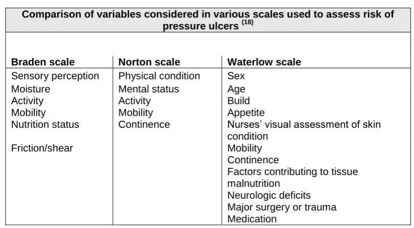
Table 2.4. Comparison of pressure ulcer risk assessment methods [28].
Comparison of variables considered in various scales used to assess risk of
pressure ulcers (18)
Braden scale Norton scale Waterlow scale
Sensory perception Physical condition Sex
Moisture Mental status Age
Activity Activity Build
Mobility Mobility Appetite
Nutrition status Continence Nurses’ visual assessment of skin condition
Friction/shear Mobility
Continence
Factors contributing to tissue malnutrition
Neurologic deficits Major surgery or trauma Medication
These categories of risk assessment are also used to categorise the different types of pressure relieving cushions.
2.2.5 Aetiology or causes
The pressure ulcers are caused by impaired blood supply and tissue malnutrition, as a
result of prolonged pressure, friction, or shear. The development occurs with the
disruption of the vascular network of arteries, arterioles and capillaries [28]. Tissue
compression exceeding the capillary filling pressure of 32 mmHg, that lasts longer than
2 hours, can cause local ischemia and necrosis. Skin overlying bony prominences (eg,
Carol Diane Hepburn 20 | P a g e
As regards the pressure, the intensity, duration, and the tissue's tolerance for pressure,
must be considered [30]. Not only the intense pressure, but also moderate pressure
acting continuously over a long period of time can result in ulcer formation [30]. A
theory was put forward, that the duration of the pressure was more important than the
amount of pressure sustained by the capillaries [31]. This being the case, the pressure
is one of the most important factors in the formation of pressure ulcers, but not limited
to only this component or factor [30, 32, 33, 34].
There are different types of pressure which can cause a pressure ulcer and these are:
Interface pressure – which occurs with the pressure of the body pressing the
skin down onto a firm surface, which can be either, a bed, a wheelchair, or
cushion etc.
Shear – this is pressure which occurs, when layers of skin are forced to slide
over one another, or deeper layers of tissue slide over one another. Shear can
occur when a person slides down or is pulled up, out of a bed or out of a
wheelchair
Friction– this pressure is caused by something rubbing against the surface of
the skin, such as a mattress, cushion or clothing [35].
In an alert person, the body’s motor and sensory systems are responsible for relieving
the effects of a continuous load or pressure, which usually results in the initiation of
frequent small body movements and periodic changes to their posture to relieve the
load and restore tissue perfusion [36]. This form of subconscious postural shifts or
fidgeting ensures that we move when needed.
Many people at risk of developing pressure ulcers are either, unable to effectively
reposition themselves, or are not provided with the sensory feedback that prompts the
required subconscious movements. In the case of patients who are unconscious,
sedated, anaesthetised, have limited mobility, or are paralysed and therefore cannot
sense or respond to these signals, cannot initiate these spontaneous movements. This
can result in the skin and soft tissues being subjected to, prolonged and unrelieved
pressures if no intervention is put in place. Therefore, people with medical condition
that limits their ability to change positions, requires them to use a wheelchair or
Carol Diane Hepburn 21 | P a g e
angle to a surface. Where the same amount of force is applied to both a small and
large area, a greater pressure is exerted on the smaller area than the larger, see
Figure 2.13 [38, 39].
Figure 2.13. Diagrammatic definition of pressure [38, 39]
When the pressure over the bony prominences distorts the skin and underlying tissues,
internal stresses have also been found to be ‘tensile’ due to stretching and ‘shear’
caused by the distortions. This can mean that even when pressure is applied
perpendicular, tensile and shear can also occur within the underlying soft tissues near
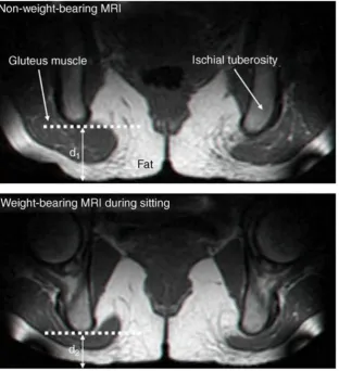
bony prominences, see Figure 2.14 [38, 39, 40]. The MRI (Magnetic resonance
imaging) studies shown in Figure 2.15 also illustrates significant distortion during
loading [41]. The pressure exhibited at the intersection between the skin (or skin &
clothing) and a support surface (bed, mattress, wheelchair, wheelchair cushion) is
Carol Diane Hepburn 22 | P a g e
Figure 2.14. Tissue distortion due to pressure.
Bending of the lines in (b) shows that when external pressure is applied over a bony prominence, compressive, shear (distorting) and tensile
(stretching) stresses occur. [38, 40]
Figure 2.15. MRI showing the effect on the tissues during the application &
Carol Diane Hepburn 23 | P a g e
Shear stress is the application force parallel or tangential to the surface of an
object with the base of the object remaining stationary. This causes the object to
change shape or deform. Shear stress is expressed in similar terms as pressure, most
frequently as pascals (Pa), or sometimes as newtons/square metre (N/m2).
Shear stress = Tangential force applied (N)
(pascals or Area of application of force (m2)
N/m2)
1Pa = 1N/m2 1kPa = 1000N/m2 --- Equation 2.1
The effect of shear stress on the internal tissues can be seen in Figure 2.16 [38, 42].
With the application of shear force, friction between the skin and the support surface
has the tendency to hold the skin in place, while deeper tissues are displaced. The
amount of displacement, i.e. shear strain, is greater in the vicinity of the bone than in
the superficial tissue layers.
Figure 2.16. Effect of shear stress on body tissue layers [38, 42]. .
The areas of greatest shear stress are near the bony prominences, where interface
pressures tend to be the highest. This can mean patients with slender body types have
a tendency to have higher shear stress in the coccyx and sacrum, than do obese body
types. This effect prevents the blood flow within the blood vessels in these areas, by
several mechanisms, such as; direct compression and occlusion of blood vessels,
stretching and narrowing of the dermal capillary beds – when sufficiently high shear
stresses are applied, the internal diameter of the capillaries becomes inadequate for
blood flow [43, 44]; bending and pinching the blood vessels running perpendicular to
Carol Diane Hepburn 24 | P a g e
2.2.5.3. Friction
Friction is the force that counters the relative motion of two objects that are touching
and is measured in Newtons (N). However, the term 'friction' as mentioned before, is
also frequently used to mean the action of one object rubbing against the other. Friction
helps in the development of shear stresses with the human body, by keeping the skin
in place against a support surface while the rest of the body moves towards the foot of
a bed or the edge of a seat. The relative movement of the skin and underlying tissues
causes shear stresses to develop in the soft tissues overlying the bony prominences
such as the sacrum [38, 39].
2.2.5.4. Additional causes for pressure ulcers
As mentioned previously the pressure is not the only factor in the causes of pressure
ulcers. Other extrinsic or external factors, in addition to pressure, friction and shear, are
moisture and heat (sometimes encompassed as the microclimate). The intrinsic factors
are, reduced mobility, impaired sensation, acute, chronic or terminal illness, pyrexia
(high temperature), dehydration, incontinence/other moisture sources, vascular
disease, malnutrition, a history of pressure ulcers, pain effecting the desire to reposition
themselves, some types of medication (e.g. steroids), old age, levels of consciousness
and cognitive status [46, 47]. This list is not exhaustive.
2.2.5.4.1 The microclimate
The microclimate, identified as the environment near the interface with skin/clothing
and the support surface, has a direct relationship with some of the extrinsic factors,
such as heat and moisture. Many studies have stated that the microclimate includes
the skin temperature and skin moisture between the patient’s interface with the skin
and the support surface, this sometimes includes air movement.
An increase in skin moisture contributes to a series of damaging forces, such as
maceration leading to skin breakdown, it weakens the stratum corneum (outer layer of
the epidermis or skin), leading to skin damage [48, 49, 50]. Equally, excessive dryness
can lead to skin damage by cracking. A reduction in skin resilience and an increase in
the skin’s coefficient of friction due to skin moisture from increased perspiration, will
also give rise to an increase in shear stresses and friction, making the individual prone
Carol Diane Hepburn 25 | P a g e
also giving raise to the ideal environment for the development of a pressure ulcer.
Other factors that can increase excessive moisture on the skin surface as well as
perspiration are, urinary or faecal incontinence, wound/fistula drainage or vomit. All
these components increase the risk of pressure ulcerations by weakening the cross
linkages between the collagen and damaging the epidermis.
Another significant risk factor for pressure ulcers is an increased body temperature
(pyrexia). An increased skin temperature is related to pressure ulceration by increasing
susceptibility to the ischaemic effects of the pressure and shear stresses and by
weakening the stratum corneum.
Whether a support surface will have an impact on the microclimate will depend on the
characteristics of the support surface. For materials, such as foam, the surfaces have
poor heat transfer properties, gel-filled products have a cooling effect, however, these
wear off after time exceeding 2 hours [38] and can increase the humidity in that area.
Fluid-filled products and alternating pressure air mattresses, both reduce skin
temperatures [38, 50, 51]. Fabrics, such as 3D knitted spacer fabrics have the
properties which dissipate heat and help to evaporate moisture [52, 53, 54, 84].
2.3 Prevention
The solutions to combat pressure ulcers come in a variety of combinations, in which no
one solution can be used in isolation.
The solutions to pressure ulcers come in the form of ‘Interventions’, pressure-relieving
cushions, beds and mattresses can either mould around the shape of a patient to
distribute the patient’s weight over a larger contact area (using constant low-pressure
devices) (CLP); or vary the pressure beneath the patient mechanically, resulting in a
reduction in the time pressure is applied in one area (alternating-pressure devices)
(AP). These are mainly classified as being of a lower technological specification (i.e.
“low-tech”).
By comparison, air-fluidised devices, where warmed air circulates through fine ceramic
beads covered by a permeable sheet, and low air-loss beds, where patients are
supported on a series of air sacs through which warmed air passes, are
high-specification CLP devices. Alternating-pressure devices generate alternating high and
![Figure 2.2. Potential areas of for pressure ulcers in lateral, seated and supine positions [14]](https://thumb-us.123doks.com/thumbv2/123dok_us/9689952.1951999/28.595.195.397.92.474/figure-potential-pressure-ulcers-lateral-seated-supine-positions.webp)
![Figure 2.3 Anatomy of the female and male pelvis [15]](https://thumb-us.123doks.com/thumbv2/123dok_us/9689952.1951999/29.595.145.490.85.415/figure-anatomy-female-male-pelvis.webp)
![Figure 2.7. Category I Pressure ulcer [24]](https://thumb-us.123doks.com/thumbv2/123dok_us/9689952.1951999/32.595.123.490.103.306/figure-category-pressure-ulcer.webp)
![Figure 2.9. Category III Pressure ulcer, with curled under wound edges (with Epibole)[24]](https://thumb-us.123doks.com/thumbv2/123dok_us/9689952.1951999/33.595.163.508.318.526/figure-category-pressure-ulcer-curled-wound-edges-epibole.webp)
![Figure 2.10. Category IV Pressure ulcer [24]](https://thumb-us.123doks.com/thumbv2/123dok_us/9689952.1951999/34.595.138.459.201.407/figure-category-iv-pressure-ulcer.webp)
![Figure 2.11. Unstageable/Unclassified Pressure ulcer [24]](https://thumb-us.123doks.com/thumbv2/123dok_us/9689952.1951999/35.595.127.532.96.335/figure-unstageable-unclassified-pressure-ulcer.webp)
![Table 2.1. The Braden Scale: a score of 16 or below indicates that the patient is AT RISK of pressure sore development.; the lower the score the higher the risk [18, 25, 26]](https://thumb-us.123doks.com/thumbv2/123dok_us/9689952.1951999/37.595.112.547.102.801/table-braden-scale-indicates-patient-pressure-development-higher.webp)
![Table 2.2. The Waterlow Scale [18, 25, 26].](https://thumb-us.123doks.com/thumbv2/123dok_us/9689952.1951999/38.595.116.534.284.589/table-waterlow-scale.webp)