Jufri et al. World Journal of Pharmaceutical and Life Sciences
FORMULATION AND PHYSICAL STABILITY TEST OF MICROEMULSION GEL OF
KETOCONAZOLE
Hilda Nur Aziza and Mahdi Jufri*
Faculty of Pharmacy, Universitas Indonesia, Depok, Indonesia.
Article Received on 27/08/2018 Article Revised on 17/09/2018 Article Accepted on 08/10/2018
INTRODUCTION
Ketoconazole is an imidazole antifungal agent and a synthetic broad spectrum antifungal agent. Ketoconazole will inhibit cytochrome p450 enzymes and interferes with ergostrerol synthesis.[1] Target sites of ketoconazole are basal epidermis and stratum corneum which aim to treat superficial and systemic fungal infections.[2]
Ketoconazole most common adverse effects are nausea, hepatic and cardiovascular effect. Topical application is recommended to restrict the therapeutic effect to the affected area and to reduce systemic incrimination.[3] Ketoconazole which has 4.4 as its log P shows that the drug is lipophilic and has a poor solubility.[4] Microemulsion has ability to enhance drug delivery, efficacy, and bioavailability of several drug. Antimicrobial activity increases when the microemulsion system with Model ofloxacin drug used,[5] because of its poor solubility. The novelty of this research ketoconazole is formulated into microemulsion to enhance its solubility and effectiveness.
METHODS
Construction of Pseudo-Ternary Phase Diagram Pseudo-Ternary Phase Diagram was constructed to determine regions of microemulsion formation, using water titration method. Surfactant was blend with surfactant, with 1:1 ratio, each surfactant and
co-surfactant was mixed with oil phase, and isopropyl myristate concentration variant were made with the ratio of 9:1, 8:2, 7:3, 6:4, 5:5, 4:6, 3:7, 2:8, and 1:9 (% w/w). Water was added in each mixture and stirred with a magnetic stirrer. All mixture was observed visually. Clear and homogenous visualization were classified as microemulsion. The Pseudo-Ternary Phase Diagram was prepared on the basis of point which form the microemulsion region in the diagram phase.
Preliminary experiments
Microemulsion was selected at desired component ratio, different concentration oil, surfactants, and cosurfactant were determined for the best preparation of microemulsion. The concentration of isopropyl myristate used were 5%, 8%, 9%, then concentration of tween 80 used were 20%, 25%, 30%, 35% and propylene glycol concentration used were 20%, 25%, 30% mixture was added and the stirring continues, water was added and mixed with homogenizer. Prepared microemulsion ketoconazole was stored for 24 hours. The best formulation was a formulation which gave clear and homogenous microemulsion.
Microemulsion gel of ketoconazole
Microemulsion gel was prepared by mixing the clear microemulsion ketoconazole was resulted from the preliminary experiments. Ketoconazole was mixed into the oil phase that contain isopropyl myristate, and for the
World Journal of Pharmaceutical and Life Sciences
WJPLS
www.wjpls.org SJIF Impact Factor: 5.088
*Corresponding Author:Mahdi Jufri
Faculty of Pharmacy, Universitas Indonesia, Depok, Indonesia.
ABSTRACT
Objective: The purpose of this study was to formulate and evaluate the physical stability of ketoconazole microemulsion gel. Method: Pseudoternary phase diagrams were constructed using tween 80 as surfactant and propylene glycol as co-surfactant. The optimized microemulsion formula containing 5% isopropyl myristate as oil phase, 25% Tween 80 as surfactant, and 25% propylene glycol as co-surfactant. Result: The formulation showed clear and transparent yellowish microemulsion, while the microemulsion gel showed cloudy yellowish color. Globule size in each microemulsion and microemulsion at were 13.38 nm and 184.7 nm; respectively. The flow diagram of the microemulsion gel properties showed the plastic flow properties. The centrifugation and cycling test result showed an unchanged organoleptic, no phase separation occured between microemulsion and gel, no syneresis occured. The result of cycling test and centrifugation test was physically stable. Conclusion: Ketoconazole microemulsion gel show a good stability at temperature 30°C ± 2°C and 40°C ± 2°C.
water phase contain demineralize water, tween 80. Oil and water phase were mixed using homogenizer at 2000 rpm. The propylene glycol was added to the oil and water phase mixture and continue mix with homogenizer. Prepared microemulsion ketoconazole was stored for 24 hours. The best formulation was a formulation which gave clear and homogenous microemulsion.
Gel base was prepared by using carbopol 940 as gelling agent. The gel bases were prepared by dispersing carbopol 940 in distilled water with constant stirring at 2000 rpm speed using homogenizer. The dispersion was left overnight. The pH of all formulation was adjusted to neutral using triethanolamine (TEA). The obtained microemulsion and gel bases with 3:1 ratio were mixed using homogenizer at 2000 rpm speed to obtain microemulsion gel.
Evaluation microemulsion gel of ketoconazole preparations
The prepared microemulsion gel of ketoconazole were evaluated to the following test:
Physical appearance
Microemulsion gel formulas were inspected for their colour, odor, transparency, homogeneity and phase separation microemulsion gel.
pH measurement
The pH value of the microemulsion gel of ketoconazole was measured using pH meters. The microemulsion gel preparation was measured by dissolving 1 gram of microemulsion gel sample with 10 mL of aquadest. pH meters of which the electrode was calibrated with standard buffer pH 4 and 7, then the calibrated electrode was inserted into the dissolved preparation, and a pH value appeared on the screen.
Particle size measurement
Particle size measurement was carried out with dynamic light scattering with zetasizer (Malvern instruments ltd., Malvern, U.K). The polydispersity index and zeta potential of the formulation was determined by the same instrument.
Transmission Electron microscopy
TEM was used to visualize globules of microemulsion ketoconazole using TEM JEOL JEM 1400. Microemulsion was dried on a carbon coated copper grid. After drying, the morphology of samples globules were observed under microscope 100 kV – 200 kV.
Rheology properties
The rheology and viscosity of the microemulsion gel formula was determined using Brookfield viscometer attached with spindle number 4. The spindle was set to rotate from 0.5, 2.0, 5.0, 10.0, 20.0 and changed to 20.0, 10.0, 5.0, 2.0, 0.5 rpm. It was used for dial the reading at each rpm. The rheological behavior of each system were
evaluated by plotting the shear stress versus the shear rate values. The viscosity values were recorded.
Physical stability study microemulsion gel of ketoconazole
To observe any change in microemulsion gel stability, it were stored at 4°C ± 2°C for 24h and followed by 24h at 40°C ± 2°C. The cycle was repeated six times. Microemulsion gel was loaded into centrifugation tube and was centrifuged at 3750 rpm for 5 hours, then it was observed for any change in homogeneity. Becher in Lachman and Lieberman indicates that centrifugation at 3750 rpm for 5 hours is equivalent to gravity effect for approximately one year.
Samples of microemulsion ketoconazole were stored in different temperature condition, which are room condition 27°±2°C; high temperature 40°±2°C; low temperature 4°±2°C every 2 weeks for 2 months. This was done to evaluate its physical stability through visual inspection by physical change such as color and phase separation. pH values were measured every 2 weeks.
RESULTS AND DISCUSSION
Construction of Pseudo-Ternary Phase Diagram Pseudo-Ternary Phase Diagram was used to determine clear microemulsion using CHEMIX School version 3.60 (demo), In Pseudo-Ternary Phase Diagram (Fig. 1) one axis represent oil phase, second axis represent the aqueous phase and third axis represent fixed weight ratio of surfactant and co-surfactant.
The colored area Fig. 1, refers to microemulsion region, from this diagram the best microemulsion zone was found on 1:1 ratio. Microemulsions consist of small oil droplets dispersed in water (o/w microemulsion).[6] The greater the concentration of isopropyl myristate added, the smaller the mixture of surfactant and co-surfactant. The formula used is a formula marked by a red dot located in the microemulsion area.
Preliminary experiments
Preparation of microemulsion ketoconazole system aimed to obtain a clear microemulsion formulation.
Microemulsions begin to form at 5% oil concentration with 25% surfactant, 25% co-surfactant. The prepared microemulsion formulas are cited in Table 1.
Table 1: Preliminary experimental results of prepared microemulsion formulae.
Formula Isopropyl Myristate (%) Tween 80 (%) Propylene glycol % Demineralize water (%) Result
F1 5
20 20 55 Cloudy
25 20 50 Cloudy
25 25 45 Clear
30 30 35 Clear
F2 8
20 20 52 Cloudy
25 20 47 Cloudy
25 25 42 Cloudy
30 30 32 Clear
F3 9
20 20 51 Cloudy
25 20 46 Cloudy
30 30 31 Cloudy
35 35 21 Clear
Microemulsion is liquid dispersions of water and oil that are made homogeneous, transparent, and stable by adding a large amount surfactant and co-surfactant. Due to its thermodynamically stable characteristic, microemulsion formed spontaneously when all component mixed together.[7] Microemulsion oil in water is formed when the oil concentration is low.[8] In preparing the microemulsion ketoconazole systems, the smaller the amount of oil phase used, the formation of microemulsions will be easier because the oil phase formed into micelles is less, and surfactant adsorbed on the surface of the micelles will be greater.
The larger used of oil phase will make it required more surfactant and co-surfactant to reduce interfacial tension between an oil phase and water phase, because microemulsion will form if the interfacial tension between an oil phase and water phase is low close to zero.[8]
Table 2: Microemulsion gel of ketoconazole formula.
Composition Content in formula (%)
Ketoconazole 1.5
Isopropyl Myristate 3.75
Tween 80 18.75
Propylene glycol 18.75
Carbopol 940 0.5
Triethanolamine 0.75 Aquademineralisata 56
Preparation of microemulsion gel of ketoconazole systems
The optimized microemulsion contain 5% isopropyl myristate, 25% tween 80, and 25% propylene glycol, while gel base was prepared using carbopol 940 as gelling agent. Triethanolamine was added to form a thick gel bases, while triethanolamine was used to achieve maximum thickness from Carbopol.[9]
Evaluation microemulsion gel of ketoconazole preparations
Physical appearance
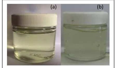
Microemulsion gel of ketoconazole (Fig. 2) showed a clear and transparent yellowish microemulsion (Pantone
11-0617 TPX), while the microemulsion gel showed a
cloudy yellowish color (Pantone 0131 C). Two prepared microemulsion and microemulsion gel have a tween 80 characteristic odor.
Fig. 2: Physical appearance (a) microemulsion and (b) microemulsion gel.
pH measurement
The pH values of microemulsion were found to be in the range of 6.27–5.81 and microemulsion gel were found to be in the range of 6.87–6.78. The results showed that all pH values of microemulsion are acceptable because adult human skin pH is 5.5–6.4. Thus, skin irritation can be avoided, meanwhile all pH values of the microemulsion gel are below 8.0 which can be due to a very alkaline pH (above 8.0). This can affect the skin physiology and interfere with normal flora that protect the skin from pathogenic bacteria.[10]
Particle size measurement
Table 3: Size measurement, polydispersity index and zeta potential result.
Weeks Globul size (nm) Polydispersity index (PDI) Zeta Potensial (mV)
Microemulsion 0 13.38 ± 0.12 0.128 -12.5 ± 0.13
8 16.93 ± 0.11 0.303
Microemulsion gel 0 184.7 ± 0.14 0.295 -29.3 ± 0.15
8 112.6 ± 0.15 0.304
Z-Average size or Z-Average mean used in dynamic light scattering is a parameter which also knew as the cumulants mean. It is the primary and most stable parameter produced by the technique. Z - Average is the main and most stable parameter. Measurements with Zaverage can be based on Polydispersity index (PDI). If the PDI value is more than 0.7 it could mean that the sample has a very wide distribution and is not suitable for measurement with Zaverage.
In table 3, the globul size of the microemulsion obtained at week 0 is 13.38 nm. this result meets the size criteria of 5 - 200 nm.[11] Compared with microemulsions, microemulsion gel are larger (with 184.7 nm) because of the addition of gel bases that increase the globules size of the microemulsion.
PDI is a measurement of particle homogeneity and it varies from 0.0 to 1.0. If PDI value closer to 0.0 it indicates narrow size distribution of the formulation. PDI of optimized microemulsion and microemulsion gel was found to be 0.128 and 0.303 at week-0; 0.295 and 0.304 at week-8. Decreasing particle size followed by a change in Polydispersity index (PDI) signifies reduced homogeneity, and decreased uniformity of globular size. This may be explained by the Ostwald ripening phenomenon in which droplets will change in size and an indication of the instability of the preparation.[12]
Transmission Electron Microscopy
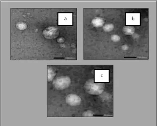
Microemulsion was shown a structure formed a spherical shape and there is a boundary between the water and the oil particles (Fig. 3).
Fig. 3: TEM image of microemulsion.
Viscosity
Rheological behavior of microemulsion gel was performed with a Brookfield viscometer with spindle No 4. The flow diagram of the microemulsion gel properties showed the plastic flow properties (Fig. 4 and Fig. 5). The viscosity value of the microemulsion gel ketoconazole at 0.5 rpm spindle number 4 on week-0 is 136000 cp (centipoise). At week-8 the viscosity was re-examined to see a change in rheological behavior. The microemulsion showed the properties of plastic flow.
The viscosity value of microemulsion gel of ketoconazole preparation at a speed of 0.5 rpm spindel 4 on week-8 is 144000 cps (centipoise).
The viscosity was increased between 0 until week-8. It is caused by addition of gel bases to the microemulsion gel preparation. For 8 weeks storage gel structure can be changed to the original state that has a tight formation so viscosity higher. Plastic flow is related to the presence of flocculate particles, since there is a
b a
yield value caused by contacts between particles and must be broken before flow occurs.[11] The result of rheogram microemulsion gel preparation at week-0 and
week-8 did not change and showed the plastic flow characteristic.
Fig. 4: Rheograms of Microemulsion gel of ketoconazole gel week-0.
Fig. 5: Rheograms of Microemulsion gel of ketoconazole gel week-8.
Cycling test
Cycling test results of microemulsion gel ketoconazole showed an unchanged organoleptic, no phase separation occured between microemulsions and gel, no syneresis occured, and microemulsion gel did not show a crystal formation. Result of cycling test was physically stable.
Centrifugation test
The result of centrifugation test showed that no separation occured separation between microemulsion and gel, and no color changes occured. The formula were found to be stable after 5 hours of centrifugation at 3750 rpm.
Physical Stability Test of microemulsion gel at temperature of 27° ± 2°C and 40 °±2 °C
Microemulsion gel of ketoconazole gel did not show any change in color and odor. Separation did not occured and microemulsion gel is still homogeneous from week-0 to week-8. The pH value at 27° ± 2°C range from 6.87 to 6.78. and pH value at 40° ± 2°C ranges from 6.87 – 6.76.
The pH value of microemulsion gel at 27° ± 2°C and 40 °±2 °C decreased in each measurement. The decreased pH value is the result of Tween 80 hydrolisis releasing fatty acids and made the pH become more acid.[13]
Physical Stability Test of microemulsion gel at temperature of 4°±2°C
After 8 weeks of storage, the microemulsion gel formula showed a change in color from cloudy yellowish to white. The microemulsion gel visualized a thick and frozen form. The gel will melt and the color will return to cloudy yellowish color when the formula was taken to room temperature. pH value at week-8 of storage showed a decreased and a range from 6.87 - 6.69.
CONCLUSION
Microemulsion ketoconazole was prepared by Pseudo-Ternary Phase Diagram. The optimized microemulsion contains 5% isopropyl myristate, 25% Tween 80, and 25% propylene glycol, while gel base was prepared using Carbopol 940 as gelling agent. Microemulsion ketoconazole showed a clear and transparent yellowish microemulsion (Pantone 11-0617 TPX), while the microemulsion gel showed a cloudy yellowish colour
(Pantone 0131 C).
Tests on several physical stability parameters showed an unchanged organoleptic, no phase separation occured between microemulsions and gel, and no syneresis occured. The result of cycling test and centrifugation test was physically stable. Ketoconazole microemulsion gel showed a good stability at temperature 30°C ± 2°C and 40°C ± 2°C.
REFERENCES
1. Badawi AA, Sakran WS, Ramadan MA, El-Mancy SMS. Improvement of the microbiological activity of topical ketoconazole using microemulsion systems. J Drug Deliv Sci Technol, 2012; 22(6): 473–8.
2. Jacobs GA, Gerber M, Malan MM, Preez JL, Lizelle T, Plessis J, et al. Topical delivery of acyclovir and ketoconazole. Drug Deliv, July 2017; 2016: 7544. 3. Patel MR, Patel RB, Parikh JR, Solanki AB, Patel
BG. Investigating effect of microemulsion components: In vitro permeation of ketoconazole. Pharm Dev Technol, 2011; 16(3): 250–8.
4. Moffat AC, Galichet LY, Osselton MD, Widdop B, editors. Clarke’s Analysis of Drugs and Poisons. 3rd ed. London: Pharmaceutical Press, 2004.
5. Sowmya, Suryaprakash Reddy, Prasad S, Kumar B. Preparation and Evaluation Of Ofloaxacic Microemulsion Gel. Int J Pharm Ind Res., 2012; 2(3): 228–34.
6. Eccleston GM. Encyclopedia of Pharmaceutical Technology. 3rd ed. Swarbrick J, editor. New York: Informa Healthcare USA, 2007.
7. Felton AL. Remington Essentials of Pharmaceutics. Pharm Press, 2013.
8. Manoharan C, Basarkar A, Singh J. Pharmaceutical Suspensions. Kulshreshtha AK, Wall GM, Singh ON, editors. New York: Springer, 2010.
9. Rowe RC, Sheskey PJ, Quinn ME. Handbook of Pharmaceutical Excipients. 6th ed. London: Pharmaceutical Press and American Pharmacists Association, 2009.
10. Barel AO, Maibach HI, Paye M. Handbook of Cosmetic Science and Technology. 3rd ed. New York, 2009.
11. Sinko PJ. Physical Pharmacy and Pharmaceutics 5th ed. Jakarta: EGC Medical Books, 2006.
12. Eccleston GM. Encyclopedia of Pharmaceutical Technology. 3rd ed. Swarbrick J, editor. New York: Informa Healthcare USA, 2007.