PURIFICATION AND CH ARACTERIZATION OF
CELL WALLMANNOPRO TEINS OF CANDIDA
ALB/CANS USING INTACT CELL METHOD
Z. FARAHNEJAD,* M.J. RASAEE,** H. YADEGARI,*
AND M. F ROUZANDEH MOGHADAM***
From the *Deparlmenl 0/ Medical Mycology, the **Department o/Clinical Biochemist/Yo and the ***Department o/Medical Biotechnology, School o/Medical Sciences,
Tarbiat Modarres University, Tehran. I.R. Iran.
ABSTRACT
Virulence of the opportunistic yeast, Candida albieans, involves the interplay of many complex changes including the yeast-hyphae transition, which mainly involves protein changes. Cell wall mannoproteins are found to be the main cause of adherence of C. albieans to epithel ial cells in the first step of an infection process. In the present study, cell wall mannoproteins of intact yeast were purified using a simple treatment of yeast with mercaptoethanol and sodium dodecyl sulfate followed by Concanavalin A chromatography. Both electrophoretic analysis of the column effluent and Western blot analysis using polyclonal and monoclonal antibodies showed the presence of mannoproteins with molecular weight in the range of30-50 kDa. Dot blot analysis of the purified antigen with the polyclonal and monoclonal antibodies prepared in this study showed that outer membrane mannoprotein antigens were obtained successfully following the above simple purification strategy.
MJIRl, Vol. 18, No.2, 167-172,2004.
Keywords: Candida a/hicans, Mannoproteins, Purification, Characterization.
Summer 1383 August 2004
INTRODUCTION
Candida albicans is a dimorphic opportunistic patho gen that grows as a yeast or as a mycelial fungus depend ing on the environmental conditions. I The major compo nents (80-90%) of cell wall of C albicans are carbohydrates, mannose or polymers of mannose covalently associated with proteins to form glycoproteins, also referred to as mamlO-proteins. These proteins contain �-glucans that are branched polymers of glucose containing �-1,3 and �-1,6 linkages and chitin, which is an unbranched homopolymer ofN-acetyl-D-glucoseamine containing �-1,4 bonds. P ro teins (6-25%) and lipids (1-7%) are also present as minor wall components.2.3 �-Glucans and chitin (0.6-9%) are also present as structural components of the wall (47-60% by weight).
Mannoproteins are considered as the most important antigenic component of Candida strains composing 10-30% of the cell wall. I This group of proteins are mainly com posed of carbohydrate polymannose containing more than
ISO strongly bonded mannosyl units.
Address of Correspondence: Mohammad Javad Rasaee, Tel.: +98-21-8013030; Fax: +98-21-8006544. E-mail: rasaee [email protected].
Mannoproteins play an important role in the process of adherence of Candida strains to mucosal surfaces which allows the organism to cause infection. Therefore the study of the cell wall proteins ofC albicans is of immense impor tance in order to understand the actual mechanism of infec tion leading to probable prevention or treatment of the dis ease.
In several studies, cell wall polymers have been extracted under controlled degradation of whole cells or of isolated walls using various enzymes for digestion.4-6 As an altema tive method, solubilization of cell wall materials using chemi cals such as mercaptoethanol (ME), dithiothreitol (DTT), and ethylene diamine has also been reported.1-9
Analysis of antigen expression with polyclonal and monoclonal antibodies has revealed the complex antigenic
Purification of C. albicans Cell Wall Mannoproteins
composition of the surface of e. albicans cells. These stud ies suggested that mannoproteins are the main antigenic cell wall components,1O which may subsequently be used as basic antigen during antibody development. In this case, it is obvious that natural conformation of dominant anti gens such as surface antigens is of extreme importance.
[n this study, we have attempted to purifY the surface mannoprotein ofe. albicans using intact cells with chemi cal treatments (sodium dodecyl sulfate (SDS) and ME) fol lowed by Concanavalin A affinity chromatography. In this way, the major confomlational characters of mannoprotein were left unaltered and hence the mannoproteins were puri fied in their natural conformation.
MATERIAL AND METHODS
Organism isolation
Candida albicans was isolated, cultured, and maintained from patients with vaginitis. The isolated strain was identi fied by using Candida check (latron laboratories, Tokyo, Japan) with an additional genn tube test and by examining morphological characteristics.
Microbiological observations of pseudo hyphae, hyphae and chlamydospores were made on cornmeal Tween 80 agar incubated at 35°C for 3 days. Culture medium GYEP con taining 2% glucose, 0.3% yeast extract and 0. 1 % peptone (supplemented with penicillin 100 IU/mL and streptomycin 100 lJg!mL) were used for e. albicans.
Glucose and peptone solutions were autoclaved (Solu tion A) while water-dissolved yeast extract was filtered through 0.2lJm filters (Solution B). Solutions A and B were mixed and reconstituted to 2 L. The resulting solution was divided into 4 Erlenmeyers and 2 mL of e. albicans suspen sion was added to each container. The containers were in cubated and shaken (100 rpm) at 29°C for 48 h.
At the end of incubation time, samples were centrifuged (x800 g, 5°C), the pellet was collected, washed three times with D.D. water, centrifuged three days after each washing step (x800 g, 5°C), and finally it was stored at -20°C. Out of 2 L of culture medium about 22 g of e. albicans was ob tained.
Antiserum preparation
Candida albicans serotype A from vaginitis cultures was used. These isolates were grown on Sabouraud's dex trose agar plates, washed using saline, harvested by cen trifugation and washed three times in sterile saline.
Balb/c mice were inoculated subcutaneously with a I: I
emulsion of 106 cell suspension in saline and complete Freund's adjuvant. Two weeks later the mice received an other inoculation IP with a I: I emulsion of I 06 cell suspen sions in saline and incomplete Freund's adjuvant.
Booster injections were given every IS days for two months. The mice were checked for antibody production and titer after each bleeding episode which followed after each injection by ELISA method.
ELISA for detection and titration of antibody
An ELISA method was developed and used for detec tion of antibody. In this method, the crude extract, purified mannoprotein and irrelevant antigens (such as BSA) were coated onto the wells of a microtiter plate in similar concen trations. Wells were washed (PBS, 10 mM, containing 0.05 Tween 80), blocked with PBS containing 0.3% gelatin and added willi dilutions of antibody in duplicates (1 :500, I: I 000,
I :2000) and incubated for 2 h. A dilution of normal mouse serum, I : 300, was used to indicate nonspecific binding.
At the end of incubation time, wells were emptied, washed four times and added with a I :2000 dilution of anti-mouse immunoglobulin labeled willi HRP (Sigma Chemical Co., St. Louis, MO, USA), incubated for I h, and washed with Tween-20 containing PBS buffer (0.05%). Finally tetramethyl benzedene substrate solution was added and the optical density was detected at 450 nm.
Preparation of cell wall extracts
This was perfonned according to the method of Casson et al.11 with minor modifications as follows. e. albicans sus pension was treated with lysis buffer (containing 2% SDS and 5% ME dissolved in water). The resulting suspension was vortexed vigorously, incubated in boiling water for 5 min and cooled in ice bath for 2 min.
The resulting solution was centrifuged at 14,000 g for 10
Table I. Analysis of the protein content of the crude and purified mannoprotein from Candida albicans.
Serial Steps of sample Amount of protein Condition of Sample used in
number preparation extract extract each step
1 2 L culture 22 g Wet C. albicans
2 Freeze dry 13 g Dry C. albicans
3 Crude extract 30 mg - Cell wall extract
4 Concana va lin A 1.Smg - Puritied protein
250
160
lOS
75
50
3S
30
25
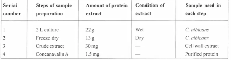
Fig. 1. SDS-PAGE (12% agar) analysis of whole cell wall extract of Candida albicans (20 bands are present). Molecular weight markers are shown in the right side of the electrophoretic pattern.
min, the supe111atant was removed and measured for protein content using Bradford protein assay procedure. The re sulting solution was dialyzed against 0.1 M acetate buffer solution, pH=6.0 and the clear solution was freeze dried and finally electrophoresed on polyacrylamide gel.
Affinity chromatography
In order to further purity the crude extract, a Sepharose 4B Concanavalin A packing material was used. Ten milliliters of Concanavalin A beads was prepared (as the instruction procedure provided by the manufacturer, Sigma Chemical Co.), and were packed in a lOx 1.5 cm column, washed and equilibrated with acetate butfer (0.1 M, pH=6 containing sodium chloride 1 M, calciwn chloride, manganese chlOlide and magnesium chloride, 1 mM). The crude extract (400 mg) was dissolved in a minimum quantity of equilibration buffer (I mL), loaded onto the colwnn and washed extensively with acetate buffer (0.1 M, pH=6) until no protein was detected in the column effluent.
Mannoproteins were then eluted using acetate buffer containing methyl a-D-mannopyranoside (0.3 M) with a slow rate of 15 m Lih. Samples were collected in 2 mL increments and were analysed by SDS polyacrylamide gel
electrophore-250
160
105
75
50
35
30
25
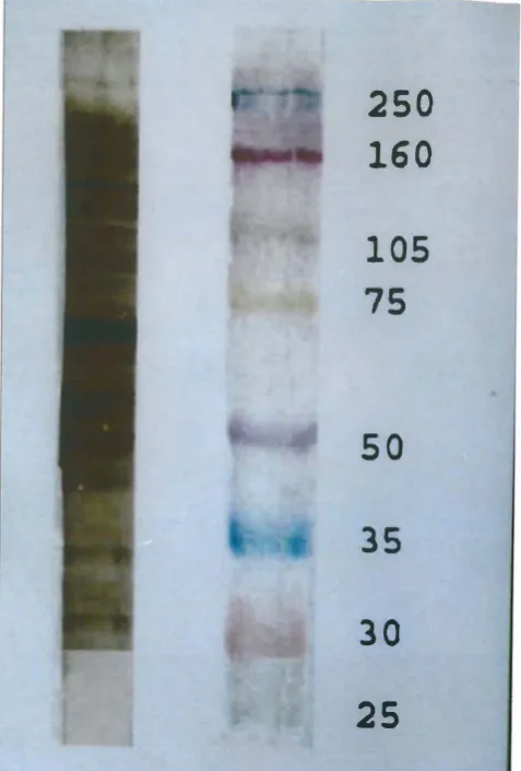
Fig. 2. Western blot analysis of cell wall extract of Candida albicans,
lIsing whole cell wall proteins.
sis (SDS-PAGE) followed by Western blotting analysis us ing polycional antibody prepared against cell wall proteins. Further characterization of mannoprotein prepared in the above procedures was perfonned by dot blot analysis.
RESULTS
Table I indicates the amount of crude and purified mannoprotein obtained in different steps. It was found that out of 2 L of liquid culture containing 22 g of wet yeast, 1.5 mg of mannoprotein could be obtained. Figure I shows the result of SDS-PAGE of the crude extract (after SDS and 2 ME treatment), where around 20 bands (from 25 kDa up to 250 kDa) were obtained. When these extracts were blotted and detected with polyclonal antibodies, 19 bands (from 30 kDa t0250 kDa) were observed (Fig. 2).
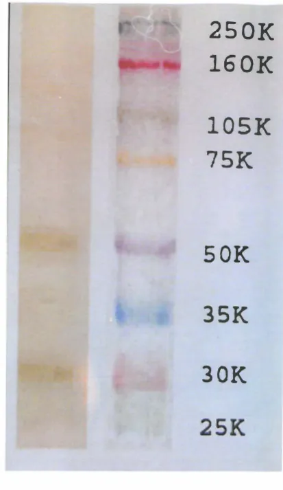
Figures 3A and 38 are SDS-PAGE results for crude ex tract being further purified using Concanavalin A colwnn. Here the result indicated that only two bands (50 kDa and 30 kDa) both in Coomassie blue staining (Fig. 3A) and silver
Purification of C. albicans Cell Wall Mannoproteins
nitrate staining (Fig. 3B) were presented. When these pro teins were transferred to nitrocellulose paper and stained with polyclonal antibody, the same two bands (a strong 50 kDa and a relatively weak band at 30 kDa) were observed (Fig.3C).
A.
B.
250 160
105
75
50
35
30 :15
250K 160K
l05K
75K
SOK
3SK
30K 25K
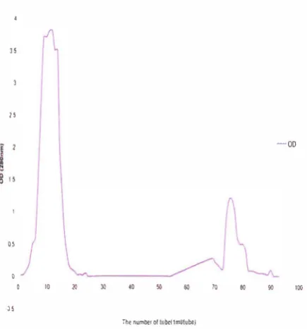
Figure 4 shows the chromatographic pattern of Con canavalin A purification of mannoprotein. Here it was found that only one peak containing mannoprotein (a 50 kDa and a 30 kDa protein as was shown in electrophoresis) was eluted after methyl a-D-mannopyranoside (0.3 M) treatment of the column between tubes 55 to 75.
The results of dot blot analysis are shown in Figure 5. In these results it was indicated that the crude extract reacted
with monoclonal antibodies offactor 6 (specific for C. albicans
obtained from Iatron laboratories, Tokyo, Japan) and polyclonal antibodies prepared in this study (Figs. 5A and
c.
250K
160K
lOSK
75K
S OK
3SK
30K
2SK
Fig. 3. SDS-PAGE of purified C. albicans extract after Concanava lin A chromatography stained with Coomassie blue (A), silver nitrate (8) and then transferred to nitrocellulose paper (C). ln every case only two bands in 50 kDa and 30 kDa regions were observed.
5B).
F urther, it was shown that purified antigen (mannoprotein) reacted with the same combination of anti bodies mentioned as above in a comparably lesser extent (Figs. 5C and 5D) when an equal amount of antigen was blotted. However no reaction was detected between the
antibodies when BSA or crude extract of Aspergillus were
used as antigen. These results indicated that the antigens prepared in this study only reacted with monoclonal and
polyclonal antibodies prepared for C. albicans.
DISCUSSION
We have shown that the extraction of mannoprotein from the intact cell wall of yeast using chemical method and af finity chromatography with Concanavalin A has more
/1
-00
II
01
L
0
0 10 20 30 '0 50 60 10 eo 9D 100
·Jl
The number or lubct1mUtube)
Fig. 4. Chromatographic pattern of column effluent after methyl O:-D-mannopyranoside treatment. Only one broad band between tubes 55-75 was observed.
vantages than enzyme digestion. Using this procedure, we expected that purified antigens should retain main epitope features and conformational characters such that they may be successfully used as immunogen and antigen base for assay development. The simple and gentle treatment of C. albicans with SDS and 2 ME released a varied array of ma terial from the cell wall. Many reports on analysis of cell wall extracts of Candida albicans are available. P onton and Jones (1986) used DTT, DTT with protease, �-glucuronidase and chitinase to release the wall components.8
Casanova et al. (1989) used SDS followed by zymolyase
treatment. 12 Yadegari et al. (200 I) reported the use of SDS
followed by ConcanavalinAand DEAE ion exchange chro matography.'3 Casanova and Chaffin ( 199 1) examined dif
ferent methods for cell wall release of glycoproteins of Can
dida albicans." In their experiments they used 2 ME , zymolyase, SDS, boiling and a combination of these reagents and physical conditions.
In all these procedures an array of proteins detected by SDS-PAGE starting from 20 kDa up to 400 kDa have been reported. 14
After perfonning Concanavalin A affinity chromatogra phy we found just two bands in the region of 55 kDa (a predominant band) and 30 kDa (a weak band). This showed that mannoprotein may be purified in a simple procedure as explained.
Western blot analysis confirmed this finding and the antibodies prepared against whole yeast extract, although reacting with 2 1 bands in crude extract, reacted with 2 bands
Fig. 5. Dot blot analysis of crude extract detected with polyclonal antibody (A), crude extract detected with monoclonal antibody (B), purified mannoprotein detected with polyclonal (C) and mono clonal antibody (D). A negative control BSA (E) and crude extract of Aspergillus (F) was tested to show that the antigen is specific in C. albicans.
after the purification procedure was carried out. Using mono clonal antibody prepared against epitope 6 with purified antigen showed that the antigens were reactive towards this antibody in dot blot analysis. We therefore concluded that mannoproteins of low molecular weight (30-55 kDa) may be purified by a two-step treatment explained in this work.
REFERENCES
I. Elorza MV, Marcilla A, Rafael S: Wall mannoproteins of the
yeast and mycelial cells of Candida albicans: nature of the
glycosidic bonds and polydispersity of their mannan moieties. Journal of General Microbiology 134: 2393-2403, 1988. 2. Calderone RA, Braun PC: Adherence and receptor relationships
of Candida albicans. Microbiol Rev 55: 1-20, 1991.
3. Cassone A: Cell wall of Candida albicans: its function and its
impact on the host. Top Med Mycol 3: 248-314, 1989. 4. Kitamura K: A protein that participates in yeast cell wall lysis
during zymolyase digestion. Agric BioI Chem 46: 2093-9, 1982.
5. Marcilla A, Elorza MV, Mormenco S, Rico H, Sentandreu R: Candida albicans mycelial wall structure: supermolecular com plexes released by zymolyase chitinase and �-mercaptoethanol. Arch Microbiol 155: 312-9, 1991.
6. Van Rinsum J, Klis FM, Van den Ende H: Cell wall glucomannoproteins of Saccharomyces cerevisiae. Yeast 7: 717-726, 1991.
7. Chaffin WL, Stocco DM: Cell wall mannoproteins of Candida albicans. Canad J Microbiol 29: 1436-44, 1983.
8. Ponton J, Jones 1M: Analysis of cell wall extracts of Candida albicans by sodium dodecyl sulfate-polyacrylamide gel elec trophoresis and Western blot techniques. Infect Immun 53: 565-572, 1986.
Purification of C. albicans Cell Wall Mannoproteins
9. Mormenco S, Riro H, Aguadoe IY, Sentandreu R: Study of
supermolecular structures released from the cell wall of Can
dida albiCClI1s by ethylene diamine treatment. Arch Microbiol 166: 327-335, 1996.
10. Brauner DL Cutler JE: Variability in expression of a cell sur face determinant on Candida albicans as evidenced by an ag glutinating monoclonal antibody. Infection and Immunity 43: 966-972, 1984.
I I. Casanova M, Chaffin WL: Cell wall glycoproteins of Candida albicans as released by different methods. Journal of General Microbiology 137: 1045-50, 1991.
12. Casanova M, Cail ML, Lardenoso L, Martinez JP, Sentandreu R: Identification of wall-specific antigens synthesized during
germ tube formation by C albicans. Infection and Immunity
57: 262-271,1989.
13. Yadegari MH, Moazeni M, Zavaran-Hoseini A, Khosravi A:
Extraction of Candida albicans cell wall mannoprotein. Modarres Journal of Medical Science 4(2): 207-218, 2002.
14. Sundstrom PM, Tam MR, Nichols LJ, Kenny ZL: Antigenic difference in the surface mannoproteins ofC albicans revalied by monoclonal antibodies. Infection and Immunity 56: 601-6,
1989.