RESEARCH ARTICLE
Synthesis of lignin based composites of TiO
2
for potential application as radical scavengers
in sunscreen formulation
Mohamad Nasir Mohamad Ibrahim
1*, Anwar Iqbal
1, Chai Chuan Shen
1, Showkat Ahmad Bhawani
2and Farook Adam
1Abstract
Titanium dioxide (TiO2) is added in sunscreens due to its ability to absorb ultraviolet (UV) light. However, upon irradia-tion of UV light, reactive oxygen species particularly hydroxyl radical which can damage human skin will be gener-ated. In this study, lignin/TiO2 composites were employed to quench the hydroxyl radicals generated by the TiO2. The lignin was extracted from oil palm empty fruit bunch (OPEFB) via kraft and soda pulping processes. The kraft lignin composite was labelled as KL/TiO2 whereas the soda lignin composite was labelled as SL/TiO2. The lignins and the composites were characterized by FTIR, UV spectroscopy, 13C NMR, SEM, EDX, and XRD. The relative hydroxyl radical production of composites and TiO2 were compared through photo-oxidation of coumarin to 7-hydroxycoumarin as a test medium. The effect of types and amounts of lignin used were studied. The KL/TiO2 composite showed the least radical production due to higher phenolic hydroxyl content of kraft lignin. The activity of the hydroxyl radicals will be quenched when it abstract hydrogen atoms from the phenolic hydroxyl groups.
Keywords: TiO2, Hydroxyl radical, Lignin, Oil palm empty fruit bunch, Lignin/TiO2 composite
© The Author(s) 2019. This article is distributed under the terms of the Creative Commons Attribution 4.0 International License (http://creat iveco mmons .org/licen ses/by/4.0/), which permits unrestricted use, distribution, and reproduction in any medium, provided you give appropriate credit to the original author(s) and the source, provide a link to the Creative Commons license, and indicate if changes were made. The Creative Commons Public Domain Dedication waiver (http://creat iveco mmons .org/ publi cdoma in/zero/1.0/) applies to the data made available in this article, unless otherwise stated.
Introduction
Titanium dioxide (TiO2) is used as an inorganic agent in sunscreens due to its ability to reflect, scatter and absorb a wide range of ultraviolet radiation in sunlight [1]. The maximum loading of TiO2 in sunscreens is 25%, and the crystalline form of TiO2 that mostly used for this applica-tion is anatase [2]. The TiO2 is also employed as opacifi-ers and pigments in paints and paper coatings due to the whiteness and opaque characteristics [3]. Moreover, TiO2 possesses excellent photocatalytic capability which makes it suitable for removal of organic compounds in con-taminated water [4]. However, this capability of TiO2 is a double-edged sword. The photocatalytic activity of TiO2 can generate superoxide and hydroxyl radicals by the irradiation of sunlight. This is unfavorable for the appli-cation in sunscreens. These reactive oxygen species with
cytotoxic and genotoxic characteristics can react with biomolecules such as protein and DNA upon formation on the surface of TiO2 [5]. This may result in carcinogen-esis, enzyme inactivation and potential damage of bio-molecules [5]. The reactive oxygen species will cause the degradation of other organic agents in sunscreen [6].
A variety of methods have adopted by many research-ers to reduce the photocatalytic activity of TiO2. The alu-mina was used to coat TiO2 by Picatonotto et al. [7]. The inorganic surface coating minimizes the photocatalytic activity by reducing holes and electrons generation and enhancing electron–hole recombination which leads to quenching of photocatalytic activity. The encapsulation of TiO2 in zeolites reduced its photocatalytic efficiency by increasing the band gap so that excitation can only take place under ultraviolet radiation with the wavelength below 265 nm (ultraviolet C (UVC) region) [8]. The UVC can be absorbed by the ozone layer and will not reach the surface of the earth. Moreover, instead of inhibiting the photocatalytic activity of TiO2, Chen et al. [9] utilized
Open Access
*Correspondence: [email protected]
1 School of Chemical Sciences, Universiti Sains Malaysia, 11800 Gelugor,
Penang, Malaysia
the antioxidant properties of chitosan to capture the free radicals generated by the photocatalytic activity of TiO2.
Chemically, lignin is a natural, amorphous and cross-linked phenolic biopolymer with very complex structure [10]. Lignin provides structural rigidity for plants and supports transport of nutrients and water in plants. It can be extracted from the black liquor which is always considered as waste in pulping and paper industries [11]. Lignin is made up of three major phenylpropanoid units that differ in the amount of methoxyl groups namely
p-hydroxyphenyl (H), guaiacyl (G) and syringyl (S) as shown in Fig. 1 [10]. It can be extracted from different biomass materials such as oil palm ligocellulosic waste, and kenaf through soda, kraft, ethanol or other pulp-ing processes [10, 12]. The composition and properties of lignin vary for different pulping processes, types, and parts of plants.
The oil palm empty fruit bunch (OPEFB) is one of the agriculture wastes generated by the oil palm industry. The oil palm agriculture wastes are normally burned in incinerators and thus lead to environmental pollutions such as haze which affect human health [13]. In the pre-vious studies, OPEFB lignin has been employed in several applications such as food emulsifying agent, heavy metal adsorption for waste water treatment, wood adhesive, cor-rosion inhibitor and additives in mud drilling [14]. The spe-cial advantage of OPEFB lignin is that it can be obtained from agriculture wastes instead of other plants such as pine tree and acacia tree which may involve deforestation.
The previous study showed that oil palm empty fruit bunch lignin exhibited antioxidant properties and acted as free radical scavenger due to the presence of phenolic hydroxyl group [15]. The phenolic hydroxyl groups act as a proton donor and can stabilize the resulting radicals by substituents and delocalization. Besides the antioxidant properties, the cytotoxic effects of lignin from different sources have been studied and lignin from all sources are proved safe to the human cell [16]. This previous research has removed the doubts about the safety concerns of lignin. Lignin can be a natural sun blocker for broad spectrum since it contains a large amount of ultraviolet absorbing functional groups such as phenolic, ketone and other chromophores as reported by Qian et al. [17].
The antioxidant properties and UV absorption capa-bility of lignin had motivated us to employ lignin to scavenge the hydroxyl radicals generated from the pho-tocatalytic activity of TiO2 by forming a lignin/TiO2 com-posite. The effects of types and amounts of lignin were investigated. Although lignin has been used for reduc-ing phototoxicity of TiO2 by some researches [2, 18], the lignin extracted from oil palm empty fruit bunch for scavenging free radical generated by TiO2 has not been explored yet. Therefore, this study also intends to recy-cle the oil palm lignocellulosic waste into a useful prod-uct for potential cosmetic application. Thus, a disposal alternative of agriculture wastes in the oil palm industry is suggested.
Experimental
Materials
The oil palm empty fruit bunch (OPEFB) fiber used in this study was supplied by Sabutek (M) Sdn. Bhd., Malaysia, a local company specializing in recycling oil palm ligno-cellulosic waste. All the chemicals used are analytical rea-gent grade. Most of them are from Qrec (Asia) Sdn. Bhd., Malaysia and Sigma-Aldrich Chemie GmbH, Germany.
Kraft and soda pulping processes
Both kraft and soda pulping processes were carried out in a 10 L stainless steel rotary digester. The OPEFB (500 g) fiber was used for both pulping processes. For kraft pulp-ing, 19% of active alkali and 25% of sulfidity with water to fiber ratio of 8 were added to the digester. The digester was heated from room temperature to 170 °C for 1 h and maintained at 170 °C for another 3 h. For soda pulping, 25% of active alkali with no percentage of sulfidity was added into the digester and similar conditions as kraft pulping were used [15]. The black liquor produced from the pulping processes was separated from the pulp by fil-tering with screening through a filfil-tering sieve twice and collected. The black liquor was filtered again via vacuum filtration for further removing the pulp from black liquor.
Preparation of OPEFB Kraft and Soda Lignin
The pH of kraft and soda black liquors measured were 13.75 and 13.49, respectively. The kraft and soda lignins
were precipitated from concentrated black liquors by acidifying them until pH 2.0 using 20% (v/v) sulfuric acid. The suspensions were centrifuged at 3500 rpm for 10 min. The liquid layer was discarded while the precipi-tate was washed with acidified distilled water (pH 2.0). The kraft and soda lignins were dried in an oven at 55 °C for 3 days [15].
Synthesis of lignin/TiO2 composites
The lignin/TiO2 composites were synthesized by using the previously reported method [18]. The kraft lignin (1.0 g) was mixed with 70 mL of ultrapure water under stirring. The solubilization of kraft lignin was carried
out by adding 30 mL of 1 M NaOH dropwise followed by the addition of 0.1 g of anatase TiO2. The mixture was homogenized at 900 rpm for 40 min and sonicated at 40 kHz for 150 min. The mixture was cooled down to 0 °C in an ice bath and diluted with 200 mL of ultrapure water. The kraft lignin/TiO2 composite was precipitate by adding 300 mL of 0.25 M. The kraft lignin/TiO2 suspen-sion was centrifuged at 3500 rpm for 10 min. The liquid layer was discarded. The precipitate was washed with ultrapure water twice and dried in an oven at 80 °C for 6 h. The dried kraft lignin/TiO2 composite was ground into powder and labeled as KL/TiO2-1.0. The proce-dures were repeated by using soda lignin to produce soda
Fig. 2 FTIR spectra of kraft lignin and soda lignin with wavenumber of 4000 cm−1 to 400 cm−1
Table 1 FTIR absorption bands and assignments for kraft and soda lignins
Frequency
(cm−1) [10] Assignments Frequency(cm−1)
Kraft Soda
3450–3400 O–H stretching (phenolic OH and aliphatic OH)` 3426 3411
2960–2925 C–H stretching (CH3 and CH2 groups) 2925 2938
2855–2840 C–H stretching (OCH3) 2851 2842
1715–1705 C=O stretching (unconjugated ketone, carbonyl and ester groups) 1711 1708
~ 1600 C–C stretching (aromatic skeleton) 1606 1604
1513 C–C stretching (aromatic skeleton) 1512 1513
1460 C–H deformation (asymmetric in –CH3 and –CH2) 1462 1459
1425 C–C stretching (aromatic skeleton) with C-H in-plane deformation 1424 1425
1270 C–O stretching vibration of secondary alcohol 1274 1267
~ 1220 C–O(H) + C–O (Ar) (phenolic OH and ether in syringyl and guaiacyl) 1217 1213
1115 Ar–CH in plane deformation (syringyl) 1122 1117
~ 1030 C–O(H) + C–O(C) (first order aliphatic Oh and ether 1035 1030
lignin/TiO2 composite. The composite was labelled as SL/TiO2-1.0. The other three composites were prepared by varying the weight of kraft lignin added (0.5 g, 1.5 g, and 2.0 g). The kraft lignin/TiO2 composites formed were labeled as KL/TiO2-0.5, KL/TiO2-1.5 and KL/TiO2-2.0, respectively.
Characterization of Lignin and Lignin/TiO2 Composite FTIR analysis
The FTIR analysis was performed for lignins, lignin/ TiO2 composites and anatase TiO2 by using Perkin Elmer model System 2000 instrument. The FTIR spectra were recorded in a direct transmittance mode and a frequency range of 4000 to 400 cm−1 with a resolution of 4 cm−1 and 16 scans. The KBr pellet for FTIR analysis was pre-pared by mixing the solid sample with KBr in a ratio of 1:100 (w/w).
UV spectroscopy
The UV spectrometric analysis of kraft and soda lignins in dioxane solution and alkaline solution were conducted. In brief, 7 mg of lignin was dissolved in 5 mL of dioxane-water solution (9:1, v/v). Similarly, 7 mg of lignin was dis-solved in 5 mL of pH 12 NaOH solution. Subsequently, 50-fold dilution was carried out for both solutions [10]. The UV spectra were recorded by using UV Perkin Elmer Lambda 35.
13C NMR analysis
Since lignin is difficult to dissolve in most deuterated sol-vents [12], acetylation was done to enhance the results of the analysis. 1.0 g of lignin, 7.5 mL pyridine and 7.5 mL
acetic anhydride were mixed in 250 mL conical flask and stirred for 24 h at room temperature. The mixture was cooled down to 0 °C in an ice bath. Into the solu-tion, 150 mL of 1% HCl (w/v) was then added dropwise. The formed precipitate was filtered using vacuum filtra-tion. The resulting precipitate was washed with distilled water and dried in an oven at 50 °C for 48 h. The acety-lated lignin (150 mg) was dissolved in 0.4 mL d6-DMSO and injected into the NMR tube. The analysis was con-ducted at 50 °C with 22,000 scans by using Bruker Avance 500 MHz.
XRD analysis
The XRD analysis was performed for KL/TiO2-1.0 and SL/TiO2-1.0 using Bruker D8 Advance equipped with Cu Kα radiation, λ of 0.154 nm, the voltage of 40 kV and current of 40 mA. The XRD diffractogram were recorded with 2 theta degree ranged from 10°to 90° at a continuous scanning rate of 3° min−1.
SEM and EDX analysis
The SEM analysis was carried out for kraft lignin, KL/ TiO2-1.0 and SL/TiO2-1.0 and anatase TiO2 with Leo Supra 50 VP Field Emission Scanning Electron Micro-scope. The magnification was set as 10,000 for each sam-ple. The EDX analysis was conducted for KL/TiO2-1.0 and SL/TiO2-1.0 using Oxford-Instruments INCA 400 with X-Max Detector.
Detection of hydroxyl radicals
Coumarin solution was used to detect the presence of hydroxyl radicals following the method reported by
Ishibashi et al. [19] using Perkin Elmer LS 55 Fluores-cence Spectrophotometer with the excitation wavelength of 332 nm. Coumarin powder (0.15 g) was dissolved in 1
L distilled water and sonicated at 40 kHz for 2 h to pre-pare 1 × 10⁻³ M coumarin solution. A total of five cou-marin solutions were prepared by adding kraft lignin, soda lignin, KL/TiO2-1.0, SL/TiO2-1.0, and anatase TiO2, respectively. All the coumarin solutions were shaken at 500 rpm for 15 min before irradiated under sunlight. The sample was withdrawn from the coumarin solution and filtered with a 0.45 µm syringe filter every 15 min inter-val. The sample (0.5 mL) was diluted to 10 mL with dis-tilled water. The diluted sample was used to determine the fluorescence intensity which indirectly corresponds to the amount of hydroxyl radicals produced. The proce-dures were repeated with KL/TiO2-0.5, KL/TiO2-1.0, KL/ TiO2-1.5, and KL/TiO2-2.0.
Results and discussion
FTIR analysis
Figure 2 shows the FTIR spectra for kraft and soda lignins. The corresponding assignments and bands for both lignins are presented in Table 1. The assignments of the bands are reported according to previous literature finding [10]. It was revealed that kraft and soda lignins had similar functional groups. This finding is in agree-ment with the results reported by Ibrahim et al. [10]. A wide absorption band which appeared around 3400 cm−1 is assigned to aromatic and aliphatic OH groups. The absorption bands in the region between 3000 and 2840 cm−1 are attributed to C-H stretching in methyl, methylene and methoxyl groups. The absorption band between 1715 and 1705 cm−1 presented in the spectra of both lignins can be related to the unconjugated carbonyl Fig. 4 FTIR spectra of kraft lignin, KL/TiO2-1.0 and anatase TiO2 with wavenumber of 4000 cm−1 to 400 cm−1
O H OH Lignin
TiO2 surface
O Lignin condensation
TiO2 surface
H2O
+
Scheme 1 The condensation reaction betwen hydroxyl groups of lignin and surface hydroxyl groups of TiO2
stretching. Both kraft and soda lignins showed absorp-tion bands around 1600 cm−1, 1515 cm−1, and 1425 cm−1 which are attributed to typical aromatic ring vibrations of the phenylpropane (C9) skeleton. The absorption band around 1460 cm−1 attributes to C–H deformation in methyl, methylene and methoxyl groups. The absorp-tion band around 1270 cm−1 and 1117 cm−1 in the spec-tra can be attributed to C–O stretching vibration of a secondary alcohol and aromatic C–H in plane deforma-tions of syringyl, respectively. The absorption band pre-sented around 1220 cm−1 is assigned to C–O stretching in syringyl (S) and guaiacyl (G) which also indicates the presence of phenolic hydroxyl group and ether in syrin-gyl and guaiacyl. The absorption band around 1030 cm−1 corresponds to the aromatic C–H in-plane deformation of guaiacyl. The aromatic C–H out of plane bending appears at 796 cm−1 and 814 cm−1 in the spectra for kraft lignin and soda lignin respectively.
The absence of absorption band at 1166 cm−1 in the spectra which is normally assigned to p-hydroxyl phe-nylpropane indicates that OPEFB lignin is more similar to wood lignin instead of plant lignin which is normally HGS lignin. The FTIR spectra of lignin/TiO2 composites are shown in Figs. 3 and 4. Both composites, KL/TiO2 -1.0, and SL/TiO2-1.0 have far more absorption bands between 4000 and 700 cm−1 compared to anatase TiO
2 due to different functional groups and complex cross-linked phenolic structure in lignin. Besides, both com-posites showed typical sharp peaks around 663 cm−1 and 521 cm−1. These peaks are attributed to Ti–O-Ti stretch-ing bonds [2]. The composites exhibited the absorption bands corresponding to both lignin and TiO2 indicating that lignin has successfully formed composite with TiO2.
The surface of TiO2 and lignin molecules have abun-dant hydroxyl groups. Through condensation reaction, these hydroxyl groups can react with each other and form linkages as shown in Scheme 1. The bond formation is possible since the synthesis is carried in one pot route. Water is removed as the second product.
UV spectroscopy
The non-etherified phenolic group is the most important functional group in lignin. It is because the phenylpro-pane units are most likely to be attacked in the chemi-cal reactions of lignin. In this study, the phenolic groups
Table 2 λmax around 280 nm for soda lignin and kraft
lignin
Lignin λmax in dioxane-water
solution (nm) λNaOH solution max in pH 12 (nm)
Kraft 282 291
Soda 283 292
presented in both kraft and soda lignins was determined by UV spectroscopy and 13C NMR [10, 12]. Figure 5 shows the UV spectra of kraft lignin and soda lignin in neutral solution and alkaline solution which was a diox-ane-water solution (9:1, v/v) and pH 12 NaOH solu-tion respectively. The absorpsolu-tion around 280 nm can be attributed to non-conjugated phenolic units [19]. Both lignins showed first maximum absorption around 220 nm and second maximum absorption around 280 nm in dioxane-water solution. However, in pH 12 NaOH solu-tion, the two lignins showed two maximum absorption
around 220 and 290 nm. The maximum absorption of kraft lignin and soda lignin were shifted to 291 nm and 292 nm respectively in pH 12 NaOH solution as shown in Table 2.
This is mainly due to the dissociation of phenolic group in alkaline solution increases the conjugation of the oxy-gen atom with the benzene ring and thus shifts the maxi-mum absorption to a higher wavelength. The absorbance of kraft lignin was higher than that of soda lignin in both dioxane-water solution and pH 12 NaOH solution. This can be explained by the fact that kraft lignin con-tains higher phenolic hydroxyl content than soda lignin that increases the electron density in benzene ring and absorbance around 280 nm.
13C NMR spectroscopy
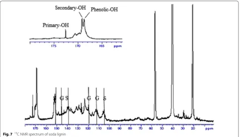
The results of UV analysis was supported by 13C NMR analysis. The estimation of lignin moieties was carried out by referring to Capanema et al. [20]. The 13C NMR spectra of acetylated kraft and soda lignins are shown in Figs. 6 and 7 respectively. The integral of the 155– 102 ppm region was set as a reference and assumed that it includes six aromatic carbons and 0.12 vinylic carbons. The integral values were divided by 6.12 which is equiva-lent to one aromatic ring (Ar). It was observed that kraft lignin contained higher amount of secondary aliphatic –OH (0.35 Ar−1) than that of soda lignin (0.26 Ar−1) Fig. 7 13C NMR spectrum of soda lignin
which was determined from the integration from 170 to 169 ppm region respectively. It was revealed that the amount of primary aliphatic –OH for kraft lignin (0.30
Ar−1) was higher than soda lignin (0.28 Ar−1) according to the integration from 173 to 170 ppm. On the other hand, kraft lignin consisted of a larger quantity of phe-nolic –OH (0.67 Ar−1) than soda lignin (0.46 Ar−1) based on the integration from 169 to 167 ppm. This can be con-tributed to the more intense cleavage of alkyl–aryl ether linkages during the kraft pulping process which lead to the formation of more phenolic end groups and thus increased the phenolic hydroxyl content in kraft lignin [16]. The higher phenolic –OH content enabled more hydroxyl radicals can be neutralized by the hydrogen atom abstraction from phenolic hydroxyl group.
Both soda and kraft lignins showed signals at 104 ppm, 138.5 ppm and 152 ppm which can be attributed to C-2 or C6, C-4, and C-3 or C5 in S unit. Besides, both lignins also gave signals at 112 ppm and 119 ppm which can be assigned to C-2 and C-6 in G unit. The higher S unit signals in both lignins are owning to OPEFB lignin contained more S unit compared to G unit. The signals at 123.5 ppm and 131 ppm can be referred to pyridine which was used for acetylation of lignin [21].
XRD analysis
The crystal structure of KL/TiO2-1.0 and SL/TiO2-1.0 were investigated through XRD analysis. The XRD dif-fractograms in Fig. 8 reveal that both composites had similar XRD patterns to that of pure anatase TiO2 [22]. The diffraction peaks at 2θ = 25.4°, 37.9°, 48.1°, 53.9°, 55.1°, 62.7°, 68.8°, 70.4°, 75.1° and 82.7° can be indexed to (101), (004), (200), (105), (211), (204), (115), (220), (215) and (224) crystal planes of anatase TiO2, respec-tively. This indicates that the initial crystal cell structure of TiO2 is maintained and no other crystalline by-prod-ucts are formed even after the formation of lignin/TiO2 composite. Since lignin is amorphous in nature and lack of ordered structure [10, 12], the XRD diffractograms of both composites did not show any diffraction peak that can be referred to lignin.
Fig. 9 SEM image of anatase TiO2 at 10,000× magnification
Fig. 10 SEM image of kraft lignin at 10,000× magnification
Fig. 11 SEM image of KL/TiO2-1.0 at 10,000× magnification
SEM and EDX analysis
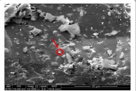
The SEM analysis was carried out to study the surface morphologies of kraft lignin, KL/TiO2-1.0, SL/TiO2-1.0, and anatase TiO2. The micromorphology of TiO2 was presented as many small spherical particles tended to aggregate to form a large sphere as shown in Fig. 9. Fig-ure 10 shows that kraft lignin possessed typical rough surface morphology of lignin [11]. Figures 11 and 12 depict the surface of KL/TiO2-1.0, and SL/TiO2-1.0 were rougher than and significantly different from the surface morphology of pure lignin and crystal mor-phology of naked TiO2. Based on Figs. 11 and 12, it was found that TiO2 particles were well dispersed in the lignin matrix indicating TiO2 is incorporated into lignin successfully. Besides, the tendency of TiO2 particles to
aggregate was reduced in both composites compared to that in pure anatase TiO2. This is mainly because of the sonication used in the formation of composite prevents the aggregation of TiO2 particles.
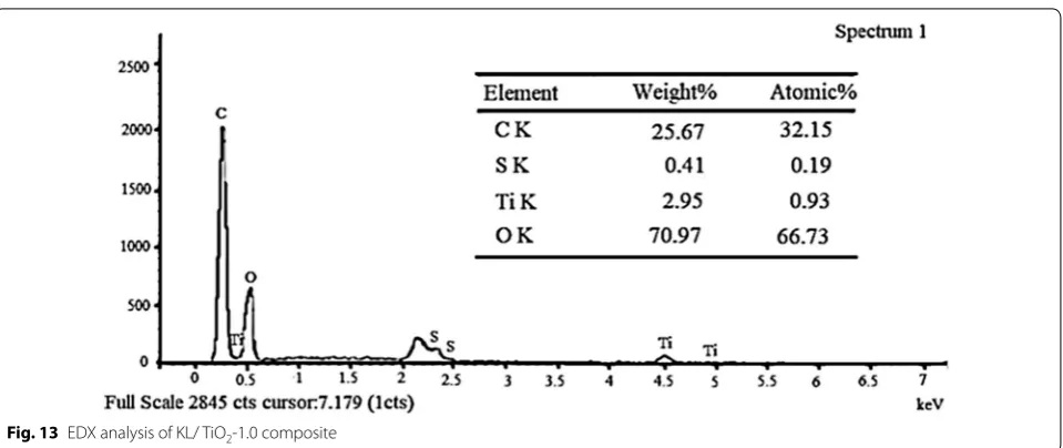
The element composition and distribution in the composites were investigated by EDX analysis. The EDX spectrum for KL/TiO2-1.0 and SL/TiO2-1.0 are shown in Figs. 13 and 14 respectively. Both composites were composed of the expected elements such as car-bon, oxygen, and titanium. The carbon and oxygen can be assigned to lignin while titanium and oxygen can be ascribed to TiO2. It further indicates that TiO2 has been incorporated into lignin. The presence of sulfur in KL/ TiO2-1.0 is owning to the hydrosulfide anions which are derived from the kraft pulping process [10].
Fig. 13 EDX analysis of KL/ TiO2-1.0 composite
Relative production of hydroxyl radicals
The relative production of hydroxyl radicals by kraft lignin, soda lignin, KL/TiO2-1.0, SL/TiO2 and anatase TiO2 were investigated through the photo-oxidation of coumarin to 7-hydroxycoumarin as a test reaction under the sunlight exposure. Coumarin reacts directly with hydroxyl radical to produce a fluorescing compound
7-hydroxycoumarin which can be detected by spectro-fluorometer as depicted in Fig. 15. The highly fluorescent 7-hydroxycoumarin can be detected at 460 nm in the flu-orescence spectrum [23].
Comparison between lignin/TiO2 composite and pure anatase TiO2
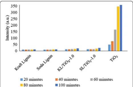
Fluorescence spectra of the coumarin solution irradiated under sunlight in the presence of kraft lignin, soda lignin, KL/TiO2-1.0, SL/TiO2-1.0, and anatase TiO2 are shown in Fig. 16. As shown in Figs. 16a–e and 17, coumarin solu-tion in the presence of TiO2 exhibited the highest emis-sion peak intensity throughout the experiment indicating Fig. 15 The photo-oxidation of coumarin to 7-hydroxycoumarin [24]
the highest concentration of hydroxyl radicals being pro-duced followed by SL/TiO2-1.0 and KL/TiO2-1.0. This can be attributed to the role of lignin as radical scaven-ger or antioxidant. Under the same sunlight irradiation, the peak intensity of coumarin solution in the presence of soda lignin and kraft lignin remained at the same level over irradiation time. This can be explained that both soda and kraft lignins possess no photocatalytic activity under sunlight irradiation. The result also indicated that the lignin does not completely quench the photocatalytic activity of TiO2 since hydroxyl radical generations still occur in the presence of both composites. The quench-ing process takes place when hydrogen atoms were abstracted from phenolic hydroxyl group by hydroxyl radical to form phenoxyl radical as proposed by Barclay et al. [24]. The presence of phenolic hydroxyl group in soda and kraft lignins has been proved by the UV and
13C NMR analyses. Moreover, according to the FTIR and 13C NMR spectra, both OPEFB lignins contained two main phenylpropanoid units which were guaiacyl (G) and syringyl (S). The methoxyl groups at the ortho posi-tion in G and S units aid in stabilizing phenoxyl radicals by resonance and hindering them from propagation. Besides, the interaction between the hydroxyl groups of phenolic compounds and the π-electrons of the benzene ring allows the phenolic compounds to produce radicals that can be stabilized by extended delocalization. Thus, the phenoxyl radical produced has much greater chemi-cal stability than the initial radichemi-cal.
Comparison between types of lignin
Figure 18 shows the fluorescence intensity of coumarin solution with KL/TiO2-1.0 and SL/TiO2-1.0 at 460 nm. The fluorescence intensity of both coumarin solutions increased with sunlight irradiation time. However, the fluorescence intensity of the solution with SL/TiO2-1.0 was higher than that of. KL/TiO2-1.0. The result indicates that kraft lignin has better hydroxyl radical quenching performance than soda lignin. This is highly possible due to higher phenolic hydroxyl group content of kraft lignin as mentioned in the UV and 13C NMR analysis. The higher phenolic hydroxyl content is caused by the severe process of kraft pulping which lead to extensive depolymerization of lignin such as cleavage of alkyl-aryl ether linkages. The hydroxide and hydrosulfide anions reacted with lignin causing the poly-mer to break down into smaller molecular weights frag-ments during the kraft pulping process and eventually the formation of new free phenolic hydroxyl group [10]. Hence, the low molecular weight fraction of kraft lignin possessed more non-etherified phenolic hydroxyl group than the high molecular weight fraction of soda lignin. It was suggested that the kraft lignin with higher content of non-etherified phenolic hydroxyl groups allows more hydroxyl radicals to be neutralized by the hydrogen atom abstraction from phe-nolic hydroxyl groups.
Comparison between amounts of lignin
Since kraft lignin showed better performance in quench-ing hydroxyl radicals than soda lignin, it was used to pre-pare three other composites with different amount of lignin.
Figures 19a–e and 20 show that fluorescence intensity of coumarin solution in the presence of KL/TiO2-0.5 was the highest throughout the experiment compared to the solutions with other three composites. This may be due to the low amount of lignin used. The amount of lignin is not sufficient to form a neat coating layer around TiO2 particles and quench the hydroxyl radicals generated. According to Fig. 20, there was not much difference in Fig. 17 Fluorescence intensity of coumarin solution with kraft lignin,
soda lignin, KL/TiO2-1.0, SL/TiO2-1.0 and TiO2 at 460 nm against sunlight illumination time
the fluorescence intensities of the coumarin solutions with KL/TiO2-1.0, KL/TiO2-1.5 and KL/TiO2-2.0 under the same sunlight illumination. It was suggested that 1.0 of lignin is enough to cover TiO2 particles and quench hydroxyl radicals before they diffuse away from TiO2 which may cause damages to biomolecules or other sun-screen ingredients. Besides, the excess amount of lignin will also make the color of composite becomes darker which is unfavorable for the appearance of sunscreens. Although the fluorescent intensities of all the three cou-marin solution increased slightly over the sunlight irra-diation time, it does not mean that the lignin lost the capability to capture the free radicals. It could be attrib-uted to the adsorption of coumarin molecules on vacant sites available on the surface of lignin. This will increase the competition between coumarin molecules and lignin
in reacting with hydroxyl radicals. Thus, more coumarin adsorbed on the lignin surface over illumination time and produced more 7-hydroxycoumarin which lead to increasing of fluorescence intensity.
Conclusion
13C NMR analysis. The higher phenolic hydroxyl con-tent provides more hydrogen atoms to quench the activ-ity of the hydroxyl radicals. The excess amount of lignin did not improve the radical scavenging activity but gave a darker color of the composite which is unfavorable for sunscreens and cosmetic products.
Abbreviations
TiO2: titanium II dioxide; OPEFB: oil palm empty fruit bunch; KL: kraft lignin; SL: soda lignin.
Authors’ contributions
All authors have equally contributed to the paper and have given approval to the final version of the paper. All authors read and approved the final manuscript.
Author details
1 School of Chemical Sciences, Universiti Sains Malaysia, 11800 Gelugor,
Penang, Malaysia. 2 Department of Chemistry, Faculty of Resource Science
and Technology, Universiti Malaysia Sarawak (UNIMAS), 94300 Kota Samara-han, Sarawak, Malaysia.
Acknowledgements
This project was financially supported by Universiti Sains Malaysia (USM) through Short Term Research Grant (304/PKIMIA/6316174) and (304/ PKIMIA/6313215), Research University Grant (RUI) (1001/PKIMIA/8011083) and Trans-disciplinary Research Grant Scheme (TRGS) (203/PKIMIA/6769001).
Competing interests
The authors declared that they have no competing interests.
Availability of data and materials Not applicable.
Funding Not applicable.
Publisher’s Note
Springer Nature remains neutral with regard to jurisdictional claims in pub-lished maps and institutional affiliations.
Received: 7 November 2018 Accepted: 16 January 2019
References
1. Sambandan DR, Ratner D (2011) Sunscreens: an overview and update. J Am Acad Dermatol 64(4):748–758
2. Morsella M, d’Alessandro N, Lanterna AE, Scaiano JC (2016) Improving the sunscreen properties of TiO2 through an understanding of its catalytic
properties. ACS Omega 1(3):464–469
3. Ayed S, Belgacem RB, Zayani JO, Matoussi A (2016) Structural and optical properties of ZnO/TiO2 composites. Superlattices Microstruct 91:118–128
4. Barnes RJ, Molina R, Xu J, Dobson PJ, Thompson IP (2013) Comparison of TiO2 and ZnO nanoparticles for photocatalytic degradation of methylene
blue and the correlated inactivation of gram-positive and gram-negative bacteria. J Nanopart Res 15(2):1432
5. Carlotti ME, Ugazio E, Sapino S, Fenoglio I, Greco G, Fubini B (2009) Role of particle coating in controlling skin damage photoinduced by titania nanoparticles. Free Radical Res 43(3):312–322
6. Mao L, Meng C, Zeng C, Ji Y, Yang X, Gao S (2011) The effect of nitrate, bicarbonate and natural organic matter on the degradation of sunscreen agent p-aminobenzoic acid by simulated solar irradiation. Sci Total Envi-ron 409(24):5376–5381
7. Picatonotto T, Vione D, Eugenia Carlotti M (2002) Effect of some addi-tives used in the cosmetic field on the photocatalytic activity of rutile. J Dispers Sci Technol 23(6):845–852
8. Shen B, Scaiano JC, English AM (2006) Zeolite encapsulation decreases TiO2-photosensitized ROS generation in cultured human skin fibroblasts. Photochem Photobiol 82(1):5–12
9. Chen X, Liu Y, Shi H, Wang X, Qi K, Zhou X, Xin JH (2010) Carboxymethyl chitosan coating to block photocatalytic activity of TiO2 nanoparticles.
Text Res J 80(20):2214–2222
10. Ibrahim MNM, Zakaria N, Sipaut CS, Sulaiman O, Hashim R (2011) Chemi-cal and thermal properties of lignins from oil palm biomass as a substi-tute for phenol in a phenol formaldehyde resin production. Carbohyd Polym 86(1):112–119
11. Ibrahim MNM, Ngah WW, Norliyana MS, Daud WW, Rafatullah M, Sulaiman O, Hashim R (2010) A novel agricultural waste adsorbent for the removal of lead (II) ions from aqueous solutions. J Hazard Mater 182(1–3):377–385
12. Hussin MH, Zhang HH, Aziz NA, Samad NA, Faris AH, Ibrahim MNM, Iqbal A, Latip AFA, Haafiz MM (2017) Preparation of environmental friendly phenol-formaldehyde wood adhesive modified with kenaf lignin. Beni-Suef Univ J Basic Appl Sci 6(4):409–418
13. Abdullah N, Sulaiman F (2013) The oil palm wastes in Malaysia. Biomass now-sustainable growth and use. InTech, pp 75–100
14. Ibrahim MNM, Salleh NM, Yusof NNM, Ghani AM (2010) Keistimewaan Lignin Daripada Kelapa Sawit. Penerbit Universiti Sains Malaysia, Pulau Pinang, pp 157–160
15. Salleh NM, Ismail S, Ibrahim MNM (2015) Radical scavenging activ-ity of lignin extracted from oil palm empty fruit bunch and its effect on glutathione-S-transferase enzymes activity. Asian J Pharm Clin Res 8:81–87
16. Ugartondo V, Mitjans M, Vinardell MP (2008) Comparative antioxidant and cytotoxic effects of lignins from different sources. Biores Technol 99(14):6683–6687
17. Morsella M, Giammatteo M, Arrizza L, Tonucci L, Bressan M, d’Alessandro N (2015) Lignin coating to quench photocatalytic activity of titanium dioxide nanoparticles for potential skin care applications. RSC Adv 5(71):57453–57461
18. Ishibashi KI, Fujishima A, Watanabe T, Hashimoto K (2000) Detection of active oxidative species in TiO2 photocatalysis using the fluorescence
technique. Electrochem Commun 2(3):207–210
19. Zhao X, Dai L, Liu D (2009) Characterization and comparison of Acetosolv and Milox lignin isolated from crofton weed stem. J Appl Polym Sci 114(2):1295–1302
20. Capanema EA, Balakshin MY, Kadla JF (2004) A comprehensive approach for quantitative lignin characterization by NMR spectroscopy. J Agric Food Chem 52(7):1850–1860
21. Fulmer GR, Miller AJ, Sherden NH, Gottlieb HE, Nudelman A, Stoltz BM, Bercaw JE, Goldberg KI (2010) NMR chemical shifts of trace impurities: common laboratory solvents, organics, and gases in deuterated solvents relevant to the organometallic chemist. Organometallics 29(9):2176–2179 Fig. 20 Fluorescence intensity of coumarin solution in the presence
•fast, convenient online submission •
thorough peer review by experienced researchers in your field • rapid publication on acceptance
• support for research data, including large and complex data types •
gold Open Access which fosters wider collaboration and increased citations maximum visibility for your research: over 100M website views per year •
At BMC, research is always in progress.
Learn more biomedcentral.com/submissions
Ready to submit your research? Choose BMC and benefit from:
22. Muniandy L, Adam F, Mohamed AR, Ng EP, Rahman NRA (2016) Carbon modified anatase TiO2 for the rapid photo degradation of methylene
blue: a comparative study. Surf Interf 5:19–29
23. Barclay LRC, Xi F, Norris JQ (1997) Antioxidant properties of phenolic lignin model compounds. J Wood Chem Technol 17(1–2):73–90



![Fig. 15 The photo-oxidation of coumarin to 7-hydroxycoumarin [24]](https://thumb-us.123doks.com/thumbv2/123dok_us/377079.2034851/10.595.59.289.88.134/fig-photo-oxidation-coumarin-hydroxycoumarin.webp)