Original Article
Comparison of Osteogenic and Chondrogenic Differentiation Ability of Buccal
Fat Pad Derived Mesenchymal Stem Cells and Gingival Derived Cells
Hamid Ghaderi 1, Mahboobeh Razmkhah 2, Farin Kiany 3, Nooshafarin Chenari 2, Mohammad Reza Haghshenas 2, Abbas Ghaderi 4
1
Dentist, Private Practice, Shiraz, Iran.
2
Institute for Cancer Research, School of Medicine, Shiraz University of Medical Sciences, Shiraz, Iran.
3
Oral and Dental, Disease Research Center, School of Dentistry, Shiraz University of Medical Sciences, Shiraz, Iran.
4 Institute for Cancer Research, Dept. of Immunology, School of Medicine, Shiraz University of Medical Sciences, Shiraz, Iran.
KEY WORDS
Mesenchymal stem cell;
Buccal fat pad;
Gingiva;
Chondrocyte;
Osteocyte;
Received January 2017;
Received in Revised form August 2017; Accepted September 2017;
ABSTRACT
Statement of the Problem: One major goal of tissue engineering and regenerative medicine is to find an appropriate source of mesenchymal stem cells (MSCs) with
higher differentiation ability.
Purpose: In this experimental study, the osteogenic and chondrogenic differentiation ability of buccal fat pad derived MSCs (BFP-MSCs) with gingival derived cells
(GDCs) were compared.
Materials and Method: BFP-MSCs and GDCs were cultured enzymatically and expanded. The expanded cells were analyzed for membrane-associated markers,
using flow cytometry. Then the ability of these cells to differentiate into osteocyte
and chondrocyte was assessed morphologically and by mRNA expression of
colla-gen I (COLL), BGLA and bone morphocolla-genetic protein 2 (BMP2) using qRT-PCR.
Results: Flow cytometry analysis showed that both BFP-MSCs and GDCs expressed the characteristic stem cell markers such as CD73, CD44, and CD90, whereas they
did not express hematopoietic markers. Mineralized calcium deposition was
ob-served apparently in BFP-MSCs cultured in osteogenic medium but GDCs showed
fewer mineralized nodules. The mRNA expression levels of BGLA and BMP2
showed 7×10⁵ and 733-fold more mRNA expression in BFP-MSCs treated with
differentiation media compared to the control group. In chondrogenic differentiation,
BFP-MSCs transformed from a spindle to a cuboidal shape while GDCs showed
only a slight transformation. In addition, mRNA expression of COLL showed
282-fold higher expression in BFP-MSCs in comparison to the control group.Such
sig-nificant difference in mRNA expression of BGLA, BMP2, and COLL was not
ob-served in GDCs compared to their corresponding controls.
Conclusion: Based on the present results, BFP yields a greater proportion of stem cells compared to gingiva. Therefore, this tissue can be introduced as an easily
avail-able source for the treatment of periodontal defects and other maxillofacial injuries.
Corresponding Author: Kiany F., Dept. of Periodontics, Oral and Dental Disease Research Center, School of Dentistry Shiraz Dental School, Shiraz University of Medical Sciences, Shiraz, Iran. Email: [email protected] Tel: +98-71-36263193-4
Cite this article as: Ghaderi H., Kiany F., Razmkhah M., Chenari N., Haghshenas MR., Ghaderi A. Comparison of Osteogenic and Chondrogenic Differentiation Ability of Buccal Fat Pad Derived Mesenchymal Stem Cells and Gingival Derived Cells. J Dent Shiraz Univ Med Sci., 2018 June; 19(2): 124-131.
Introduction
Mesenchymal stem cells (MSCs) are a heterogeneous
population of non-hematopoietic stem cells with a high
capacity for self-renewal and regeneration. These cells
are capable of differentiating into various lineages
line-ages in the presence of a series of stimulations. [1]
Sev-eral studies have reported the successful treatment of
bone and cartilage defects, vascular ischemia, and
coro-nary artery disease upon local administration of MSCs
to the sites of injury. [2-3]
MSCs are present in many adult tissues, such as
synovium, muscle, adipose tissue, and bone marrow. [4]
Adipose (fat) tissue represents an abundant and
accessi-ble source of MSCs, and deriving of these cells is
ac-companied with minimal patient discomfort. Therefore,
adipose tissue may be a practical and appealing source
of donor tissue for clinical applications. [5]
Buccal fat pad (BFP), one of the encapsulated fat
masses in the cheek, is located between the buccinator
muscle and several superficial muscles including
masse-ter, zygomaticus major, and zygomaticus minor on both
sides of the face. [6-7] BFP is a new and easily
accessi-ble source of MSCs and BFP derived MSCs
(BFP-MSCs) have virtually the same characteristics as
adi-pose derived stem cell (ASCs). It means that they have
the ability of giving rise to various cell lineages and
therefore could be suitable for clinical uses such as
peri-odontal defect treatment. [8]
Gingiva as the soft tissue surrounding the teeth
has unique structure that contributes to the resistance
against shear stress or friction. [2] Previous studies have
demonstrated that gingival tissue possess progenitors or
adult stem cells with similar properties to MSCs such as
pluripotency, self-renewal ability and
immunomodula-tory properties and play a crucial role in the repair and
regeneration of periodontal tissues. [9-11]
In this study, for the first time the osteogenic and
chondrogenic capabilities of BFP-MSCs and gingival
derived cells (GDCs) have been compared. Results of
this study may contribute to obtain a more accessible
source of stem cells for periodontal, oral implant and
maxillo-facial surgeries.
Materials and Method
Subjects
In this experimental study, BFP was obtained from five
healthy individuals undergoing elective orthognathic
surgery at Rajaee Hospital, Shiraz University of
Medi-cal Sciences, Shiraz, Iran. The age range of the
individ-uals undergoing oral surgery was 19-28 years and no
history of diabetes or other systemic complications were
reported for them.
Human gingival samples were collected from five
patients undergoing crown-lengthening surgery, with
age range of 35-45 years, and with no history of
perio-dontal or systemic diseases. The gingival tissues were
obtained as part of routine periodontal surgery at the
School of Dentistry, Shiraz University of Medical
Sci-ences, Shiraz, Iran. Informed consent was obtained from
all the patients. The informed consent and experimental
protocols in this study were reviewed and approved by
the Ethics Committee of Shiraz University of Medical
Sciences, Shiraz, Iran.
Both BFP and gingival tissue samples were
main-tained and transferred to Stem Cell and Cancer
Labora-tory of Shiraz Institute for Cancer Research (ICR),
Shi-raz University of Medical Sciences, in a media
contain-ing 50U/mL penicillin, streptomycin, amphotericin B
and DMEM culture medium (Gibco, USA) containing
10% fetal bovine serum (FBS, Gibco, USA).
Isolation and culture of BFP-MSCs and GDCs
Samples were washed several times with sterile
phos-phate-buffered saline (PBS), chopped into small pieces,
and then digested using 0.2 % collagenase type I. The
resulted soup was centrifuged and the pellet was
care-fully put on Ficoll (Biosera, UK) and centrifuged again.
The second white layer was washed with PBS and
final-ly re-suspended in DMEM containing 10% fetal bovine
serum and 1% penicillin/streptomycin (Biosera, UK)
and maintained in a humidified incubator at 37˚C and
5% CO2. The medium was changed every 4 days. After
3–4 days, individual cell colonies were visible upon
microscopic examination. The cells were analyzed by
flow cytometry and differentiation protocols in passage
3.
Cell staining, data acquisition and flow cytometry analysis
Expression of mesenchymal markers on extracted cells
was analyzed by flow cytometry. BFP-MSCs and GDCs
were detached by trypsin-EDTA (Biosera, UK),
re-suspended in PBS at a density of 1×10⁶ cells/mL. The
selected cells were then incubated with PE-conjugated
anti-CD166, anti-CD105, anti-CD44 antibodies,
FITC-conjugated anti-CD34, anti-CD45, anti-CD14, and
APC-conjugated anti-CD90, and anti-CD73, as well as
dye/isotype matched antibodies (all from BD
Biosci-ences, USA) in dark environment for 30 minutes.
Table 1: The sequences of primers used in qRT-PCR method
Forward primer Reverse primer
COLL 5'-TGCCCCATCTGCCCAACTGA -3' 5'-TGCAGGTCCCTGAGGCCC-3'
BGLA 5'-GAGCCCTCACACTCCTCGC-3′ 5'-CAGCCAACTCGTCACAGTCC-3'
BMP2 5'-GAGGCAAAGAAAAGGAACGGAC-3' 5'-GCAGCAACGCTAGAAGACAG-3'
18sRNA 5'-GTTGATTAAGTCCCTGCCCT-3' 5'-TCCGAGGGCCTCACTAAACC-3'
cell sample was assessed on a FACSCalibur flow
cy-tometer (BD Bioscience, USA).The data was analyzed
by FlowJo software package. Positive cells were
count-ed and comparcount-ed with the signals of the corresponding
antibody isotype controls.
Differentiation of human BFP-MSCs and GDCs to chondrocyte
and osteocyte
The potential of BFP-MSCs and GDCs to differentiate
into chondrogenic and osteogenic lineages was
exam-ined using the following procedures. 1×10⁵ cells were
cultured in each well of 4-well tissue culture plate for
differentiation. When the cultures were 60–80%
conflu-ent, an appropriate differentiation kit (STEMPRO
Dif-ferentiation Kit; GIBCO, USA) was used for
differenti-ating the cells regarding the manufacturer’s instructions.
On days 7, 14 and 21 post treatment, cells were stained
with Alizarin red (Merck, Germany) and Safranin
(Merck, Germany) for evaluating osteogenic and
chon-drogenic differentiation, respectively. For the control
group, 1×10⁵ cells were cultured with common media
(DMEM and 10% FBS), without differentiation agents,
and the same procedure of staining and RNA isolation
was carried out.
RNA isolation and reverse transcription
When the culture flasks were 80-90% confluent, in
pas-sage 3, cells were cultured in three separated plates and
the total RNA was extracted on 7, 14 and 21 days post
culture by RNX Plus solution (Cinnagene, Iran). During
RNA extraction, DNase I (GIBCO, USA) was added to
avoid DNA contamination.
Then cDNA was synthesized from 5µg of the total
RNA, using the ReverAid first strand cDNA synthesis
Kit Fermentas, Lithuania) according to recommended
instructions.
Quantitative Real-Time PCR (qRT-PCR)
The expression and quantity of BGLA, bone morphoge-
netic protein 2 (BMP2) (as osteogenic markers) and
collagen I (COLL) (as a chondrogenic marker) gene
transcripts were determined using an ABI thermal
cy-cler. Briefly approximately 2µl cDNA was amplified in
a final volume of 20µl including 10µl of SYBR Green I
PCR Master Mix (Fermentas, Lithuania), 0.3µl of each
forward and reverse primers and 7.4µl DEPC treated
water. 18s rRNA housekeeping gene expression was
used to normalize the level of target gene expression.
Thermal cycling for all the genes was set up
through a denaturation step at 95°C for 10 minutes,
fol-lowed by 50 cycles (denaturation at 95°C for 15 s,
an-nealing and extension at 60ºC for 60s). Table 1 shows
the forward and reverse primers for 18s rRNA, BGLA,
BMP2 and COLL genes. Primers were designed using
Primer-Blast online software (http://www.ncbi.nlm.nih.
go/tools/primer-blast).
Statistical Analysis
The relative amounts of BGLA, BMP2, and COLL tran-scripts were determined from ΔCt and 2−ΔΔCt formulas. Relative expressions were plotted and evaluated by
means of Prism 5 software (San Diego, CA, USA).
Results
Flow cytometry analysis
The BFP-MSCs and GDCs were harvested in passage 3
and identified by a FACSCalibur flow cytometer. Flow
cytometric analysis of both BFP-MSCs and GDCs is
shown in Figure 1 and Table 2. Comparison of forward
(F-) and side (S-) scatter (SC) dot plots demonstrated
that both cells were similar in granularity and size.
Table 2: Mesenchymal, lymphocyte or leukocyte antigens and hematopoietic markers on BFP-MSCs and GDCs obtained from relat-ed tissues.
MSCs type Cell surface markers(mean ±SD)
CD166 CD105 CD44 CD90 CD73 CD14 CD34 CD45
BFP- MSCs 94.4±2.2 85±4.7 99.7±0.7 93.2±6.3 97.2±1.8 0 4.7±3 0
GDCs 87.4±18.2 39.5±15.2 99.8±0.3 99.8±0.3 97.4±3 0 1 o.9
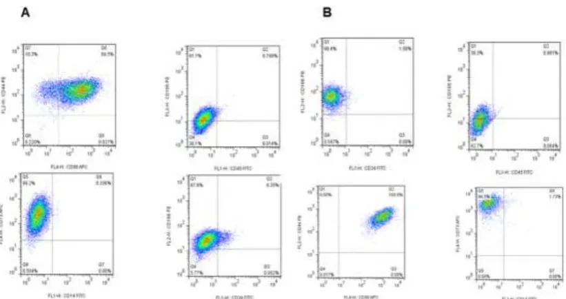
Figure 1: Flow cytometry analysis of mesenchymal specific markers on BFP-MSCs (A) and GDCs (B) obtained from BFP and gingival specimens, respectively. Both cells isolated were positive for the expression of CD90, CD73, and CD44 but negative for the expression of CD14, CD45, and CD34
Both BFP-MSCs and GDCs expressed the
charac-teristic stem cell markers such as CD73, CD166, CD44,
CD90 and CD105, whereas they did not express or
rare-ly expressed rare-lymphocyte or leukocyte antigens and
hematopoietic markers such as CD14, CD45, and CD34
(Table 2 and Figure 1). As shown in Table 2, the
ex-pression of all markers had similar mean percentage in
both cell types except for CD105 which had 2-fold
higher expression in BFP-MSCs compared to GDCs
(85±4.7% vs 39.5±15.2%).
Osteogenic and chondrogenic differentiation
Morphological changes
Under a phase contrast microscope, mineralized calcium
deposition was observed in BFP-MSCs cultured in
oste-ogenic medium (OM) on day7 until 21, which strongly
increased on day 21(Figure 2b). GDCs cultured in OM
showed some mineralized nodule formation on day 21,
but they were fewer than those observed in
differentiat-ed BFP-MSCs (Figure 2e). There was no mineralizdifferentiat-ed
nodule formation in both cell types (BFP-MSCs and
GDCs) cultured in normal media, without
differentia-tion agents, as the control group at all time periods
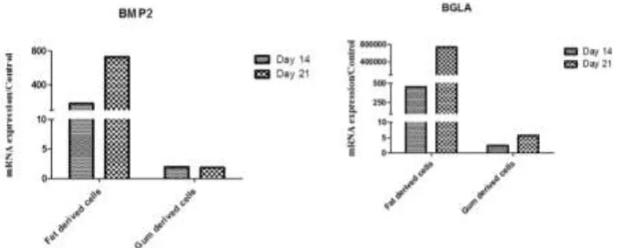
Figure 3: Evaluation of BGLA and BMP2 genes expression in BFP-MSCs and GDCs differentiated to osteocytes after 14 and 21 days post treatment of the cells. The expression and quantity of BGLA and BMP2 gene transcripts was determined by qRT-PCR
ures 2a and 2d). In chondrogenic differentiation, cells
transformed from a spindle to a cuboidal shape in
BFP-MSCs while GDCs showed only a slight transformation
(Figures 2c and 2f). In control group, no transformation
was observed.
Expressions of BGLA and BMP2 as osteogenic and COLL as
chondrogenic markers
Total RNA was isolated from both cell types during
differentiation on days 7, 14 and 21 and subsequently
mRNA level of BGLA, BMP2 and COLL were
evaluat-ed using qRT-PCR.
As depicted in Figure 3, BGLA and BMP2 gene
transcripts showed a higher expression in BFP-MSCs on
14 and 21 days post culture compared to GDCs.
Ac-cordingly, BGLA mRNA had approximately 7×10⁵-fold
more expression at day 21 compared to the control
group after culturing with osteogenic media. In addition,
BMP2 showed 733-fold more mRNA expression in
BFP-MSCs treated with differentiation media compared
to control. No obvious changes were observed in
mRNA expression of these two genes after treating
GDCs with osteogenic media.
Figure 4: Expression of COLL gene transcript 14 and 21 days after treatment of BFP-MSCs and GDCs with chondro-genic differentiation media. The expression and quantity of COLL gene transcript was determined using qRT-PCR
Similarly, differentiation to chondrocytes showed
a higher ability of BFP-MSCs for transformation
com-pared to GDCs. COLL mRNA showed a 282-fold
high-er expression at day 21 in BFP-MSCs but not in GDCs
compared to control (Figure 4).
Discussion
MSCs are present in various organs and are known as
attractive resources in regenerative medicine because of
their outstanding characteristics including extensive
expansion and differentiation into various cell types. In
engineering medicine, the availability of stem cell
source, simple isolation and the ability to expand the
cells into large numbers is desirable. [12]
BFP and gingiva are two unique oral reservoirs for
MSCs that usually yield to a high number of progenitor
cells compared to other sources such as bone marrow.
[13] Adipose tissue holds great promise in regenerative
medicine; it is accessible in sufficient quantities as
waste material, and it contains more progenitor cells
giving rise to diverse cell populations in comparison to
bone marrow. [6-7] The common anatomical areas for
obtaining fat tissue are abdomen, breast, knee, and
cheek. BFP is a deep fat pad located in cheek, which is
usually discarded during plastic surgery but because of
having a high number of stem cells, could be a suitable
candidate in facial reconstruction procedures. [6-7] As
well as BFP, gingival tissue harbors a population of
cells that have similar features compared to stem cells.
Gingiva is the most applicable stem cell source
com-pared to the other dental origins. [11, 14] Gingival
tis-sue can be simply obtained as a byproduct from the
clinically resected gingival tissues during periodontal
cells from gingiva renders it as a candidate for the
source of progenitor cells in the regenerative treatment
of periodontal defects. [11, 14] Zhang et al. [11]
isolat-ed a population of progenitor/stromal cells from
gingi-val tissues that showed stem cell characteristics.
To find a better source of MSCs for future therapi-
es of periodontal defects, we obtained and cultured cells
from two intra oral sources including adipose and
gingi-val tissues, and then compared their stemness features
and differentiation ability to osteocyte and chondrocyte.
Subsequently differentiation was confirmed by
detec-tion of COLL, BGLA and BMP2 mRNA expressions.
Based on the results, BFP-MSCs and GDCs could
easily grow and expand from small specimens of both
tissues. Surface staining of mesenchymal specific
mark-ers indicated that features of both cell types were similar
to other known MSC sources. Moreover, both types of
cells did not express or rarely expressed
non-mesenchymal markers such as CD14, CD45, and CD34.
The expression of all the investigated mesenchymal
markers were similar between these two populations
except for the expression of CD105 (Endoglin) which is
the TGF-β receptor [15] and the difference in its
expres-sion was expected, because it may have been modified
based on the tissue source.
It is reported that despite the lack of a specific
cell-surface marker for adult MSCs of distinct tissue
origins, these cells express a panel of mesenchymal cell
markers such as CD73, CD90, CD105, and CD44 but
are negative for endothelial and hematopoietic markers
such as CD31, CD34, and CD45. [16] Similar to our
study, a recent study has revealed that BFP-MSCs
ex-pressed stem cell markers such as CD73, CD90, and
CD105, whereas they did not express lymphocyte or
leukocyte antigens and hematopoietic markers such as
CD14, CD31, and CD34. [17] Similarly, it has been
shown that GDCs consistently express CD29, CD44,
CD73, and CD90 and are negative for CD34 and CD45,
but are positive for CD105, CD146, and Stro-1 in
varia-ble population subsets. [2]
Moreover, we investigated the differentiation
abil-ity of BFP-MSCs and GDCs to osteocyte and
chondro-cyte. The findings demonstrated that both cell types
could be induced to give rise to these lineages.
Our pervious study on isolated ASCs from breast
tissue showed that these cells could represent
hepato-genic and chondrohepato-genic differentiation markers on the
certain differentiating conditions. [18] In addition, a
pervious study indicated that adipose tissue isolated
from BFP contained MSCs with stemness properties
that were able to differentiate towards the adipogenic
and osteogenic lineages. [17] Like BFP-MSCs, it has
been reported that gingiva-derived MSCs can also
dif-ferentiate to osteocytes, adipocytes, and chondrocytes
when they are properly induced. [2] Our results also
indicated that cellular transformation towards
chondro-cyte and osteochondro-cyte and the expression of corresponding
genes including COLL, BGLA, and BMP2 genes were
considerably higher in BFP-MSCs than GDCs.
GDCs cultured in osteogenic media showed some
mineralized nodule formations, but fewer than those
observed within the culture of BFP-MSCs. Those
nod-ules were associated with slight transformation.
The findings of the present study imply that BFP
can be a better alternative source of stem cells than
gin-gival tissue. ASCs from BFP are easily reachable in
abundant quantities with a minimal invasive harvesting
method and allow a high number of ASCs to be derived.
Moreover, the chance of contamination during surgical
removal of gingiva increases due to the presence of oral
normal flora. Another important concern in culture of
gingival tissue is the growth of fibroblasts, because
most of the stromal cells in gingival connective tissue
are fibroblasts. Fibroblasts and stem cells share common
properties including a spindle shape, plastic adherence
and overlapping cell surface antigens but there are no
specific markers to discriminate fibroblast from stem
cells, however the differentiation ability of stem cells is
not identified for fibroblasts. [19] Fibroblasts derived
from different tissue origins can exhibit multilineage
differentiation potentials and immunomodulatory
func-tions. Nevertheless, because of the lack of definite
markers and the unidentified in vivo identity of MSCs,
the exact relationship between MSCs and fibroblasts
remains elusive. [2]
Tomasello et al. [20] showed that the chronic
in-flammatory condition that exists in periodontitis does
not negatively influence the number or the stem cell
marker profile of GDCs. Actually they confirmed that
overexpression of proinflammatory cytokines permit a
higher osteogenic differentiation potential of extracted
findings convince us for using inflamed periodontal
tissue for future researches and in tissue engineering
applications.
Conclusion
Based on the present findings, adipose tissue,
particu-larly BFP, can be introduced as a simply accessible
source for extraction of stem cells. This tissue yields
greater proportion of stem cells compared to other
sources such as gingiva. Considering these features,
BFP may be an outstanding cell source for clinical use
and regenerative treatments in maxillofacial bony and
periodontal defects.
Acknowledgements
The authors would like to thank all the participants in
the study. This work was supported by a grant from
Shiraz University of Medical Sciences [Grant No. 6265]
and Shiraz Institute for Cancer Research
[ICR-100-504]. This research was done as a requirement for the
Dentistry thesis defended by Dr. Hamid Ghaderi.
Conflict of Interest
The authors declare that they have no conflicts of
inter-est concerning this article.
References
[1] Deng J, Petersen BE, Steindler DA, Jorgensen ML, Lay-well ED. Mesenchymal stem cells spontaneously express neural proteins in culture and are neurogenic after trans-plantation. Stem Cells. 2006; 24: 1054-1064.
[2] Zhang QZ, Nguyen AL, Yu WH, Le AD. Human oral mucosa and gingiva: a unique reservoir for mesenchymal stem cells. J Dent Res. 2012; 91: 1011-1018.
[3] Docheva D, Popov C, Mutschler W, Schieker M. Human mesenchymal stem cells in contact with their environ-ment: surfacecharacteristics and the integrin system. J Cell Mol Med. 2007; 11: 21-38.
[4] Noël D, Djouad F, Jorgense C. Regenerative medicine through mesenchymal stem cells for bone and cartilage repair. Curr Opin Investig Drugs. 2002; 3: 1000-1004. [5] Cowan CM, Shi YY, Aalami OO, Chou YF, Mari C,
Thomas R, et al. Adipose-derived adult stromal cells heal critical-size mouse calvarial defects. Nat Biotechnol. 2004; 22: 560-567.
[6] Zhang HM, Yan YP, Qi KM, Wang JQ, Liu ZF. Anatom-
ical structure of the buccal fat pad and its clinical adapta-tions. Plast Reconstr Surg. 2002; 109: 2509-2518. [7] Gassner HG, Rafii A, Young A, Murakami C, Moe KS,
Larrabee WF Jr. Surgical anatomy of the face: implica-tions for modern face-lift techniques. Arch Facial Plast Surg. 2008; 10: 9-19.
[8] Farré-Guasch E, Martí-Pagè C, Hernádez-Alfaro F, Klei- n-Nulend J, Casals N. Buccal fat pad, an oral access source of human adipose stem cells with potential for os-teochondral tissue engineering: an in vitro study. Tissue Eng Part C Methods. 2010; 16: 1083-1094.
[9] Tomar GB, Srivastava RK, Gupta N, Barhanpurkar AP, Pote ST, Jhaveri HM, et al. Human gingiva-derived mes-enchymal stem cells are superior to bone marrow-derived mesenchymal stem cells for cell therapy in regenerative medicine. Biochem Biophys Res Commun. 2010; 393: 377-383.
[10]Mitrano TI, Grob MS, Carrión F, Nova-Lamperti E, Luz PA, Fierro FS, et al. Culture and characterization of mes-enchymal stem cells from human gingival tissue. J Perio-dontol. 2010; 81: 917-925.
[11]Zhang Q, Shi S, Liu Y, Uyanne J, Shi Y, Shi S, et al. Mesenchymal stem cells derived from human gingiva are capable of immunomodulatory functions and ameliorate inflammation-related tissue destructionin experimental colitis. J Immunol. 2009; 183: 7787-7798.
[12]Chen FM, Jin Y. Periodontal tissue engineering and re-generation: current approaches and expandingopportuni-ties. Tissue Eng Part B Rev. 2010; 16: 219-255. [13]Bunnell BA, Flaat M, Gagliardi C, Patel B, Ripoll C.
Adipose-derived stem cells: isolation, expansion and dif-ferentiation. Methods. 2008; 45: 115-120.
[14]Häkkinen L, Uitto VJ, Larjava H. Cell biology of gingi-val wound healing. Periodontol 2000. 2000; 24: 127-152. [15]Valluru M, Staton CA, Reed MW, Brown NJ.
Trans-forming Growth Factor-β and Endoglin Signaling Or-chestrate Wound Healing. Front Physiol. 2011; 2: 89. [16]Fournier BP, Ferre FC, Couty L, Lataillade JJ, Gourven
M, Naveau A, et al. Multipotent progenitor cells in gingi-val connective tissue. Tissue Eng Part A. 2010; 16: 2891-2899.
[18]Razmkhah M, Jaberipour M, Erfani N, Habibagahi M, Talei AR, Ghaderi A. Adipose derived stem cells (ASCs) isolated from breast cancer tissue express IL-4, IL-10 and TGF-β1 and upregulate expression of regulatory mole-cules on T cells: do they protect breast cancer cells from the immune response? Cell Immunol. 2011; 266: 116-122.
[19]Alt E, Yan Y, Gehmert S, Song YH, Altman A, Gehmert
S, et al. Fibroblasts share mesenchymal phenotypes with stem cells, but lack their differentiation and colony-forming potential. Biol Cell. 2011; 103: 197-208. [20]Tomasello L, Mauceri R, Coppola A, Pitrone M, Pizzo G,