A Real-Time Pulse Peak Detection Algorithm for
the Photoplethysmogram
Dae-Geun Jang, Sangjun Park, and Minsoo Hahn
Korea Advanced Institute of Science and Technology/Department of Electrical Engineering, Daejeon, South Korea {jangdg85, psj}@kaist.ac.kr, [email protected]
Seung-Hun Park
Kyunghee University/Department of Biomedical Engineering, Yongin, South Korea [email protected]
Abstract—In this paper, we propose a simple and low complexity pulse peak detection algorithm using cascaded recursive digital filters and a slope sum function (SSF) with an adaptive thresholding scheme. The algorithm first eliminates noises in the photoplethysmogram (PPG) using the cascaded lowpass and highpass digital filters. The filters have been designed with 3-dB cutoff frequencies of 11 Hz and 0.5 Hz, respectively. The filtered PPG signal is then transformed by the SSF. The SSF simplifies detecting the pulse peaks by enhancing the upslope of the PPG signal and suppressing the remainder. A threshold for identifying SSF peaks is updated using the median filter with an order of 5. This update makes the threshold adaptive to variations of SSF heights. The detected SSF peaks localize ranges for pulse peak detection. Finally, the pulse peak is identified by picking the local maxima within the range from an onset index of the SSF signal to the following zero index. In order to cope with over-detected and missed information, the proposed algorithm employs knowledge-based rules as post-processing. The algorithm is tested on a database where PPG waveforms are collected from 127 subjects. The results are promising, suggesting that the method provides simpler but accurate pulse peak detection in real applications.
Index Terms—photoplethysmogram, slope sum function, pulse peak detection, pulse rate detection
I. INTRODUCTION
A reliable pulse peak detection facilitates extraction of all other characteristic points such as dicrotic wave on the signal with the reference to the pulse peak [1]. This implies that a false detection of pulse peaks can adversely affect delineating the photoplethysmogram (PPG) signal. Practically, accurate detection of pulse peaks from the PPG signal is difficult, not only because of the physiological variability of pulse peaks, but also because of the respiration, motion artifacts, and electrical interference noises [2]. In particular, the real-time pulse
Manuscript received November 30, 2013; revised January 11 2014. This research was supported by the MSIP (Ministry of Science, ICT& Future Planning), Korea, under the ITRC (Information Technology Research Center) support program supervised by the NIPA (National IT Industry Promotion Agency) (NIPA-2013-(H0301-13-2001)).
peak detection for small ubiquitous or wearable application is a more strenuous task since if further requires low computational burden and low memory capacity.
The accurate pulse peak detection is required not only to delineate PPG signal, but also to analyze physiological states correctly [2], [3]. In the pulse rate variability (PRV) analysis, variation in the time interval between successive pulse peaks is investigated to assess autonomic function. Some of PRV parameters such as the Lyapunov exponent are sensitive to each variation [4] and thus the accurate pulse peak detection is a must for the PRV analysis. Further the accurate pulse peak detection is required to calculate the pulse transit time (PTT), which is defined as the time it takes a pulse wave to travel between two arterial sites, since pulse peaks are considered as the start and end points of each pulse wave travel [5]. In addition, the reliable pulse peak detection is also required for basic clinical applications such as pulse rate monitoring [2], [3].
In order to detect pulse peaks accurately, the noises including a baseline wandering component should be suppressed. For this end, a considerable number of algorithms have been proposed to eliminate the noises [6]. The algorithm basically used to suppress the noises is digital bandpass filter. This algorithm does not require detection of any reference points in the PPG signal. However, it usually increases a computational complexity caused by convolution computations since it needs a considerably high filter order in most cases. Park et al. also proposed a wavelet adaptive filter (WAF) for the noise removal [7]. The algorithm first estimates the noises using the wavelet transform and then subtracts it from the PPG signal using the least mean square adaptive filter. This approach well estimates noises since the wavelet transform is proper to analysis of inherently non-stationary PPG signals. However, the WAF method is computationally heavy due to its time-frequency transformation.
II. METHODS
A. Materials
In order to train and to test the algorithm, we utilized the HIMS (health improvement and management system) database [1]. The database includes PPG signals and their annotation information collected from 355 subjects. Details pertinent to dataset being utilized for training and testing are described below:
Training dataset: The PPG data from 228 subjects (85 male, 143 female; age range 10-88; test time 1 min.) were involved in the training dataset. Pulse peaks are manually annotated by two biomedical engineers in the database.
Testing dataset: A dataset of the DVP signal from 127 subjects (50 male, 77 female; age range 16-67; test time 1 min.) was utilized to assess the performance of the pulse peak detection algorithm. Pulse peaks are also manually annotated by two biomedical engineers in the database.
In order to acquire PPG signals, we used a computer-aided photoplethysmograph (HUBI Brain) [8]. The HUBI Brain has a red LED sensor with the wavelength of 660 nm to detect blood volume changes in microvascular bed of tissues. The detected analog signal is filtered to smooth high frequency noises such as 60 Hz power line interference. The signal is then sampled at 512 Hz with 6-bit resolution and transferred to PC via the USB communication protocol for further processing.
For the acquisition of the PPG signal, subjects were required to maintain sitting posture while holding the HUBI Brain apparatus in a natural way. The PPG signals were obtained for one minute from the left index finger.
This study was approved by the ethics committee of Inje University Sanggye Paik Hospital and recorded data were obtained with subject consent.
B. Noise Removal
The bandpass filter for the pulse peak detection algorithm reduces noises in the PPG signal by matching the spectrum of the average pulse peaks [9], [10]. Therefore, it attenuates other characteristic waves such as dicrotic wave as well as noises. The passband that maximizes the pulse peak energy is approximately 0.5-11 Hz [11], [12]. The filter implemented in this algorithm is a recursive integer digital filter in which poles are located to cancel the zeros on the unit circle of the Z-plane. The bandpass filter is formed by cascading a lowpass and a highpass filter.
Lowpass filter: The transfer function of the second order lowpass filter is defined by (1) [10], [13].
2 1 2 15
)
1
(
)
1
(
)
(
z
z
z
H
(1)
The corresponding difference equation of the filter is (2). ) 30 ( ) 15 ( 2 ) ( ) 2 ( ) ( 2 ) ( T nT x T nT x nT x T nT y T nT y nT y
(2)
where T is the sampling period and n is an arbitrary integer. The 3-dB cutoff frequency of the filter is about 11 Hz and the group delay is 14T.
Highpass filter: The highpass filter is implemented by subtracting a first order lowpass filter from an all-pass filter with a delay [10]. Further consideration is that a first order lowpass filter has a gain of m, where m is the number of zeros equally spaced around the unit circle. The transfer function of the first order highpass filter becomes (3) [10], [13].
1 774 387 387 1 1 774 1 ) ( ) ( z z z z H z z
Hhp lp
(3)
Here, z-387 indicates the all-pass filter with the delay of 387 and a constant of 774 indicates the gain of the first order lowpass filter. The difference equation of the filter is (4). ) 774 ( 774 1 ) 388 ( ) 387 ( ) ( 774 1 ) ( ) ( T nT x T nT x T nT x nT x T nT y nT y
(4)
The 3-dB cutoff frequency of the filter is about 0.5 Hz and the gain is one. The group delay is 387T.
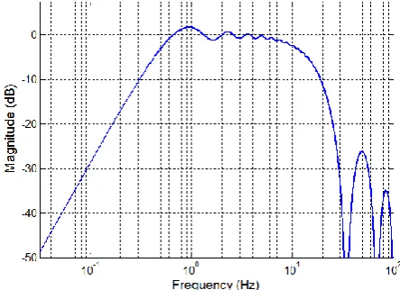
Fig. 1 shows magnitude response of the cascaded lowpass and highpass integer filters. It shows that the cascaded filter has 3-dB passband ranged from 0.5 Hz to 11 Hz.
Figure 1. Magnitude response of cascaded lowpass and highpass filters.
C. Signal Transformation
To simplify detecting pulse peaks, we used the SSF [14]. The SSF enhances the upslope of PPG waveform and suppresses the remainder of the waveform [15]. The SSF at time i, SSFi, is defined as (5).
0 : Δ 0
0 Δ : Δ Δ Δ k k k k i w i k k i s s s x where x
SSF
(5)
where w and sk are the length of the analyzing window
Fig. 2 shows that the SSF onset completely coincides with the pulse onset and the pulse peak is definitely appeared in the range between the SSF onset and the SSF offset [14]. Based on the fact, the proposed algorithm first localizes the SSF onset and the SSF offset. The pulse peak is then identified as the local maxima within the range.
Figure 2. Relationship between (a) PPG signal and (b) SSF signal [17].
D. Peak Finding with Adaptive Thresholding Scheme In order to extract pulse peaks, the algorithm first converts the PPG signal into the SSF signal by (5). In the SSF signal, the algorithm calculates the initial threshold value as 70 % of the maximum peak within the first 3-seconds interval. The threshold is then adaptively updated to float over the noises using the estimates of the SSF peak. To implement this, every time the SSF peak is estimated, it is added to the buffer containing the five most recent SSF peaks, as others usually did in ECG analysis. These processes are repeated until all the PPG samples are examined [17]. In this study, the 3-seconds interval was chosen since it covers at least one cardiac cycle not only for normal subjects but also for abnormal subjects who have cardiovascular diseases such as bradycardia arrhythmia. The ratio of 70 % was experimentally chosen using the training dataset [17].
E. Post-Processing
In order to cope with over-detected and missed pulse peaks, we applied the knowledge-based rules as previously proposed in [17]. The rules basically utilize the fact that the PPG is a slowly time varying signal and the difference between two adjacent PPG pulses cannot go beyond certain range.
For estimating missed pulse peaks, the rule first calculates differences between two adjacent pulse peaks (PP). The reference value (R) is calculated by applying the median filter with an order of 5 to the differences. If the reference and the difference values are differed larger than the half the reference, then the rule considers that the algorithm missed a pulse peak; otherwise, it considers that the algorithm correctly detects a pulse peak. If the missed peak is found, then the rule finds the local maximum PPG having a positive SSF value in a range from preceding pulse peak plus the half the reference (PP(n)+0.5R(n)) to the minimum value between the next pulse peak and the reference (MIN{PP(n+1), R(n)}.
For eliminating over-detected pulse peaks, the rule first calculates a difference between the first and the second pulse peaks as an initial reference value. The differences (D1, D2, D3, and D4) between nth and (n+1)th, nth and (n+2)th, nth and (n+3)th, and nth and (n+4)th pulse peaks are respectively calculated. The rule then calculates the differences between D1, D2, D3, D4 and the reference, and selects a pulse peak index having the minimum difference (D). If D is D1, then the rule considers it as correctly estimated pulse peak index; otherwise, the rule removes (n+1)th pulse peak, (n+1) and (n+2)th pulse peaks, and (n+1), (n+2), and (n+3)th pulse peaks for D=D2, D=D3, and D=D4, respectively.
In this study, we first estimated the missed pulse peaks and then eliminated over-detected ones.
III. PERFORMANCE EVALUATION
A. Noise Removal
Fig. 3 shows an example of the noise removal using the cascaded lowpass and highpass integer filters.
Figure 3. An example of the noise removal from the PPG signal using the cascaded lowpass and highpass integer filters: (a) noisy PPG
signal and (b) filtered PPG signal.
In Fig. 3, we can notice that the baseline wandering component and the high frequency noises are effectively suppressed by the cascaded lowpass and highpass integer filters. Further the figure shows that the filters do not significantly distort morphological information of the PPG signal.
B. Pulse Peak Detection
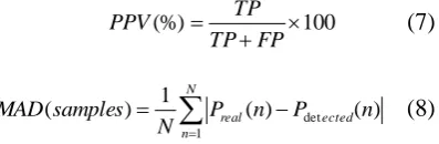
In order to verify the efficacy of the proposed algorithm, we investigate how closely the algorithm detects true position of pulse peaks and how accurately it extracts pulse peak intervals. For this end, we used two quantitative measures: sensitivity, positive predictive value (PPV), and mean absolute difference (MAD). Each measure is defined as (6) to (8), respectively.
100
(%)
FN TP
TP y
100
(%)
FP TP
TP
PPV
(7)
N
n
ected
real n P n
P N samples MAD
1
det ( )
) ( 1
)
(
(8)
where TP, FP, and FN are the true positive, the false positive, and the false negative, respectively. N is either the number of the pulse peaks or the number of pulse peak intervals. The Preal and Pdetected are the pulse peak
indices or pulse peak intervals manually annotated in the database and automatically detected by the algorithm, respectively.
In this study, true detection is defined by two rules. In the index-based rule, the true detection is defined as if the difference between manual and algorithm generated annotations is less than three samples (approximately 5 ms) intervals. Similarly, in the interval-based rule, it is considered to be true when the difference between the manually annotated and the algorithmically estimated QRS interval is within three samples. Table I shows the performance evaluation results.
TABLE I. TYPE SIZES FOR CAMERA-READY PAPERS
Rules Sensitivity PPV MAD
Index 96.45 % 60.57 % 3.34 samples
Interval 97.34 % 80.29 % 2.60 samples
In Table I, we obtained outstanding achievements in a sensitivity of 96.45 %, a positive predictive value of 60.57 %, and a mean absolute difference of 3.34 samples for interval-based rule, and a sensitivity of 97.34 %, a positive predictive value of 80.29 %, and a mean absolute difference of 2.60 samples for interval based rule, respectively. These results show that the proposed pulse peak detection algorithm recognizes well both the true positions of pulse peaks and the intervals of pulse peaks.
IV. PERFORMANCE EVALUATION
The conventional pulse peak detection algorithms require high computational complexity because of a high filter order and the time-frequency transformation. In order to reduce computational complexity while maintaining the detection accuracy, a real-time pulse peak detection algorithm has been proposed. The algorithm employed the cascaded recursive integer filters and the slope sum function with an adaptive thresholding scheme. Its efficacy and performance have been evaluated on the database where the PPG signals were practically collected. The results are promising, suggesting the proposed algorithm can provide simpler and accurate pulse peak detection in real-time environments with the reduced computational burden. Therefore, we can conclude that the proposed pulse peak algorithm can be used not only to monitor pulse rate and to delineate other characteristic points of the PPG signal with the reference to the pulse peak, but also to analyze pulse transit time and pulse rate variability in real-time.
REFERENCES
[1] D. G. Jang, U. Farooq, S. H. Park, C. W. Goh, and M. Hahn, “A knowledge-based approach to arterial stiffness estimation using the digital volume pulse,” IEEE Trans. Biomed. Circ. Syst., vol. 6, no. 4, pp. 366-374, Aug. 2012.
[2] J. Allen, “Photoplethysmography and its application in clinical physiological measurement,” Physiol. Meas., vol. 28, pp. R1-R39, Jan. 2007.
[3] A. Reisner, P. A. Shaltis, D. McCombie, and H. H. Asada, “Utility of the photoplethysmogram in circulatory monitoring,” Anesthesiology, vol. 108, no. 5, pp. 950-958, May 2008.
[4] Task Force of The European Society of Cardiology and The North American Society of Pacing and Electrophysiology, “Heart rate variability: Standards of measurement, physiological interpretation, and clinical use,” Circulation, vol. 93, no. 5, pp. 1043-1065, Mar. 1996.
[5] B. M. McCarthy, B. O’Rlynn, and A. Mathewson, “An investigation of pulse transit time as a non-invasive blood pressure measurement method,” J. Phys.: Conf. Ser., vol. 307, no. 1, pp. 1-5.
[6] S. K. Mitra, Digital Signal Processing: A Computer-Based Approach, 3rd ed., New York, USA: McGraw-Hill, 2006, pp. 427-429.
[7] K. L. Park, K. J. Lee, and H. R. Yoon, “Application of a wavelet adaptive filter to minimize distortion of the ST-segment,” Med. Biol. Eng. Comput., vol. 36, pp. 581-586, Sept. 1998.
[8] D. G. Jang, J. H. Park, U. Farooq, D. H. Son, S. H. Park, and M. Hahn, “A computer-aided design of photoplethysmography for managing cardiovascular diseases in home healthcare environments,” presented at the International Conference on Ubiquitous Healthcare, Jeju, South Korea, Oct. 28-30, 2010. [9] A. U. Rajendra, S. S. Jasjit, A. E. S. Jos, and S. M. Krishnan,
Advances in Cardiac Signal Processing, Berlin, Germany: Springer-Verlag, 2007, pp. 65-66.
[10] J. Pan and W. J. Tompkins, “A real-time QRS detection algorithm,” IEEE Trans. Biomed. Eng., vol. 32, no. 3, pp. 230-236, Mar. 1985.
[11] R. Laulkar and N. Daimiwal, “Application of finger photoplethysmography,” Int. J. Eng. Res. App., vol. 2, no. 1, pp. 877-880, Jan-Feb 2012.
[12] J. Zheng, S. Hu, S. Xin, V. P. Crabtree, and P. R. Smith, “Non-invasive photoplethysmography to assess lower limb peripheral perfusion with selected postural changes,” Physiol. Meas. [13] P. A. Lynn, “Online digital filters for biological signals: Some fast
designs for a small computer,” Med. Biol. Eng. Comput., vol. 15, no. 5, pp. 534-540, Sept. 1977.
[14] D. G. Jang, U. Farooq, J. H. Park, S. H. Park, and M. Hahn, “An adaptive SSF-based pulse peak detection algorithm for heart rate variability analysis in home healthcare environments,” in Proc. Int. Conf. Ubi. Healthcare, Oct. 28-30, 2010, pp. 70-71.
[15] W. Zong, G. B. Moody, and R. G. Mark, “Reduction of false arterial blood pressure alarms using signal quality assessment and relationships between the electrocardiogram and arterial blood pressure,” Med. Biol. Eng. Comput., vol. 42, no. 5, pp. 698-706, Sept. 2004.
[16] W. Zong, T. Heldt, G. B. Moody, and R. G. Mark, “An open-source algorithm to detect onset of arterial blood pressure pulses,” Comp. Cardio., vol. 30, pp. 259-262, 2003.
[17] D. G. Jang, U. Farooq, S. H. Park, and M. Hahn, “A robust method for pulse peak detection in the baseline wandering digital volume pulse,” IEEE Trans. Biomed. Circ. Syst.
Sangjun Park received his B.S. degree in Electrical Engineering from Korea Advanced Institute of Science and Technology (KAIST), Daejeon, South Korea, in 2011. He is currently working towards his Ph.D. degree at KAIST. His research interests include speech, audio, and biological signal processing, speech enhancement, speech recognition, and speech synthesis.
Seung-Hun Park received his B.S. and M.S. degrees in Electrical Engineering from Seoul National University, Seoul, South Korea, in 1981 and 1984, respectively. He received his Ph.D. degree in Electrical Engineering from University of Florida, in 1990. From 1985 to 1990, he was with Electronics and Telecommunications Research Institute (ETRI), Daejeon, South Korea. From 1990 to 1998, he was a faculty member of the Department of Biomedical Engineering, Konkuk
University, South Korea. From 2000, he has been a full professor in the Department of Biomedical Engineering, Kyunghee University. His research interests include u-healthcare systems, rehabilitation and wellness devices and systems, and biological signal processing.
![Figure 2. Relationship between (a) PPG signal and (b) SSF signal [17].](https://thumb-us.123doks.com/thumbv2/123dok_us/9951227.1983062/3.595.62.284.173.322/figure-relationship-ppg-signal-b-ssf-signal.webp)