University of Pennsylvania
ScholarlyCommons
Publicly Accessible Penn Dissertations
1-1-2013
Methods in and Applications of the Sequencing of
Short Non-Coding RNAs
Paul Ryvkin
University of Pennsylvania, [email protected]
Follow this and additional works at:
http://repository.upenn.edu/edissertations
Part of the
Bioinformatics Commons
,
Genetics Commons
, and the
Molecular Biology Commons
This paper is posted at ScholarlyCommons.http://repository.upenn.edu/edissertations/922
For more information, please [email protected].
Recommended Citation
Ryvkin, Paul, "Methods in and Applications of the Sequencing of Short Non-Coding RNAs" (2013).Publicly Accessible Penn Dissertations. 922.
Methods in and Applications of the Sequencing of Short Non-Coding
RNAs
Abstract
Short non-coding RNAs are important for all domains of life. With the advent of modern molecular biology
their applicability to medicine has become apparent in settings ranging from diagonistic biomarkers to
therapeutics and fields ranging from oncology to neurology. In addition, a critical, recent technological
development is high-throughput sequencing of nucleic acids. The convergence of modern biotechnology with
developments in RNA biology presents opportunities in both basic research and medical settings. Here I
present two novel methods for leveraging high-throughput sequencing in the study of short non-coding
RNAs, as well as a study in which they are applied to Alzheimer's Disease (AD). The computational methods
presented here include High-throughput Annotation of Modified Ribonucleotides (HAMR), which enables
researchers to detect post-transcriptional covalent modifications to RNAs in a high-throughput manner. In
addition, I describe Classification of RNAs by Analysis of Length (CoRAL), a computational method that
allows researchers to characterize the pathways responsible for short non-coding RNA biogenesis. Lastly, I
present an application of the study of non-coding RNAs to Alzheimer's disease. When applied to the study of
AD, it is apparent that several classes of non-coding RNAs, particularly tRNAs and tRNA fragments, show
striking changes in the dorsolateral prefrontal cortex of affected human brains. Interestingly, the nature of
these changes differs between mitochondrial and nuclear tRNAs, implicating an association between
Alzheimer's disease and perturbation of mitochondrial function. In addition, by combining known genetic
factors of AD with genes that are differentially expressed and targets of regulatory RNAs that are differentially
expressed, I construct a network of genes that are potentially relevant to the pathogenesis of the disease. By
combining genetics data with novel results from the study of non-coding RNAs, we can further elucidate the
molecular mechanisms that underly Alzheimer's disease pathogenesis.
Degree Type
Dissertation
Degree Name
Doctor of Philosophy (PhD)
Graduate Group
Genomics & Computational Biology
First Advisor
Li-San Wang
Keywords
Alzheimer's disease, machine learning, non-coding RNA, RNA, RNA modification, sequencing
Subject Categories
Bioinformatics | Genetics | Molecular Biology
METHODS IN AND APPLICATIONS OF THE
SEQUENCING OF SHORT NON-CODING
RNAS
Paul Ryvkin
A DISSERTATION
in
Genomics and Computational Biology
Presented to the Faculties of the University of Pennsylvania
in
Partial Fulfillment of the Requirements for the
Degree of Doctor of Philosophy
2013
Supervisor of Dissertation
_________________
Li-San Wang, Ph.D.
Assistant Professor of Pathology and Laboratory Medicine
Graduate Group Chairperson
_____________________
Maja Bucan, Ph.D. Professor of Genetics
Dissertation Committee: James Eberwine, Ph.D. (Chair)
Professor of Pharmacology Brian Gregory, Ph.D.
Assistant Professor of Biology F. Bradley Johnson, M.D. Ph.D.
Associate Professor of Pathology and Laboratory Medicine Tandy Warnow, Ph.D.
METHODS IN AND APPLICATIONS OF THE
SEQUENCING OF SHORT NON-CODING RNAS
COPYRIGHT
2013
Paul Ryvkin
This work is licensed under the Creative Commons Attribution- NonCommercial-ShareAlike 3.0
License
To view a copy of this license, visit
iii
Dedication
This work is dedicated to my loving parents, Mark and Yelena Ryvkin, to whom I owe everything
iv
Acknowledgements
First I must thank my thesis advisor, Li-San Wang, whose careful guidance and perseverance
show through in this work. Thanks also go to my thesis committee who graciously took the time to
provide useful input throughout the process.
This section would not be complete without thanking all of my former and current
labmates, particularly: Fan Li for transforming my ugly hacks into useful apps, Kajia Cao for
keeping my head out of the clouds, Fanny Leung for helping with the benchwork and the machine
learning algorithms, Otto Valladares for keeping the servers humming, Micah Childress for putting
a public face on my software, and everyone else in the Wang lab.
My appreciation goes to the staff at the Institute for Biomedical Informatics, particularly
Hannah Chervitz and Tiffany Barlow for their organizational prowess. I’d also like to thank all the
GCB students I’ve known; the elder for their sage advice, the co-matriculating for their
commiseration, and the younger for excellent times had.
In the course of my time at Penn I’ve worked with many, many other researchers, without
whom this work would not have been possible. I’d like to thank Brad Johnson, who taught me so
many important molecular biology techniques ranging from nucleic acids extraction to keeping the
supernatant. Also to thank is Brian Gregory and everyone in his lab, particularly Isabelle Dragomir
for her help with the sequencing library preparation and Lee Vandivier and Ian Silverman for their
help with validating experiments. Thanks also go to Alice-Chen Plotkin for her help with tissue
processing, Vivianna Van Deerlin for her help with large-scale RNA extraction, Theresa Schuck
for her help with tissue dissection, and Virginia Lee for her ever insightful input. I particularly
appreciate everyone at the Center for Neurodegenerative Disease Research and everyone in
Gerard Schellenberg’s lab for being gracious hosts for much of this work.
A special thank-you goes to John Trojanowski and the Institute on Aging for providing the
funding for the Alzheimer’s study, which yielded almost all of the data necessary for this work.
v
Medical Sciences, the National Human Genome Research Institute, the National Institute on
Aging, Penn Alzheimer’s Disease Center, and the National Science Foundation.
Finally I’d like to thank my office in Blockley Hall for sheltering my computer from the
elements, my bicycle for faithfully transporting me from point A to point B, my two cats for being
endlessly fascinating felids, the never-boring city of Philadelphia, the food and company at Grace
Tavern, the scenery of Rittenhouse Park, and the game of Bridge (special thanks to Kathleen
Sprouffske, Miler Lee, Rumen Kostadinov, and Aaron Goodman). I also thank my wonderful
girlfriend Chrystelle Browman for supporting me despite the unique challenges of dating a PhD
vi
ABSTRACT
METHODS IN AND APPLICATIONS OF THE SEQUENCING OF SHORT NON-CODING RNAS
Paul Ryvkin
Li-San Wang
Short non-coding RNAs are important for all domains of life. With the advent of modern molecular
biology their applicability to medicine has become apparent in settings ranging from diagonistic
biomarkers to therapeutics and fields ranging from oncology to neurology. In addition, a critical,
recent technological development is high-throughput sequencing of nucleic acids. The
convergence of modern biotechnology with developments in RNA biology presents opportunities
in both basic research and medical settings. Here I present two novel methods for leveraging
high-throughput sequencing in the study of short non-coding RNAs, as well as a study in which
they are applied to Alzheimer’s Disease (AD). The computational methods presented here
include High-throughput Annotation of Modified Ribonucleotides (HAMR), which enables
researchers to detect post-transcriptional covalent modifications to RNAs in a high-throughput
manner. In addition, I describe Classification of RNAs by Analysis of Length (CoRAL), a
computational method that allows researchers to characterize the pathways responsible for short
non-coding RNA biogenesis. Lastly, I present an application of the study of non-coding RNAs to
Alzheimer’s disease. When applied to the study of AD, it is apparent that several classes of
non-coding RNAs, particularly tRNAs and tRNA fragments, show striking changes in the dorsolateral
prefrontal cortex of affected human brains. Interestingly, the nature of these changes differs
between mitochondrial and nuclear tRNAs, implicating an association between Alzheimer’s
disease and perturbation of mitochondrial function. In addition, by combining known genetic
factors of AD with genes that are differentially expressed and targets of regulatory RNAs that are
differentially expressed, I construct a network of genes that are potentially relevant to the
non-vii
coding RNAs, we can further elucidate the molecular mechanisms that underly Alzheimer’s
viii
CONTENTS
DEDICATION ... III
ACKNOWLEDGEMENTS... IV
ABSTRACT ... VI
CONTENTS ... VIII
LIST OF TABLES ... XI
LIST OF ILLUSTRATIONS ... XII
1.
INTRODUCTION... 1
1.1. RNA Biology ... 1
1.1.1. The Central ―Dogma‖ ... 1
1.1.2. Protein-coding RNAs ... 6
1.1.3. Non-coding RNAs ... 7
1.1.4. Short non-coding RNAs (small RNAs) ... 14
1.2. Measuring the transcriptome ... 16
1.3. Alzheimer’s Disease ... 20
1.4. Outline of dissertation ... 22
2.
HIGH-THROUGHPUT ANNOTATION OF MODIFIED RIBONUCLEOTIDES
(HAMR) ... 24
2.1. Introduction ... 24
2.2. Methods... 25
2.2.1. RNA extraction and sequencing... 25
2.2.2. tRNA locus clustering ... 26
2.2.3. Detecting candidate RT misincorporation sites ... 27
2.2.4. tRNA modification identification ... 28
2.2.5. Software ... 29
2.3. Results ... 29
ix
2.3.2. Detecting modified sites by mismatch rates ... 30
2.3.3. Calling modification types by incorporation patterns in RT ... 37
2.3.4. Expanding the tRNA modification annotation ... 41
2.3.5. Validation in S. cerevisiae small RNA dataset ... 46
2.3.6. Validation in human rRNA(-)-seq dataset ... 48
2.3.7. Detecting modifications in other RNAs ... 50
2.3.8. Software ... 50
2.4. Discussion ... 51
2.5. Acknowledgements ... 51
3.
CLASSIFICATION OF RNAS BY ANALYSIS OF LENGTH (CORAL) ... 53
3.1. Introduction ... 53
3.2. Methods... 55
3.2.1. Processing of small RNA-seq data ... 55
3.2.2. Labelling training data ... 56
3.2.3. Feature generation ... 59
3.2.4. Feature selection and classification framework ... 60
3.2.5. Evaluation of performance ... 60
3.3. Results ... 61
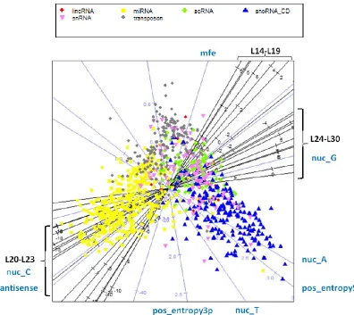
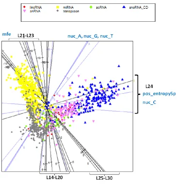
3.3.1. Visualization of the length features ... 61
3.3.2. Discriminative power of features ... 68
3.3.3. Comparison with existing classification approaches – DARIO and miRDeep ... 69
3.3.4. Building a classification model using 6 classes of ncRNAs ... 70
3.3.5. Features that can discriminate between classes of small RNAs ... 72
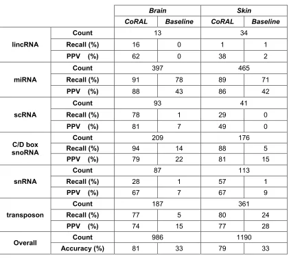
3.3.6. Validation of the classification models between datasets ... 74
3.4. Conclusions ... 77
3.4.1. Software Availability ... 78
4.
CHARACTERIZING THE NON-CODING TRANSCRIPTOME OF ALZHEIMER’S
DISEASE ... 79
4.1. Introduction ... 79
4.2. Methods... 79
4.2.1. RNA-sequencing ... 79
4.2.2. Calling small RNA loci and building smRNA locus families ... 79
4.2.3. Predicting the impact of tRNA activity changes on protein translation ... 80
4.2.4. Building a network of AD-related genes ... 81
4.3. Results ... 81
4.3.1. Sample characteristics and RNA-seq processing statistics ... 81
4.3.2. Global changes in non-rRNA transcription in the AD brain ... 82
4.3.3. Global changes in small RNA biogenesis in the AD brain ... 84
4.3.4. Differentially expressed small RNAs in the AD brain ... 91
4.3.5. tRNAs are differentially expressed and processed in the AD brain ... 98
x
4.3.7. Building an integrative network ... 104
4.4. Discussion ... 106
5.
CONCLUSION ... 107
xi
LIST OF TABLES
Table 1.1 – A compendium of non-coding RNAs found in animals. ... 9
Table 1.2 – The eukaryotic nuclear genetic code. ... 12
Table 1.3 – Genes implicated in LOAD by genome-wide association in Caucasian populations. 22 Table 2.1 – Selected RNA modifications and their known and predicted effects on RT ... 35
Table 2.2 – All tRNA sites predicted to be modified by HAMR ... 42
Table 2.3 – Comparison of novel sites in smRNA data to same loci in an rRNA(-) libraries. ... 49
Table 2.4 – Comparison of seminovel sites to rRNA(-) libraries. ... 49
Table 2.5 – Candidate sites of modification across the entire small RNAome ... 50
Table 3.1 – Number of reads and loci at each stage of smRNA-seq processing ... 56
Table 3.2 – Comparison of a 3-class CoRAL model to DARIO ... 70
Table 3.3 – Cross-tissue comparison of a 6-class CoRAL classifier ... 71
Table 3.4 – Four-way independent cross-validation of the 3-class classifier ... 77
Table 4.1 – RNA classes defined as incompatible when clustering loci. ... 80
Table 4.2 – Summary of samples and RNA-seq data processing ... 81
Table 4.3 – Top 10 AD-downregulated transcripts in the rRNA(-) libraries. ... 89
Table 4.4 – Top 10 AD-upregulated transcripts in the rRNA(-) libraries ... 91
Table 4.5 – Differentially expressed small RNAs derived from mRNAs or antisense transcripts . 93 Table 4.6 – Differentially expressed snoRNAs in rRNA(-) and smRNA libraries ... 95
Table 4.7 – Differentially expressed microRNAs. ... 96
Table 4.8 – Experimentally validated targets of the D.E. miRNAs. ... 97
Table 4.9 – Downregulated tRNAs and tRNA fragments in the AD brain with expression fold-changes. ... 99
Table 4.10 – Upregulated tRNAs and tRNA fragments in the AD brain with expression fold-changes. ... 99
Table 4.11 – Top 10 brain-expressed genes predicted to be down-translated due to tRNA changes. ... 100
Table 4.12 - Top 10 brain-expressed genes predicted to be up-translated due to tRNA changes. ... 101
Table 4.13 – KEGG pathways enriched for putative down-translated genes ... 101
Table 4.14 - Top downregulated functional categories in AD... 103
xii
LIST OF ILLUSTRATIONS
Figure 1.1 – The central hypothesis of molecular biology. ... 2
Figure 1.2 – The central hypothesis revised. ... 2
Figure 1.3 – The structure of RNAs. ... 5
Figure 1.4 – Strand-specific polyA(+) RNA-sequencing. ... 17
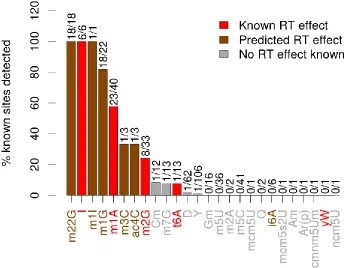
Figure 2.1 – Mismatch rates in small RNA reads mapping to three types of RNA ... 31
Figure 2.2 - Locations of known tRNA modifications predicted to affect RT incorporation ... 33
Figure 2.3 - Modification sites predicted by HAMR ... 33
Figure 2.4 – HAMR’s sensitivity for detecting different types of RNA modification ... 36
Figure 2.5 – HAMR’s sensitivity under the loose model H01 ... 36
Figure 2.6 – Observed nucleotide frequencies in cDNA for different modification types and in different organisms ... 38
Figure 2.7 – Sequenced nucleotide frequencies at known tRNA m3C sites in the human brain .. 39
Figure 2.8 – Sequenced nucleotide frequencies at known modified tRNA uridines in the human brain ... 39
Figure 2.9 – Sequenced nucleotide frequencies at guanosines when using the loose model H0140 Figure 2.10 – HAMR’s sensitivity in an independent S. cerevisiae dataset using the strict model H02 ... 47
Figure 2.11 - HAMR’s sensitivity in an independent S. cerevisiae dataset using the loose model H01 ... 47
Figure 3.1 – The effect of read count thresholds on the ability to detect smRNA loci ... 57
Figure 3.2 – Summary of RNA classes in the brain smRNA-seq ... 58
Figure 3.3 – Summary of RNA classes in the skin smRNA-seq ... 58
Figure 3.4 – Read length spectrum for brain miRNAs ... 62
Figure 3.5 – Read length spectrum for skin miRNAs ... 62
Figure 3.6 – Read length spectrum for brain C/D box snoRNAs ... 62
Figure 3.7 – Read length spectrum for skin C/D box snoRNAs ... 62
Figure 3.8 – Read length spectrum for brain transposon-derived smRNAs ... 63
Figure 3.9 – Read length spectrum for skin transposon-derived smRNAs ... 63
Figure 3.10 – SAVoR plot for a brain microRNA ... 64
Figure 3.11 – SAVoR plot for a brain C/D box snoRNA ... 65
Figure 3.12 – SAVoR plot for a brain transposon-derived smRNA locus ... 65
Figure 3.13 – Correlation heatmap of all the features in the brain data ... 67
Figure 3.14 – Multidimensional-scaling projection of the features in the brain data ... 68
Figure 3.15 - Multidimensional-scaling projection of the features in the skin data ... 69
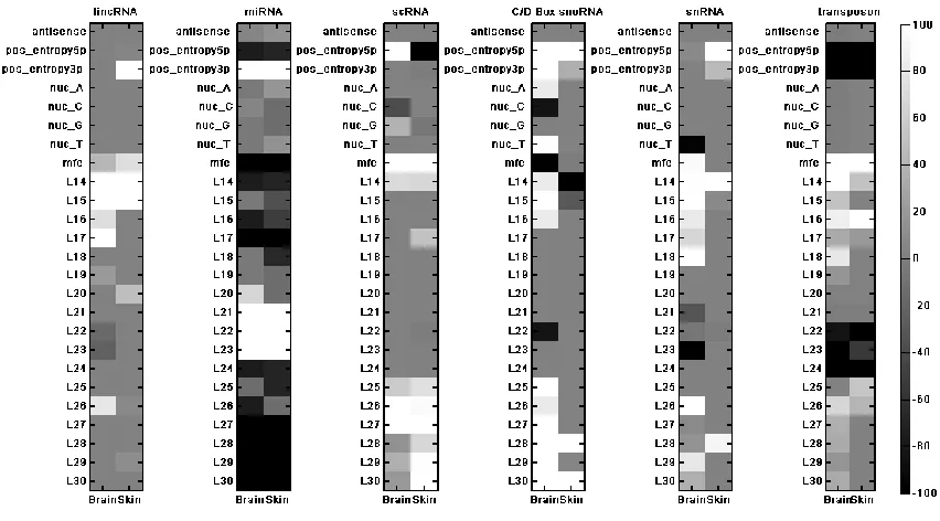
Figure 3.16 - Feature importance map of the 6-class classifier for each tissue ... 73
Figure 3.17 – lincRNA-derived smRNA locus overlap between brain and skin ... 75
Figure 3.18 - miRNA locus overlap between brain and skin ... 75
Figure 3.19 - scRNA-derived smRNA locus overlap between brain and skin... 76
Figure 3.20 – C/D box snoRNA-derived smRNA locus overlap between brain and skin ... 76
Figure 3.21 - snRNA-derived smRNA locus overlap between brain and skin ... 76
Figure 3.22 - Transposon-derived smRNA locus overlap between brain and skin ... 76
Figure 4.1- Summary of sequenced RNAs in the rRNA(-) libraries ... 83
Figure 4.2 – Summary of antisense transcription in the rRNA(-) libraries ... 84
Figure 4.3 – Summary of sequenced RNAs in the smRNA-seq libraries ... 86
Figure 4.4 – Summary of antisense transcription in the smRNA libraries ... 87
Figure 4.5 – Number of differentially expressed ncRNA transcripts by RNA class ... 88
Figure 4.6 - Number of differentially expressed smRNA loci by ncRNA class. ... 92
1
1. Introduction
1.1. RNA Biology
1.1.1. The Central “Dogma”
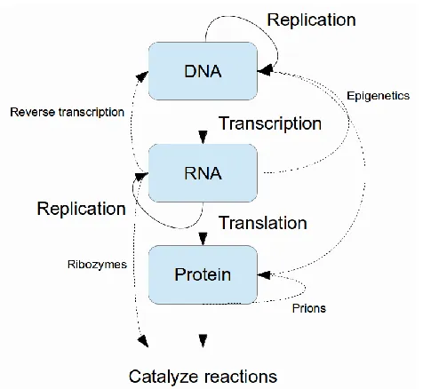
The central hypothesis (or as Francis Crick infamously and erroneously coined it, the ―central
dogma‖) [35,36] of molecular biology outlines the relationship between three important types of
organic molecules: DNA (deoxyribonucleic acids), RNA (ribonucleic acids), and proteins (Figure 1.1). The totality of each type of molecule in the cell is referred to as the genome, the
transcriptome, and the proteome, respectively. Under this framework, information flows from DNA
to RNA and then to proteins; DNA serves as a template for transcription of RNA, which in turn
serves as a template for translation into protein. Proteins form enzymes which carry out a range
of functions throughout the cell and are generally responsible for phenotype, or the appearance
and behavior of the organism. While we now know that there are many exceptions to this view
2
Figure 1.1 – The central hypothesis of molecular biology.
3
DNA can be considered a fixed information storage medium for the cell. Exceptions to
this picture of DNA include the entire field of epigenetics which seeks to describe dynamic
modifications to DNA, as well as the study of the processes of DNA replication and repair. In
general, however, DNA serves only as a template and is not responsible for catalyzing other
types of reactions.
RNA, in contrast, exists in a constant state of flux via creation (transcription from DNA)
and destruction (finely controlled turnover by enzymes). Similarly, proteins, which comprise
enzymes, exist in a constant state of flux. For many years, proteins alone were considered to be
the workhorse of the cell – after all, they catalyze nearly all of the reactions necessary to support
life while DNA and RNA ―merely‖ store information. However, with the discovery of catalytic RNAs
(ribozymes) [27,69,94], these molecules are now appreciated as more than simple ―messengers‖
between DNA and proteins. It is especially difficult to write off RNAs since the machinery that
translates RNA into protein (the ribosome) is itself made up of RNA; indeed, it has been shown
that the RNA (not the protein) component of this machinery is responsible for its activity [120].
RNA is therefore a key component of the cellular machinery and not simply a transitory
messenger.
Like the other ubiquitous organic polymers central to life (DNA and proteins), RNA
primarily stores information by way of its sequence. While DNA is a polymer of the
deoxyribonucleotides deoxyadenosine (dA), deoxycytidine (dC), deoxyguanosine (dG), and
deoxythymidine (dT), RNA is a polymer of the ribonucleotides adenosine (A), cytidine (C),
guanosine (G), and uridine (U) [6]. The key differences are RNA’s inclusion of a hydroxyl group
where DNA is missing one, the substitution of uridine for thymidine, and RNA’s propensity to exist
in a greater variety of structural forms. Analogous to DNA, it is the sequential order of the
ribonucleotides that form the primary information content of RNA. Another form of information
stored by RNA is its structure; RNAs are prone to fold into particular geometries which can be
4
its sequence. Its secondary structure is a graph whose nodes are nucleotides and whose edges
represent Watson-Crick and wobble-pairing interactions between pairs of these nucleotides. Its
tertiary structure describes long-range interactions between its base-paired and/or unpaired
sections. Finally, the quaternary structure of an RNA models its interactions with other molecules.
In addition to the folding geometry of the RNA, a third form of information is the presence of
non-canonical nucleotides formed by covalent modification of the standard four [3,4,37,166,169] – in
5
6
1.1.2. Protein-coding RNAs
RNAs can be broadly categorized into two groups: those that code for proteins (coding RNAs)
and those that do not (non-coding, or ncRNAs). The only extant class of coding RNAs is
messenger RNAs (mRNAs) – however, not all mRNAs code for proteins. In higher eukaryotes,
mRNAs are transcribed by the enzyme RNA polymerase II and undergo a sophisticated
maturation process from the original mRNA transcript [117]: they can be spliced into various
isoforms [65], they are capped by a special chemical structure on the 5’ end [133], they are
polyadenylated on the 3’ end, and their sequence can be dynamically changed [139] (RNA
editing) and chemically modified (RNA modification). The terms 5’ and 3’ correspond to the
exposed atom of the ribose sugar in the ribonucleotide – generally a 5’ triphosphate on one end
and a 3’ hydroxyl group on the other end. Since mRNAs are translated into proteins from 5’ to 3’,
these are conventionally depicted as the left and right ends of the molecule, respectively. In
eukaryotic splicing, multiple alternative forms of an RNA transcript are generated when the
cellular splicing machinery removes sections called introns and concatenates together sections
designated as exons, which usually contain the coding portion of the transcript (i.e., the sequence
that will determine the translated protein). Thus one gene may produce many distinct mRNAs
with varying sequences which are then translated into proteins with a variety of functions.
Capping, in eukaryotic organisms, refers to the addition of the ribonucleotide N7-methylguanosine
(m7G) to the 5’ carbon of the mRNA via an unconventional 5’-5’ triphosphate linkage. This cap
serves to stabilize the mRNA and promote its export from the nucleus. Polyadenylation is a
process whereby a homopoylmer of adenosines is sequentially added to the 3’ end of the mRNA.
Among other functions, this poly(A) ―tail‖ regulates enzymatic degradation of the mRNA from the
3’ end. Nearly all eukaryotic mRNAs are polyadenylated with the notable exception of the histone
genes, where the 3’ terminus is designated by a small stem-loop RNA structure. The process of
RNA editing generally consists of post-transcriptional changes in the sequence of an RNA. In the
case of eukaryotic mRNAs, this is usually a deamination of adenosine to inosine or deamination
7
far less specific in its base-pairing specificity. These changes to an mRNA’s sequence can affect
its alternative splicing, stability, and even the eventual protein sequence that is coded. Other
types of changes to an RNA’s sequence, which always produce non-canonical nucleotides, are
termed RNA modifications. Examples of RNA modifications are the methylation of guanosine at
the carbon 2 amine (producing N2-methylguanosine or m2G) and the isomerization of uridine into
its C-glycoside pseudouridine (Ψ). These types of modifications are believed to be rare in
protein-coding mRNAs, but the search for them is an active field of research. So far, it seems that the
non-canonical nucleotides 5-methylcytidine (m5C) and N6-methyladenosine (m6A) can be found
in mRNAs transcriptome-wide [115]. Furthermore, the recent discovery that a gene whose
variants are found to be associated with obesity in humans, FTO, is an adenosine
N6-methyltransferase suggests that these modifications may play a very important role in human
disease [57,84].
1.1.3. Non-coding RNAs
While protein-coding mRNAs are important for deciding the sequences of proteins, the most
abundant RNAs in the cell by far are non-coding RNAs; ribosomal RNA (rRNA) can make up over
80% of all the RNA in mammalian cells. The next most abundant class of non-coding RNAs,
transfer RNAs (tRNAs), can make up another 10%. Not only are non-coding RNAs the most
abundant RNAs in the cell, they are also the most evolutionarily conserved: all cellular life on
earth relies on ribosomes, and thus ribosomal RNA, and the similarity of its sequence among
disparate organisms is great enough for it to act a universal phylogenetic character [128]. The
universality of ribosomal RNA, combined with its sufficiency for ribosomal function is a central
piece of evidence supporting the hotly-debated ―RNA world‖ hypothesis which claims that the use
of RNA as an information storage medium preceded DNA’s on Earth [26].
Aside from their lack of protein-coding capacity, there are many fundamental differences
8
are processed. For example, while the non-canonical nucleotide modifications described in
Section 1.1.2 are thought to be rare in mRNAs, they are ubiquitous in non-coding RNAs. The
most abundant non-canonical nucleotide in the cell, pseudouridine, is commonly found in rRNA
and tRNA [73].
Unlike protein-coding mRNAs, there is great diversity in the non-coding RNA population
[83,114] (Table 1.1). Unfortunately, producing a consistent nomenclature of non-coding RNAs is a difficult task, and currently it proceeds in an ad hoc manner publication by publication. For
example, while some classes of RNA are defined by their location in the cell, others are defined
by the genomic neighborhood of their DNA template. An initial useful subdivision of non-coding
RNAs is by their size: generally ncRNAs shorter than around 50 nucleotides (nt) are considered
short non-coding RNAs, or ―small RNAs,‖ while longer ones are referred to as long non-coding
RNAs (lncRNAs). Section 1.1.4 describes the many types of short non-coding RNAs, while this
9
Table 1.1 – A compendium of non-coding RNAs found in animals.
Abbreviation Name Biological role Example(s)
rRNA Ribosomal RNA Translation 5S rRNA
tRNA Transfer RNA Translation tRNAMetCAU
snoRNA Small nucleolar RNA RNA modification SNORD115
snRNA Small nuclear RNA mRNA splicing U1
scRNA Small cytoplasmic RNA Various hY1
srpRNA Signal recognition
particle RNA Protein localization lincRNA Long intergenic
non-coding RNA Various XIST, TSIX, MALAT1
miRNA Micro RNA mRNA silencing let-7
piRNA Piwi-interacting RNA Transposon silencing piR-53941
tRF tRNA fragment Unknown tRNAMetCAU 5’ half
paRNA Promoter-associated
RNA Unknown EF1a promoter
vtRNA Vault RNA Unknown; drug resistance VTRNA1-1
aRNA Antisense RNA mRNA regulation BACE1-AS
natRNA Natural antisense transcript RNA
Unknown HAS2-AS1
- Transposable elements Self replication SINEs and LINEs
Hammerhead Hammerhead ribozyme mRNA regulation C10orf118
TERC Telomerase RNA
component Telomere extension TERC
RNase P Ribonuclease P RNA
10
Ribosomal RNA is largely transcribed by RNA polymerase I and is central to an organelle
within the cell called the ribosome [122]. Ribosomes are responsible for translating mRNAs into
proteins. In eukaryotes the ribosome is made up of a small subunit (SSU) and large subunit
(LSU). In the human genome ribosomal RNA exists in many copies (as rDNA), and often in long
tandem arrays, which have long presented an obstacle to assembly of the human genome due to
their repetitive nature. Ribosomal RNA maturation takes place in the nucleolus, a small
substructure of the nucleus, where it is spliced and modified in myriad ways by other RNAs and
ribonucleoprotein (RNP) complexes. Importantly, it is the structure of the rRNA that is responsible
for its function, not necessarily its sequence; structure-over-sequence is a common theme among
ncRNAs.
The next most abundant class of ncRNA is transfer RNA [127]. Transfer RNAs are
transcribed by RNA polymerase III and tend to be around 70 nt in length; they fold into a
distinctive ―cloverleaf‖ secondary structure with an L-shaped tertiary structure. Like rRNA, the
DNA genes from which they are transcribed (tDNA) exist with high copy number in mammalian
genomes [13]. The function of tRNA is to act as an intermediary between mRNA and the
ribosome. The acceptor arm of a tRNA is covalently bonded to a specific amino acid by a highly
conserved family of proteins called tRNA aminoacyl synthases. The anticodon loop of a tRNA
contains a three-nucleotide sequence called the ―anticodon.‖ When an mRNA is being translated
by a ribosome, the appropriate tRNA associates with the mRNA’s current codon (three-letter code
associated with an amino acid) by way of sequence complementarity. Thus a tRNA provides a
link between particular codon sequences and particular amino acids, giving rise to the genetic
code (Table 1.2). In tRNAs, both the structure and sequence are of critical importance – their structure allows for the appropriate interaction with the ribosome while their sequence provides
specificity for particular codons. Notably, there are fewer tRNA anticodons encoded in the
genome than there are complementary codons in the genetic code. This is because one tRNA
can bind to multiple codons by way of covalent RNA modifications in the anticodon loop, yielding
11
induced adjacent to the anticodon can also alter the specificity of the codon-binding. Like mRNAs
12
Table 1.2 – The eukaryotic nuclear genetic code.
RNA Codon Amino acid RNA Codon Amino acid
UUA
Serine (Ser)
CGU
Arginine (Arg)
UUG CGC
UCU CGA
UCC CGG
AGU AGA
AGC AGG
UUA
Leucine (Leu)
GGU
Glycine (Gly)
UUG GGC
CUU GGA
CUC GGG
CUA AUU
Isoleucine (Ile)
CUG AUC
GUU
Valine (Val)
AUA
GUC UUU Phenylalanine (Phe)
GUA UUC
GUG CAU
Histidine (His) CCU
Proline (Pro)
CAC
CCC CAA
Glutamine (Gln)
CCA CAG
CCG AAU
Asparagine (Asn) ACU
Threonine (Thr)
AAC
ACC AAA
Lysine (Lys)
ACA AAG
ACG GAU
Aspartic acid (Asp) GCU
Alanine (Ala)
GAC
GCC GAA
Glutamic acid (Glu)
GCA GAG
GCG UAA
Stop codon UGU
Cysteine (Cys) UAG
UGC UGA
UAU
Tyrosine (Tyr) UGG Tryptophan (Trp)
13
Small nucleolar RNAs (snoRNAs) are, as their name suggests, non-coding RNAs that are
generally localized to the nucleolus (but also Cajal bodies) [49]. There are three main subclasses
of small nucleolar RNAs, each having a different set of structural and sequence motifs: C/D box,
H/ACA box, and small Cajal body-specific (scaRNA). The main function of snoRNAs is to guide
covalent modification of other RNAs, ranging from rRNA to small nuclear RNAs (snRNAs), via
small nucleolar ribonucleoprotein (snoRNP) complexes. In general, C/D box snoRNAs guide
methylation of RNAs while H/ACA box snoRNAs guide pseudouridylation of RNAs. One notable
exception is the C/D box snoRNA SNORD115, which has complementarity to the serotonin 2 C
receptor mRNA and alters its splicing [88].
Small nuclear RNAs (snRNAs) largely comprise the RNA component of the spliceosome;
that is, they make up the machinery responsible for splicing of RNAs [51,118,154]. As their name
suggests, they are largely localized to the nucleus. There are several families of snRNAs with
names such as U1, U2, and so on. In conjunction with proteins they form small nuclear
ribonucleoprotein (snRNP) complexes, which form the spliceosome. As with the other ncRNA
types described, their genes exist in high copy number scattered throughout mammalian
genomes [107].
A somewhat mysterious and only recently described class of non-coding RNAs is that of
long intergenic non-coding RNAs (lincRNAs) [24,87]. lincRNAs look very similar to mRNAs – they
are transcribed by RNA polymerase II and tend to be polyadenylated and spliced – but they do
not code for proteins and often localize to the nucleus rather than the cytoplasm. While some
notable examples of lincRNAs, such as Xist [32] and MALAT1 [82] have been well known for
quite some time, the recent application of high-throughput RNA-sequencing has illuminated many
more lincRNAs with varying levels of abundance and tissue specificity. Their function and
14
1.1.4. Short non-coding RNAs (small RNAs)
Short non-coding RNAs, or small RNAs (smRNAs), play an important role in higher eukaryotic
transcriptomes. RNAs that are considered small RNAs tend to be less than 45 nt in length,
although there is no standard cutoff for the definition. They are almost always the product of
processing a longer transcript rather than being independently transcribed directly from the
genome. The pathways responsible for generation of smRNAs generally consist of a number of
proteins and ribonucleoprotein complexes that process the precursor transcript in tandem and in
parallel. These pathways tend not to be as conserved across evolutionary distances as some
highly conserved proteins. Plants and animals, for example, have rather distinct smRNA
pathways that behave in quite different ways as a whole.
The best characterized class of smRNA to date is the microRNA [145]. MicroRNAs are a
particular subtype of small interfering RNA (siRNA) [55]. Small interfering RNAs were first
described by Craig C. Mello, Andrew Fire, and others in their 1998 Nature article, for which Mello
and Fire won a Nobel Prize in 2006. They are short (~21 nt) double-stranded RNAs which
promote gene silencing through a variety of methods – usually by either catalyzing degradation of
an mRNA transcript or inhibition of translation of an mRNA into its concomitant protein. They
target specific mRNAs by nature of having sequence complementarity (full or partial) to a
particular site on the mRNA, usually in its 3’ untranslated region (3’ UTR). The distinguishing
features of microRNAs are that they tend to be processed either from larger transcripts called
primary miRNAs (pri-miRNAs) or from introns that have been spliced out of pre-mRNAs (so called
mirtrons). In animals, the processing of pri-miRNAs into pre-miRNAs is accomplished in the
nucleus by the microprocessor complex, a protein complex that includes the Drosha and
Pasha/DGCR8 proteins; this complex recognizes hairpins on pri-miRNAs and cleaves them out,
creating pre-miRNAs. Mirtrons bypass this processing as they originate from introns and not
pri-miRNA transcripts. The resulting pre-pri-miRNA, which generally consists of a stem and a loop
structure, is then exported to the cytoplasm by the protein Exportin-5. In the cytoplasm, an
15
miRNA:miRNA* duplex by cleaving out the loop and a part of the stem. Each strand of the duplex
forms a distinct single-stranded mature miRNA with full or near-complementarity between the
two. The convention for which one is dubbed the ―star‖ miRNA is usually set by their order of
discovery, the method by which the miRNA was discovered, and the relative expression levels of
each miRNA strand in the tissue in which it was discovered. The resulting mature miRNAs tend to
be about 22nt long in animals. It is these mature miRNAs, in conjunction with the RNA-induced
silencing complex (RISC), which form a regulatory ribonucleoprotein complex that carries out
silencing activity on mRNAs. The class of protein that is central to RISC’s silencing activity is the
Argonaute family. They are responsible for guiding the miRNA to its target mRNA. The
miRNA-RISC (mimiRNA-RISC) then silences the mRNA transcript either by inhibiting translation into protein by
the ribosome or degradation of the mRNA via cleavage.
Another type of small RNA found in animals is the Piwi-interacting RNA (piRNA), named
after the Piwi class of proteins, a subclass of the Argonaute family [64,81,132,137]. Unlike
miRNAs, piRNAs tend to be significantly longer (26-32 nt versus 22nt) and also tend to have a
uridine on their 5’ end. The process by which they are generated is not yet fully clear. However,
their functional role has been partially elucidated: they are involved in the silencing of ―selfish‖
genetic elements known as transposons as well as in the placement of epigenetic marks on
chromatin. They are also highly active in mammalian testes and are required for mammalian
spermatogenesis.
There are a variety of other types of small non-coding RNAs, and in the literature they are
generally labeled by their precursor RNA. Small RNAs can be produced from any type of
precursor, ranging from protein-coding mRNA to non-coding RNA types such as rRNA, tRNA,
snRNA, and snoRNA. There is evidence that some of these small RNAs are processed like and
behave like microRNAs: they are produced by cleavage of stem-loop structures by the Dicer
protein and go on to have regulatory effects on mRNAs [9,18,103,126]. The fact that they
16
like miRNAs. However, the vast majority of non-miRNA small RNAs that are commonly found in
small RNA-seq datasets, for example, are entirely uncharacterized other than their annotated
precursor transcript. For example, tRNA-derived smRNAs (known as tRFs, or tRNA fragments)
[99], are thought to be the result of a combination of endolytic cleavage under stress response
conditions and non-specific cleavage by Dicer – but whether they are a simply non-specific
byproduct of smRNA processing pathways or go on to have functional regulatory roles has yet to
be determined. In Chapter 3 I present a quantitative method that can help researchers
characterize these largely unstudied populations of small RNAs.
1.2. Measuring the transcriptome
The advent of high-throughput sequencing (HTS), the most common subtype of which is shotgun
sequencing, has heralded in a new age of computational biology. In current-generation shotgun
sequencing, DNA (or RNA) is fragmented into smaller pieces and then a machine produces
―reads‖ by reading the sequence of these fragments from either one end (single-end sequencing)
or both ends (paired-end sequencing). While the sequencing of genomes (DNA-seq) has gained
recent attention, researchers are starting to see the value in applying these technologies to the
sequencing of RNA (RNA-seq) [124] (Figure 1.4). In DNA-seq, researchers seek genetic variants that are uncommon, as well as types of variants that are difficult to detect with genotyping
methods. This same level of sensitivity can be applied to RNA, where the goal is to not only
determine the sequences of RNA transcripts, but also to infer changes in their abundance and
alternative splicing between experimental conditions or in disease states. In this dissertation I
present alternative facets of the transcriptome that can be measured using this data, but have not
yet been fully explored (Chapters 2 and 3). While the clinical applications of RNA-seq have yet to
be fully realized, it can already be used for biomarker discovery and in the generation of target
hypotheses for, e.g., drug discovery. Traditional RNA-sequencing focuses solely on
17
when the function of the coded protein is known. However, alternative forms of RNA-sequencing,
such as those that I present in this dissertation, are just as important in assaying the impact of the
full (coding and non-coding) transcriptome (Chapter 4). Examples of alternate forms of
RNA-sequencing are: Ribosomal RNA-depleted RNA-seq (rRNA(-) RNA-seq) [30], small RNA-seq
(smRNA-seq) [95], cross-linking immunoprecipitation-high-throughput sequencing (CLIP-seq)
[105], Bisulfite RNA-seq [143], methylated RNA immuniprecipitation RNA-seq (MeRIP-seq) [115],
double-stranded RNA-seq (dsRNA-seq) [172], single-stranded RNA-seq (ssRNA-seq), and
degradome-seq (PARE, GMUCT) [2,63,66,159].
18
In rRNA(-)-seq, the goal is similar to that in regular polyA(+) seq – measure abundance of
and detect alternative splicing of transcripts. However, instead of limiting the experiment to only
those RNAs with poly(A) tails, depleting ribosomal RNA allows one to assay a wider range of
transcripts. One downside, however, is that the presence of highly abundant non-coding, polyA(-)
RNAs can reduce the dynamic range of the estimated read counts. Although with recent
increases in sequencing depth capabilities, this disadvantage has grown considerably less
important. In Chapter 4 I describe an application of rRNA(-)-seq to a study of the differences in
non-coding RNAs in the Alzheimer’s disease brain.
Small RNA-seq is similar to rRNA(-) seq in that its intended purpose is to infer the
abundance of non-coding RNAs. However, the method focuses on a subgroup of non-coding
RNAs that are shorter than a particular length; the desired range for sequenced RNAs is usually
15-45nt. In small RNA-seq, usually the rRNA depletion is forgone and instead an additional
size-fractionation step is added: the shorter fraction of RNAs is selected by polyacrylamide gel
electrophoresis (PAGE) and subsequent gel extraction. Again, this type of RNA-sequencing is
applied to Alzheimer’s disease in Chapter 4.
While the previously described methods are used to assay the abundance and splicing
changes in RNAs, there are other aspects of the transcriptome that can be measured. For
example, in CLIP-seq, the goal is to elucidate the binding-specificity of an RNA-binding protein. In
short, the RNA and all its bound proteins are cross-linked, and an antibody specific to one protein
is used to pull-down a fraction of RNA that is enriched for the protein of interest. Then after
fragmentation and removal of the proteins, RNA-sequencing is performed on the enriched
fraction. After mapping these reads back to the genome, one can infer all of the sites in the
transcriptome where the protein has some binding affinity. This can be used to determine general
rules for the specificity of this particular protein by performing de novo sequence- or structural-
motif searches within the enriched sequences. The procedure is analogous to
chromatin-19
binding proteins. Among other studies, CLIP-seq has been applied to the study of an
RNA-binding protein called TDP-43 which is implicated in the pathogenesis of neurodegenerative
disorders such as amyelotrophic lateral sclerosis (ALS) and frontotemporal dementia (FTD).
CLIP-seq is also useful for finding in vivo sites of microRNA-mRNA binding by using an antibody
specific to proteins in the miRNA silencing machinery; such studies are extremely important for
elucidating regulatory targets of short RNAs such as microRNAs.
Another example of a DNA-sequencing protocol that has been adapted to RNA-seq is
that of bisulfite sequencing. The goal in bisulfite RNA-seq is to detect sites in the transcriptome
where a cytidine has been replaced by a 5-methylcytidine (m5C) – that is, one is searching for a
particular RNA modification in all RNAs. The protocol consists of treating the RNA with bisulfite
before sequencing. Treatment with bisulfite deaminates cytosine to uracil, but 5-methylcytidine
resists this conversion. After sequencing, one can detect conversion at cytidines and infer that the
cytidines that are not converted into uridine must be methylated at the N5 position.
Computationally, this presents issues as the conversions induce mismatches between the RNA
sequences and the genomic sequence, which complicates the process of mapping these
sequences back to the genome. However, specific alignment methods have been developed to
mitigate this particular issue.
An alternative to bisulfite sequencing is another method called MeRIP-seq. Here, instead
of using bisulfite to produce a signal at unmodified cytidines, one instead uses an m5C-specific
antibody to immunoprecipitate m5C-enriched RNA. By sequencing this fraction one can infer that
enriched sequences relative to a non-specific immunoprecipitation are likely to have m5C sites. In
addition, this method can be applied to any modification rather than just m5C. An advantage over
bisulfite RNA-seq is that it does not induce mismatches in the RNA sequences; a disadvantage is
that it may lack nucleotide-by-nucleotide resolution of the specific sites that are methylated.
Another facet of the transcriptome that is a very active research area is RNA structure
20
and expensive low-throughput process. Additionally, in silico predictions based on annotated
sequences alone have limited accuracy. Now high-throughput RNA-sequencing, in conjunction
with biochemical methods, has allowed researchers to predict RNA structures
transcriptome-wide. There are several methods for accomplishing this, but they largely rely on similar
biochemical treatments: the differences lie in the algorithms used to infer structure from
sequencing data. Briefly, RNA is digested by a structure-specific RNAse enzyme and the
remaining undigested RNA is sequenced. When the desired fraction is that of double-stranded
RNA (dsRNA), the RNA is treated with an ssRNAse (single-stranded RNAse). Similarly, when
single-stranded RNA (ssRNA) is desired, the RNA is treated with a dsRNAse. By sequencing
each of these types of libraries in parallel and using computational methods to infer base-pairing
probabilities, one can begin to infer RNA secondary structure transcriptome-wide.
Another variant on RNA-seq that is a high-throughput extension of existing
low-throughput methods is degradeome sequencing. Degradeome sequencing is a high-low-throughput
version of 5’ RACE (rapid amplification of cDNA ends). The degradome is the fraction of RNA
resulting from regulatory cleavage of transcripts – these transcripts are silenced by particular
types of cleavage. These cleavage events leave particular biochemical marks on the 5’ ends of
the resulting fragments – in particular, the lack of the 5’ cap of the original transcript. By selecting
for these types of fragments with biochemical methods, one can sequence such fragments and
infer sites where cleavages like this have occurred. This method is largely used in plant
transcriptomes, where short regulatory RNAs such as microRNAs carry out their silencing activity
largely by catalyzing endolytic cleavage of the target transcript. In doing so, one can find in vivo
target sites of these regulatory RNAs.
1.3. Alzheimer’s Disease
Alzheimer’s disease (AD), the most common form of dementia, was discovered by the German
21
US; its immense societal burden is estimated at $157-$215 billion per year [173]. The FDA
currently approves of four drugs for its treatment, all of which are cholinesterase inhibitors, and
none of which are particularly effective at treating the disease. While its prevalence increases
drastically with age (the risk doubles every 5 years after age 65), we still do not know what
fundamentally causes it. Also, while it is estimated to be around 70% heritable, the genetics of AD
have yet to be fully elucidated [8].
Alzheimer’s disease is clinically characterized by progressive memory loss, cognitive
impairment, and behavioral changes. The hallmarks of its neuropathology are structures known
as senile plaques and neurofibrillary tangles. Senile plaques are extracellular protein aggregates
consisting mainly of the pepide amyloid beta (Aβ), whose precursor protein is encoded by the
gene APP (amyloid precursor protein), and whose function is yet unclear. The neurofibrillary
tangles are composed of the hyperphosphorylated protein tau (gene: MAPT), which normally
associates with microtubules, structures that maintain the internal structure and morphology of
cells.
Broadly, AD cases can be broadly classified into two categories based on their genetic
underpinning (familial or sporadic) and also by the age of onset (early or late). The familial form of
the disease is almost always caused by autosomal dominant mutations in a small number of
genes related to production of the Aβ peptide: presenilins 1 and 2, which help process APP, and
APP itself. The onset of the disease when it is familial (before 65) tends to be much earlier than
when it is LOAD. Familial cases, however, are extremely rare: they only account for 0.5% to 2.5%
of all AD cases. The vast remainder of AD cases are of unknown genetic etiology, although
several risk factors have been identified by recent genome-wide association studies (GWAS)
[14,79,119]. What these studies have shown is that the largest genetic risk factor for sporadic AD
by far is apolipoprotein E (ApoE) on chromosome 19, and alleles in a small number of other
22
research. It is hoped that these studies will lead to earlier and more accurate diagnosis of AD and
ultimately to treatments for the disease. In Chapter 4 I integrate these known genetic factors with
RNA-sequencing data in order to increase the impact of correlative functional data by connecting
them to causative genetics data.
Table 1.3 – Genes implicated in LOAD by genome-wide association in Caucasian populations.
Gene
symbol Chromosome Gene name
ApoE 19 Apolipoprotein E
TREM2 6 Triggering receptor expressed on myeloid cells 2
TOMM40 19 Translocase of outer mitochondrial membrane 40
homolog (yeast)
BIN1 2 Briding integrator 1
CLU 8 Clusterin
ABCA7 19 ATP-binding cassette sub-family A member 7
CR1 1 Erythrocyte complement receptor 1
PICALM 11 Phosphatidylinositol binding clathrin assembly protein
MS4A6A 11 Membrane-spanning 4-domains, subfamily A
CD33 19 Myeloid cell surface antigen CD33
CD2AP 6 CD2-associated protein
EPHA1 7 Ephrin type-A receptor 1
1.4. Outline of dissertation
In Chapter 2 I present a computational method that, in conjunction with one of many types of
RNA-sequencing methods, can be used to detect modified ribunocleotides transcriptome-wide.
The method can be considered a high-throughput generalization of already-existing
low-throughput methods that capitalizes on the availability of modern RNA-sequencing technology. In
addition to detecting modified nucleotides, it can also differentiate between different types of RNA
modifications.
In Chapter 3 I describe a method for characterizing and classifying many different kinds
23
it digests RNA-sequencing into biologically relevant features, rather than black box-style features
that can hinder the interpretability of the results, particularly by domain experts. Using these more
interpretable features, which were selected based on their known relevance to RNA processing
pathways, the software can predict with a high degree of accuracy the class of small non-coding
RNA. In addition, this method has been validated by comparing across independent datasets
where different tissue types were used for sequencing.
In Chapter 4 I present an integrative analysis of the rRNA-depleted and small RNA
transcriptomes of the Alzheimer’s disease prefrontal cortex. I describe the genes that are
differentially expressed and classify them by their coding potential, their known precursor RNAs,
and their predicted and experimentally verified regulatory targets. By integrating rRNA(-) and
small RNA transcriptome data with loci known to be genetically associated with AD, we can begin
to build a network that connects AD risk-associated variants with functional genomics data from
24
2. High-throughput Annotation of Modified
Ribonucleotides (HAMR)
Appeared in: Ryvkin P*, Leung YY*, Silverman IM*, Childress M, Valladares O, Dragomir I,
Gregory BD, Wang L-S. HAMR: high-throughput annotation of modified ribonucleotides. RNA.
2013. (*Joint first authors)
2.1. Introduction
Covalent post-transcriptional modifications of specific nucleotide bases in RNA molecules are
known to be highly prevalent and physiologically important. However, their overall abundance and
biological function are not well understood. This gap is even more surprising given that RNA
modifications play a role in maintaining structure, catalytic activity, and cellular abundance of
RNAs, and that all known classes of RNA molecules harbor various levels of diverse
modifications. Additionally, the recent discovery that an RNA methyl-6 adenosine demethylase
(FTO) is a risk gene in obesity highlights the significance of RNA modifications to human biology
[57,62,84].
Methods for detecting such modifications are well established
[23,34,68,70,76,77,115,130,170]. One such method is primer extension, which relies on the
differential ability of reverse transcriptase to produce cDNAs with base-pair substitutions at
positions occupied by modified nucleotides [161]. Interestingly, all high-throughput RNA
sequencing library preparation protocols require RNA to cDNA conversion by reverse
transcription (RT), thus we reasoned it is possible to identify sites of modified nucleotides in all
RNAs transcriptome-wide by uncovering nucleotides with significant sequence error rates. Using
25
identification of modified nucleotides at single-nucleotide resolution in all RNA classes
transcriptome-wide through the analysis of nucleotide substitutions found in various RNA-seq
datasets. This software will provide an important tool for future work on RNA modifications, which
are emerging as important regulators of human biology and physiology [43,84].
2.2. Methods
2.2.1. RNA extraction and sequencing
Frozen human brain tissue from four female patients without neurological pathology was obtained
from the Center for Neurodegenerative Disease Research. Trizol extraction was performed to
obtain total RNA. cDNA libraries for sequencing were generated following the Illumina small RNA
library preparation procedure. The libraries were sequenced on an Illumina GAIIx machine to
50bp and were submitted to NCBI GEO database (GSE43335). The reads were 3'
adapter-trimmed, requiring at least 6 bp of adapter sequence with at most a 6% mismatch rate. All
untrimmed reads and trimmed reads shorter than 14bp were discarded. The remaining reads
were mapped to the human genome (hg19) [59] using Bowtie [97] under ―-v 2‖ mode with a
maximum 6% mismatch rate and allowing up to 100 mappings per read. Any unmapped reads
were re-aligned to the set of tRNA transcripts with -CCA tails appended, and these were merged
into the final alignment. For the whole transcriptome libraries, the same extractions were
performed on brain samples from the same four patients, plus an additional male patient
(GSE46523). Instead of initial size-fractionation, RNAs were depleted by one round of Ribominus
(Invitrogen). Additionally, sequences mapping to known rRNA sequences were masked out of the
dataset, and both adapter-trimmed and untrimmed reads were used.
The alignments were also performed using a different alignment program, BWA [102].
26
modified sites versus Bowtie’s 202). Reads aligning to repeat regions or annotated RNAs other
than tRNAs were discarded. Nuclear tRNA annotations were taken from the "tRNAs" table in the
UCSC genome browser (hg19). Annotations for mitochondrial tRNAs were generated by running
tRNAscan-SE (v1.23) set to organelle mode on the mitochondrial genome (―chrM‖ in hg19).
Multi-mapping reads were partially resolved by taking those alignments whose mismatches aligned to
SNPs (dbSNP 135) as the true hits, prioritizing them over alignments whose mismatches had no
apparent explanation. The yeast data, consisting of 20.8 million reads sequenced on an Illumina
Genome Analyzer I, was obtained from from the NCBI Sequencing Read Archive (GSM775340).
2.2.2. tRNA locus clustering
tRNA loci were taken from the tRNAscan annotation at UCSC and were required to have a
tRNAscan score of 60.0. The loci were merged into families based on an empirical measure of
sequence similarity computed from the number of reads mapping across them simultaneously,
resulting in a clustering of tRNA loci that minimizes the number of cross-mapping reads. Each
ordered pair of loci (i,j) is assigned a similarity value
( )
where Nij is the number of reads mapping to both loci and the denominator is taken over all loci
k. Then the symmetric similarity is
( ) ( ) * ( ) ( )+
27
( ) ( ).
Hierarchical clustering with k=84 clusters yielded the fewest cross-mapping reads with
the fewest rogue clusters (those whose tRNAs decode to more than one amino acid). The two
rogue clusters were Gly(SMC)1 containing 1 tRNAValCAC and Cys(NVM)1 containing 6 tRNAAlaAGC,
1 tRNAAlaCGC, 3 tRNAAlaUGC, 1 tRNASerAGA, and 1 tRNAValAAC.
2.2.3. Detecting candidate RT misincorporation sites
The read alignment was converted to a pileup format and bases with quality score below 30 were
discarded. Candidate RT misincorporation sites were taken to be those covered by at least 10
reads and significantly enriched (FDR<5%) for mismatches by the binomial test, assuming a base
call error rate of 1%. We tested two null hypotheses. The first, H01, consists of the hypothesis that
the genotype is homozygous reference. Therefore, the probability of seeing fewer than k out of
ntot reads matching the reference nucleotide at a given site is
) , ; ( ) nucleotide reference homozygous is genotype site reads, | Pr( 1 e tot k i tot ref p n i Binom n k k
where pe is the base calling error rate. A more conservative null hypothesis, H02 assumes only
that the genotype is biallelic. It is a composite hypothesis consisting of sub hypotheses for each
of the 10 possible genotypes. HAMR tests each possible biallelic genotype and takes the
maximal p-value among all the tested genotypes. The advantage of using H02 is that it will not
falsely call significant any site that looks like a heterozygous or homozygous SNP. The main
disadvantage is that it will cause HAMR to miss simple RNA edits as well as modifications that
produce one- or two-nucleotide patterns in the cDNA. H02 is more appropriate when one wishes to
avoid false positives due to polymorphisms, but H01 can be used if corroborating DNA evidence or
28
transcriptome, the single nucleotides corresponding to the 5’ and 3’ ends of reads were discarded
to reduce false positives resulting from elevated base calling error and ligation errors on
read-ends.
2.2.4. tRNA modification identification
RNA modification data was taken from the RNA modification database [129]. Specific locations of
tRNA modifications were taken from the eukaryotic entries in tRNAdb 2009 and from the curated
S. cerevisiae data at MODOMICS [38]. The tRNAdb data were given precedence over
MODOMICS in all cases. Within the tRNAdb data, if multiple modifications were annotated for the
same site, precedence was given to the organism closest in evolutionary distance from the target
organism (either human or S. cerevisiae), using divergence time estimates as the means reported
at timetree.org [75]. For each candidate modification site, an evidence level was assigned based
on its overlap with the known modification data. The highest confidence overlap is one where a
candidate modification occurs at a particular site in a particular tRNA for both the prediction and
in the annotation. The next lowest confidence overlap is one where a known modification occurs
at that site in any isoacceptor tRNA. Finally, the lowest level of evidence is the presence of a
known modification in any eukaryotic tRNA at that site. Higher evidence data always takes priority
over lower evidence data. If multiple possible modifications of the same evidence level are
annotated at the same site, the modification data is marked as ambiguous. Modified sites were
plotted on the RFAM consensus tRNA structures using SAVoR [100]. The classifier for
identifying specific modifications by mismatch pattern is a 3-nearest-neighbor classifier in three
dimensions, with the features being the sequenced proportions of the three non-reference
nucleotides, after Laplace smoothing. For training data we only used the highest level of evidence
(same site, same tRNA) and only modifications supported by at least 3 instances in the RNA-seq
29
2.2.5. Software
The HAMR program takes as input a sequence-read alignment in BAM format (consisting of
uniquely-mapped reads) and produces a table of genome coordinates and nucleotide frequencies
at those coordinates. Given an assumed sequencing error-rate, it then performs a statistical
analysis to select those sites whose mismatch rates are higher than expected by chance. The
result is a set of sites that consist of both potential SNPs and candidate RNA modifications.
These sites may optionally be classified as particular modifications based on the models built
from no-chemical-treatment tRNA data.
The web interface allows specification of a remote, indexed BAM file and BED file with
targeted intervals for querying. The user may specify parameters for the preprocessing steps,
such as minimum base call quality score, minimum coverage at a site, assumed sequencing error
rate, and significance level. Additionally, the user may use the software to predict the modification
type based on mismatch patterns in tRNA data.
2.3. Results
Our method, HAMR, is able to detect the presence of multiple types of modifications present in
RNA sequenced only once, without chemical treatment. In addition, the signals produced by
these modifications via modulation of RT activity are present in all types of RNA sequencing
datasets, which means that HAMR could be invaluable in gleaning more data from previous
studies or publicly available data. We demonstrate that the method is able to detect modifications
in two newly generated human RNA datasets as well as a publicly available yeast dataset and
30
2.3.1. Small RNA-sequencing of tRNA families
tRNAs are the most highly modified cellular RNAs. Since they are highly represented in small
RNA sequencing libraries as tRNA fragments [22]we developed our approach on this type of
data, although in principle our method can be applied to any type of RNA-seq dataset. We
analyzed small RNA-seq data obtained using the dorsolateral prefrontal cortex of four deceased
human patients who showed no signs of neuropathology. We found that the majority of reads
(57%) mapped to known microRNAs, 23% to tRNAs, and the rest to other types of known RNAs
and intergenic regions.
Since tRNA loci exist in multiple copies across the human genome, their associated short
RNA-seq reads will often map to multiple loci. Simply eliminating the ambiguously mapped reads
would greatly reduce our data. We reasoned that the exact identity of the tRNA locus was not as
important as the family producing each read with regards to RNA modification specificity. Given
that isoacceptor tRNAs (those accepting the same amino acid) tend to have similar sequences
and isodecoders (those with the same anticodon) even more, we were able to combine similar
tRNA loci into families and refer to them by their predicted amino acid and anticodon. The 386
high-scoring tRNA loci annotated by tRNAscan-SE [106] fell into 84 tRNA families that were
distinct enough to greatly reduce read mapping ambiguity. The post-clustering cross-mapping
rate (proportion of reads that map to one or more tRNA families) ranged from 9% for shorter
reads (18 – 20 nucleotides (nt)) down to 2% for longer reads (>31nt). Furthermore, only two
families included so-called rogue tRNAs, or tRNAs that share sequence identity with their siblings
but code for a different amino acid.
2.3.2. Detecting modified sites by mismatch rates
In order to detect true post-transcriptional RNA sequence differences, we needed to exclude