ISSN (e): 2250-3021, ISSN (p): 2278-8719
Vol. 10, Issue 1, January 2020, ||Series -I|| PP 55-60
Study on Process of Digital Image Processing
Dr. C.N.Deshmukh, Prof. S. S. Tantarpale
Electronics & Telecomm Engg PRMIT&R Badnera, Amravati, IndiaResearch Scholar PRMIT&RBadnera, Amrvati, India Received 31 December 2019; Accepted 15 January 2020
ABSTRACT: Digital image processing is the process of using computer algorithms to perform image processing on digital images. It permits to apply multiple algorithms to the input data and does not cause the problems such as the build-up of noise and signal distortion while processing. As images are defined over two or more dimensions that make digital image processing “a model of multidimensional systems”. The history of digital image processing dates back to early 1920s when the first application of digital image processing came into news. Firstly, the image is captured by a camera using sunlight as the source of energy. For the acquisition of the image, a sensor array is used. These sensors sense the amount of light reflected by the object when light falls on that object. A continuous voltage signal is generated when the data is being sensed. The data collected is converted into a digital format to create digital images. For this process, sampling and quantization methods are applied. This will create a 2-dimensional array of numbers which will be a digital image.
Keywords:
Algorithms, sensor, array, acquisition.I.
INTRODUCTION
The main purpose of Image Processing includes visualization of the hidden objects in the image, Enhancement of the image through sharpening and restoration, Seek valuable information from the images, Measuring different patterns of objects in the image and Distinguishing different objects in the image.
PROCESS OF DIGITAL IMAGE PROCESSING 1. Image Acquisition:
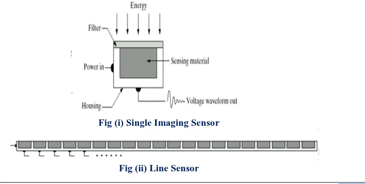
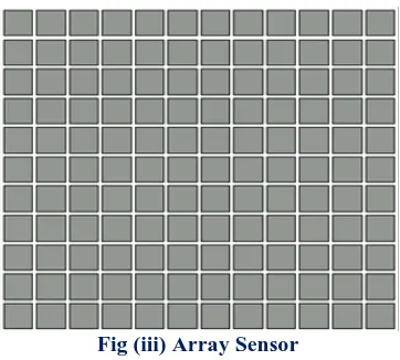
Image Acquisition is the first and important step of the digital image of processing. Its style is very simple just like being given an image which is already in digital form and it involves preprocessing such as scaling etc. It starts with the capturing of an image by the sensor (such as a monochrome or color TV camera) and digitized. In case, the output of the camera or sensor is not in digital form then an analog-to-digital converter (ADC) digitizes it. If the image is not properly acquired, then you will not be able to achieve tasks that you want to. Customized hardware is used for advanced image acquisition techniques and methods. 3D image acquisition is one such advanced method image acquisition method. Most of the images which are generated by the combination of an “illumination” source and the reflection or absorption of energy from that source of elements of the “scene” being imaged. Fig. below shows the three principal sensor arrangements used to transform illumination energy into digital images. (i)Single imaging Sensor (ii)Line sensor (iii)Array sensor. The idea is simple: Incoming energy is transformed into a voltage by the combination of input electrical power and sensor material that is responsive to the particular type of energy being detected. The output voltage waveform is the response of the sensor(s), and a digital quantity is obtained from each sensor by digitizing its response[1].
Fig (i) Single Imaging Sensor
Fig (iii) Array Sensor
2. Image Enhancement:
Image enhancement is one of the easiest and the most important areas of digital image processing. The core idea behind image enhancement is to find out information that is obscured or to highlight specific features according to the requirements of an image. Such as changing brightness & contrast etc. Basically, it involves manipulation of an image to get the desired image than original for specific applications. Many algorithms have been designed for the purpose of image enhancement in image processing to change an image’s contrast, brightness, and various other such things. Image Enhancement aims to change the human perception of the images. Image Enhancement techniques are of two types: Spatial domain and Frequency domain. Image enhancement techniques have been widely used in many applications of image processing where the subjective quality of images is important for human interpretation. Contrast is an important factor in any subjective evaluation of image quality. Contrast is created by the difference in luminance reflected from two adjacent surfaces. In other words, contrast is the difference in visual properties that makes an object distinguishable from other objects and the background. In visual perception, contrast is determined by the difference in the colour and brightness of the object with other objects. Our visual system is more sensitive to contrast than absolute luminance; therefore, we can perceive the world similarly regardless of the considerable changes in illumination conditions. Many algorithms for accomplishing contrast enhancement have been developed and applied to problems in image processing[2]. If the contrast of an image is highly concentrated on a specific range, e.g. an image is very dark; the information may be lost in those areas which are excessively and uniformly concentrated. The problem is to optimize the contrast of an image in order to represent all the information in the input image. There are two categories of Spatial domain processing: 1) Intensity Transformations : operate on single pixel, contrast manipulation, and image thresholding. 2) Spatial Filtering : work on a neighborhood of every pixel in an image, image smoothing and image sharpening[1].
3. Image Restoration:
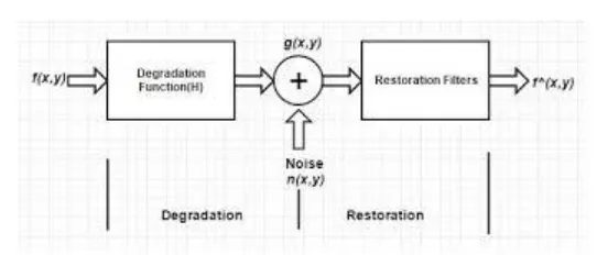
Image restoration involves improving the appearance of an image. In comparison to image enhancement which is subjective, image restoration is completely objective which makes the sense that restoration techniques are based on probabilistic or mathematical models of image degradation. Image restoration removes any form of a blur, noise from images to produce a clean and original image. The image information lost during blurring is restored through a reversal process. This process is different from the image enhancement method. The main defects that degrade an image are restored in this. The objective of image restoration techniques is to reduce noise and recover resolution loss. The most straightforward and a conventional technique for image restoration is deconvolution,[30] [31] which is performed in the frequency domain and after computing the Fourier transform of both the image and the PSF and undo the resolution loss caused by the blurring factors. This deconvolution technique, because of its direct inversion of the PSF which typically has poor matrix condition number, amplifies noise and creates an imperfect deblurred image. Also, conventionally the blurring process is assumed to be shift-invariant. Hence more sophisticated techniques, such as regularized deblurring, have been developed to offer robust recovery under different types of noises and blurring functions. It is of 3 types: 1. Geometric correction 2. radiometric correction 3. noise removal[1].
A model of the Image Degradation/Restoration Process
Fig. A model of the image degradation/restoration process.
4. Color Image Processing
Color image processing has been proved to be of great interest because of the significant increase in the use of digital images on the Internet. It includes color modeling and processing in a digital domain etc. There are various color models which are used to specify a color using a 3D coordinate system. These models are RGB Model, CMY Model, HSI Model, YIQ Model. The color image processing is done as humans can perceive thousands of colors. There are two areas of color image processing full-color processing and pseudo color processing. In full-color processing, the image is processed in full colors while in pseudo color processing the gray scale images are converted to colored images. The use of color is important in image processing because: 1) Color is a powerful descriptor that simplifies object identification and extraction. And 2)Humans can discern thousands of color shades and intensities, compared to about only two dozen shades of gray. Color image processing is divided into two major areas:
•Full-color processing: Images are acquired with a full-color sensor, such as a color TV camera or color scanner.
•Pseudocolor processing: The problem is one of assigning a color to a particular monochrome intensity or range of intensities[1].
5. Image Compression:
Image compression is defined as the process of reducing the amount of data needed to represent a digital image. This is done by removing the redundant data. The objective of image compression is to decrease the number of bits required to store and transmit without any measurable loss of information. Compression involves the techniques that are used for reducing storage necessary to save an image or bandwidth to transmit it. If we talk about its internet usage, it is mostly used to compress data. Algorithms acquire useful information from images through statistics to provide superior quality images. Compression can reduce the file sizes up to 60-70% and hence many files can be combined into one compressed document which makes the sendingeasier. It helps to reduce the consumption of excessive resources such as hard disc space and transmission bandwidth. Compression can fit more data in small memory and thus it reduces the memoryspacerequired as well as the
cost of managing data. Two types of digital image compression are 1). Lossless (or) Error – Free Compression and 2). Lossy Compression [1]
6. Morphological Processing:
Morphology a branch of biology that deals with the form and structure of animals and plants. Morphological image processing is used to extract image components for representation and description of region shape, such as boundaries, skeletons, and the convex hull connectivity analysis, blob analysis etc[1]. It is used to develop methods (region filling, thinning, thickening, and pruning) that are frequently used in conjunction with the algorithms as pre or post processing steps[33].
7. Segmentation:
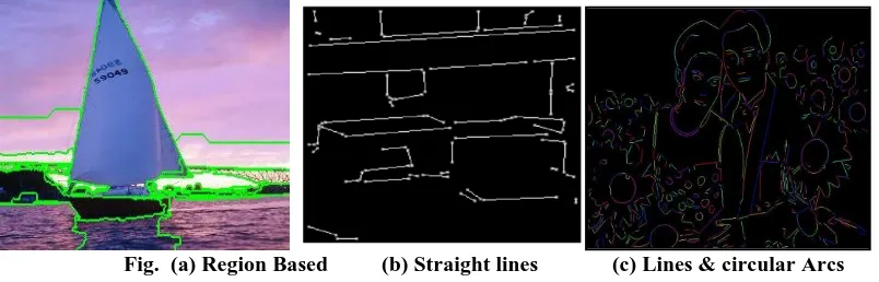
2) linear structures, such as line segments and curve segments
3) 2D shapes, such as circles, ellipses and ribbons (long, symmetric regions). As shown below
Fig. (a) Region Based (b) Straight lines (c) Lines & circular Arcs
8. Object recognition:
An object recognition system finds objects in the real world from an image of the world, using object models which are known a priori. This task is surprisingly difficult. Humans perform object recognition effortlessly and instantaneously. Recognition involves assigning of a label, such as, “vehicle” to an object completely based on its descriptors. It is a method of recognising a specific object in an image or video. There are certain techniques and models for object recognition like deep learning models, bag-of-words model etc.[1]
REFERENCES
[1]. Rafael C. Gonzalez and Richard E. Woods, “Digital Image Processing”, Indian edition published by Dorling Kindersley India Pvt. Ltd. 2009, Third Edition.
[2]. A. Deda, A. Samojedny, R. Koprowski, S. Wilczyoski, Z. Wróbel, (2013, Jun) Image analysis and processing methods in verifying the correctness of performing low-invasive esthetic medical procedures [3]. A. El-Baz, A. Farag, M. El-Ghar, R. Fahmi, S. Yuksel, T. Eldiasty, W. Miller. A new CAD system for
the evaluation of kidney diseases using DCE-MRI
[4]. A.I. El-Fallah, G.E. Ford (1997, May) Mean curvature evolution and surface area scaling in image filtering.
[5]. A. Hayat Gondal and M.N.A. Khan, A Review of Fully Automated Techniques for Brain Tumor Detection from MR Images [PDF].Available: http://www.mecs-press.org/ijmecs/ijmecs-v5-n2/IJMECS-V5-N2-8.pdf.
[6]. A. Korzyoska, A. Witkowska, R. Koprowski , J. Małyszek, W. Zieleźnik, W. Wójcik and Z. Wróbel. (2012, Nov) Influence of the measurement method of features in ultrasound images of the thyroid in the diagnosis of Hashimoto’s disease. *online+. Available: http://www.biomedical-engineering-online.com/content/11/1/91
[7]. A. Kumar, A. Yezzi, Jr., A.Tannenbaum, P. Olver, and S. Kichenassamy (1997, April) A Geometric Snake Model for Segmentation of Medical Imagery [PDF] Available: http://ieeexplore.ieee.org/xpl/articleDetails.jsp?arnumber=563665
[8]. A.K. Sinha, N.Pradhan. (2010, Dec). Development of a composite feature vector for the detection of pathological and healthy tissues in FLAIR MR Images of brain
[9]. A. Tristan, E. Mu˜noz-Moreno, L. Cordero-Grande, M. Martın-Fernandez, R. Cardenes. UsimagTool: an Interactive Research Tool for Ultrasound Image Processing
[10]. B. Fei, H. Akbari. (2012, Feb).Automatic 3D Segmentation of the Kidney in MR Images Using Wavelet Feature Extraction and Probability Shape Model
[11]. C. Chang, C. Chen, E. Chen, P. Chung and H. Tsai (1998, Jun) An Automatic Diagnostic System for CT Liver Image Classification. [PDF] Available: http://www.umbc.edu/rssipl/pdf/BME_6_98.pdf.
[12]. C. Li, C. Xu, C. Gui, and M.D. Fox: Distance regularized level set evolution and its application to image segmentation
[13]. D. Atkinson, G.Scidmore, M.B. Zawadzki, M. Detrick, and W.G. Bradley. (1996, July) Fluid-Attenuated Inversion Recovery (FLAIR) for Assessment of Cerebral Infarction
[14]. D. A. Dahab, S. S. A. Ghoniemy, G. M. Selim. (Oct, 2012). Automated Brain Tumor Detection and Identification Using Image Processing and Probabilistic Neural Network Techniques. [PDF] Available: http://www.ijipvc.org/article/IJIPVCV1I201.pdf
[16]. F. Visser, J. Hendrikse, J.J.M. Zwanenburg, P.R. Luijten, and T. Takahara. (2009, Oct) Fluid attenuated inversion recovery (FLAIR) MRI at 7.0 Tesla: comparison with 1.5 and 3.0 Tesla
[17]. F.Yang, J. Gu, T. Wen, W. Qin, Y. Xie (2012, Oct), A Shape-Optimized framework for kidney in ultrasound images NLTV Denoising and DRLSE
[18]. G. Wang, H. Yin, J. Wang, L. Sun, M. Vannier, T. Yamada. (2004, Oct) ImageParser: a tool for finite element generation from three-dimensional medical images
[19]. H. Li, H. Zhang, J. Liu, Z. Zhu. (2011, Sep) An automated and simple method for brain MR image extraction http://en.wikipedia.org/wiki/Tomography
[20]. http://www.math.uni-hamburg.de/projekte/shape/curve_evolution.html
[21]. J. Huang, F. Jian, H. Wu, H. Li. (2013, May). An improved level set method for vertebra CT image segmentation. [online]. Available: http://www.biomedical-engineering-online.com/content/12/1/48 [22]. M. Acheroy, W. Philips, A. Pizurica, and I. Lemahieu (2003 Mar) A Versatile Wavelet Domain Noise
Filtration Technique for Medical Imaging [PDF]. Available:
http://telin.ugent.be/~sanja/Papers/TMI0161.pdf.
[23]. J.R. Jensen, K.L. Gammelmark, M.H. Pedersen, and S.I. Nikolov (2006, December). Synthetic Aperture Ultrasound Imaging
[24]. M.H.F. Zarandi, M. Izadi, M. Zarinbal. Systematic image processing for diagnosing brain tumors: A Type-II fuzzy expert system approach.
[25]. S.S. Asole and V.J. Nagalkar (2012, May) Brain tumor detection using digital image processing based on soft computing [PDF] Available: www.bioinfo.in/uploadfiles/13366343493_3_1_JSIP.pdf
[26]. U.S. Department of Health and Human Services. Annual report of the U.S. scientific registry of transplant recipients and the organ procurement and transplantation network: transplant data: 1990-1999. Bureau of Health Resources Department, Richmond, VA; 2000
[27]. V.P.G.P. Rathi and S.Palani, Brain tumor MRI image classification with feature selection and extraction using linear decrement analysis [PDF].Available: http://arxiv.org/ftp/arxiv/papers/1208/1208.2128.pdf. [28]. Gurpreet kaur , Rajdavinder Singh, Image Enhancement And Its technique- A review. International
Journal of Computer Trends and Technology (IJCTT) – volume 12 number 3 – Jun 2014.
[29]. Snehal O.Mundhada, Prof. V. K. Shandilya” Image Enhancement and Its Various Techniques “International Journal of Advanced Research in Computer Science and Software Engineering” Volume 2, Issue 4, April 2012 ISSN: 2277 128X
[30]. Jinshan Tang Eli Peli, and Scott Acton, “Image Enhancement Using a Contrast Measure in the Compressed Domain”, IEEE Signal processing Letters , Vol. 10, NO. 10, October 2003.
[31]. Rakesh Soni ,Dr. Vibha Tiwari, “ A review on Digiatl Image Restoration Process” . International Research Journal of Engineering and Technology (IRJET) e-ISSN: 2395 -0056.
[32]. Navdeep Kaur and Dr. Divya Garg, “Image Restoration and De-Blurring Using Various Algorithms”,International Journal of Computer Science Trends and Technology (IJCST) – Volume 4 Issue 5, Sep - Oct 2016.
[33]. Sargun and Shashi B. Rana, “A Review of Medical Image Enhancement Techniques for Image Processing”, https://www.researchgate.net/publication/276155321.
[34]. Petros Maragos, “Tutorial on Advances in Morphological Image processing and Analysis”, SPIE Vol. 707 Visual Communications and Image Processing .
[35]. Shengyong Chen, Mingzhu Zhao, GuangWu, Chunyan Yao, and Jianwei Zhang , “Recent Advances inMorphological Cell Image Analysis”, Hindawi Publishing Corporation Computational and Mathematical Methods in Medicine Volume 2012, Article ID 101536, doi:10.1155/2012/101536.
[36]. Tri Daryanto, Sheeraz Arif and Shu Yang, “Survey: Recent Trends and Techniques in Image Co Segmentation Challenges, Issues and Its Applications”, International Journal of Computer Science and Software Engineering (IJCSSE), Volume 6, Issue 5, May 2017 ISSN (Online): 2409-4285.
[37]. Yu-Jin Zhang, Tsinghua University, Beijing, China, “An Overview of Image and Video Segmentation in the Last 40 Years”.
[38]. Mukesh Tiwari and Dr. Rakesh Singhai, “A Review of Detection and Tracking of Object from Image and Video Sequences” , International Journal of Computational Intelligence Research ISSN 0973-1873 Volume 13, Number 5 (2017), pp. 745-765.
[39]. Kuldeep Narayan Shukla, Anjali Potnis and Prashant Dwivedy, “A Review on Image Enhancement Techniques” , International Journal of Engineering and Applied Computer Science (IJEACS) Volume: 02, Issue: 07, ISBN: 978-0-9957075-8-0, July 2017.
International organization of Scientific Research
60 | Page
[41]. Versha Rani, Priyanka Kamboj, “Image Enhancement using Hybrid Filtering Technique”, InternationalJournal of Science and Research (IJSR), India Online ISSN: 2319-7064.
[42]. Ridho Dwisyah Putra, Tito Waluyo Purboyo and Anggunmeka Luhur Prasasti, “ A review of Image Enhancement Methods” , International Journal of Applied Engineering Research ISSN 0973-4562 Volume 12, Number 23 (2017) pp. 13596-13603.
[43]. Ravinder Kaur, Taqdir, “ Image Enhancement Techniques- A Review” , International Research Journal of Engineering and Technology (IRJET) e-ISSN: 2395 -0056.
[44]. Gabriel Iwasokun, “Image Enhancement Methods: A Review”,
https://www.researchgate.net/publication/271263292.
[45]. R.Himabindu, “ Implementation of efficient Image Enhancement Factor using Modified Decision Based Unsymmetric Trimmed Median Filter”, International Journal of Engineering Research & Technology (IJERT) ISSN: 2278-0181.Vol. 2 Issue 9, September – 2013.
[46]. Chao Zuo∗, Qian Chen, Xiubao Sui, “Range Limited Bi-Histogram Equalization for image contrast enhancement”, National Defense Key Laboratory of Optoelectronic Engineering, Nanjing University of Science and Technology, Nanjing, Jiangsu Province.
[47]. Soong Der Chen, “Minimum mean brightness error Bi-histogram equalization in contrast Enhancement” , https://www.researchgate.net/publication/3180783.
[48]. Bhanudas Sandbhor, G. U. Kharat, “ A Review on Underwater Image Enhancement Techniques”, International Journal of Advanced Research in Computer Science and Software Engineering Volume 5, Issue 5, MAY 2015, ISSN: 2277 128X.
[49]. Shilpa Rani, Sonika Jindal, and Bhavneet Kaur, “A Brief Review on Image Restoration Techniques”, International Journal of Computer Applications (0975 – 8887) Volume 150 – No.12, September 2016. [50]. Min Wang, Shudao Zhou, Wei Yan, “Blurred image restoration using knife-edge function and optimal
window Wiener filtering”.
[51]. Ishfaq Bashir, Adil Majeed, Owais Khursheed, “Image restoration and the various restoration techniques used in the field of digital image processing”, International Journal of Computer Science and Mobile Computing, International Journal of Computer Science and Mobile Computing, Vol.6 Issue.6, June- 2017, pg. 390-393.
[52]. Shengyong Chen, Mingzhu Zhao, GuangWu,, Chunyan Yao, and Jianwei Zhang, “Recent Advances inMorphological Cell Image Analysis”.
[53]. Min-Song Wei, Fei Xing and Zheng You, “A real-time detection and positioning method for small and weak targets using a 1D morphology-based approach in 2D images” , Light: Science & Applications (2018) .
[54]. Antonio Plaza , Jon Atli Benediktsson, “Recent advances in techniques for hyperspectral image processing”, Remote Sensing of Environment.
[55]. Nida M. Zaitoun, Musbah J. Aqel, “Survey on Image Segmentation Techniques” , International Conference on Communication, Management and Information Technology (ICCMIT 2015)
[56]. Swati Matta, “Review: Various Image Segmentation Techniques” , International Journal of Computer Science and Information Technologies, Vol. 5 (6) , 2014, 7536-7539.
[57]. Manraj, Amitpal Singh, “Current Image Segmentation Techniques-A Review”, International Journal of Computer Science and Information Technologies, Vol. 6 (2) , 2015, 1940-1942.
[58]. Sukanya C.M, Roopa Gokul, Vince Paul, “A Survey on Object Recognition Methods”, IJCSET(www.ijcset.net) | January 2016 | Vol 6, Issue 1, 48-52.
[59]. Khushboo Khurana, Reetu Awasthi, “Techniques for Object Recognition in Images and Multi-Object Detection”, International Journal of Advanced Research in Computer Engineering & Technology (IJARCET) Volume 2, Issue 4, April 2013.
[60]. A.C.Phadke, J.Joshi. Feature Extraction and Texture Classification in MRI.