Received: May 10, 2016; Revised: July 26, 2016; Accepted: July 28, 2016
© The Author 2016. Published by Oxford University Press. All rights reserved. For Permissions, please email: [email protected]. doi:10.1093/carcin/bgw077
Advance Access publication August 1, 2016 Original Manuscript
951
original manuscript
Genetic variations in the Hippo signaling pathway and
breast cancer risk in African American women in the
AMBER Consortium
Jianmin Zhang
*
, Song Yao
1, Qiang Hu
2, Qianqian Zhu
2, Song Liu
2,
Kathryn L.Lunetta
3, Stephen A.Haddad
4, Nuo Yang, He Shen, Chi-Chen Hong
1,
Lara Sucheston-Campbell
1, Edward A.Ruiz-Narvaez
4, Jeannette T.Bensen
5,
Melissa A.Troester
5, Elisa V.Bandera
6, Lynn Rosenberg
3, Christopher A.Haiman
7,
Andrew F.Olshan
5, Julie R.Palmer
4and Christine B.Ambrosone
1Department of Cancer Genetics, Roswell Park Cancer Institute, Buffalo, NY 14263, USA, 1Department of Cancer Prevention
and Control, Roswell Park Cancer Institute, Buffalo, NY 14263, USA, 2Department of Biostatistics and Bioinformatics,
Roswell Park Cancer Institute, Buffalo, NY 14263, USA, 3Department of Biostatistics, Boston University School of Public
Health, Boston, MA 02118, USA, 4Slone Epidemiology Center at Boston University, Boston, MA 02215, USA, 5Department of
Epidemiology, Gillings School of Global Public Health, University of North Carolina at Chapel Hill, Chapel Hill, NC 27599, USA, 6Cancer Prevention and Control Program, Rutgers Cancer Institute of New Jersey, The State University of New Jersey,
New Brunswick, NJ 08901, USA, and 7Department of Preventive Medicine, Keck School of Medicine, University of Southern
California/Norris Comprehensive Cancer Center, Los Angeles, CA 90089, USA
*To whom correspondence should be addressed. Department of Cancer Genetics, Roswell Park Cancer Institute, Elm & Carlton Streets, Buffalo, NY 14263, USA. Tel: +1 716 845 5929; Fax: +1 716 845 1698; Email: [email protected]
Abstract
The Hippo signaling pathway regulates cellular proliferation and survival, thus exerting profound effects on normal cell fate and tumorigenesis. Dysfunction of the Hippo pathway components has been linked with breast cancer stem cell regulation, as well as breast tumor progression and metastasis. TAZ, a key component of the Hippo pathway, is highly expressed in triple negative breast cancer; however, the associations of genetic variations in this important pathway with breast cancer risk remain largely unexplored. Here, we analyzed 8309 germline variants in 15 genes from the Hippo pathway with a total of 3663 cases and 4687 controls from the African American Breast Cancer Epidemiology and Risk Consortium. Odds ratios (ORs) were estimated using logistic regression for overall breast cancer, by estrogen receptor (ER) status (1983 ER positive and 1098 ER negative), and for case-only analyses by ER status. The Hippo signaling pathway was significantly associated with ER-negative breast cancer (pathway level P = 0.02). Gene-based analyses revealed that
CDH1 was responsible for the pathway association (P < 0.01), with rs4783673 in CDH1 statistically significant after gene-level adjustment for multiple comparisons (P = 9.2 × 10⁻⁵, corrected P = 0.02). rs142697907 in PTPN14 was associated with
Introduction
Functional screens in Drosophila identified the Hippo signaling pathway, which regulates organ size by modulating cell growth, proliferation and apoptosis (1–3). The majority of the Hippo path-way components are highly conserved from Drosophila to mam-malian species, and dysregulation of this pathway is widely observed in cancer (4–6). The core of this pathway in mammals is composed of a kinase cascade wherein the STE20-like kinase 1/2 (MST1/2), in complex with its regulatory protein salvador 1 (SAV1), phosphorylates and activates large tumor suppressor kinase 1/2 (LATS1/2) in complex with its regulatory protein MOB kinase acti-vator 1A (MOB1A). This in turn phosphorylates and inactivates the transcriptional co-activators, yes-associated protein (YAP)/ transcriptional coactivator with PDZ-binding motif (TAZ). When YAP/TAZ translocate to the nucleus, they induce expression of cell-proliferative and anti-apoptotic genes, mainly through inter-actions with transcription factors, such as: TEA domain family members (TEADs) (3). In recent years, knowledge of the com-plexity of YAP/TAZ regulation has expanded considerably. The G protein-coupled receptors (GPCRs) and the cytokine receptor leukemia inhibitory factor receptor (LIFR) are associated with the activation of LATS kinases (7,8). In addition, YAP/TAZ are also directly regulated by the extracellular matrix (9), mechanotrans-duction (10,11), actin cytoskeleton and Rho GTPases (11,12).
We and others have previous shown that TAZ is overex-pressed in breast cancers, especially in triple negative breast cancer (13–15). The expression levels and activity of TAZ are frequently upregulated in high-grade metastatic breast cancer (16–18). Activation of YAP induces epithelial to mesenchymal transition and promotes breast tumor metastasis (19,20). Both YAP and TAZ have been shown to be involved in breast cancer stem cell regulation (16,17,21,22) and mediated drug resistance in breast cancer (19,23). In addition, it has been demonstrated that hypermethylation of the promoter regions of LATS1/2 occurred in breast cancers and the decreased expression of LATS1/2 was significantly associated with large tumor size and high lymph node metastasis (24).
Epidemiological studies indicate that African-American (AA) women are more likely to be diagnosed with more aggressive breast cancer, including estrogen receptor (ER)-negative and TN breast cancer, and have higher cancer mortality than European American (EA) women (25–27). Although the mechanisms underlying these disparities are largely unknown, emerging evidence supports that cancer biology may be different across patients of different ances-tral background (25,28). Considering the critical role of Hippo signal-ing pathway played in triple-negative breast cancer, we hypothesize that this pathway may contribute in part to the biological difference in breast cancer between AA and EA women. The African American Breast Cancer Epidemiology and Risk (AMBER) Consortium was estab-lished to investigate potential genetic and non-genetic risk factors for aggressive breast cancer in AA women. Here, we comprehen-sively examined genetic variations in Hippo signaling pathway with breast cancer risk in this large AA breast cancer consortium.
Study population and methods
The AMBER consortium is a large collaborative effort to aggregate an adequate sample size to study epidemiology of breast cancer subtypes in AA women. Established in 2011, the
consortium consists of two case-control studies, the Women’s Circle of Health Study (WCHS) and the Carolina Breast Cancer Study (CBCS), and two prospective cohort studies, the Black Women’s Health Study (BWHS) and the Multiethnic Cohort (MEC). A detailed description of the consortium and the four contributing studies can be found elsewhere (29–34).
The WCHS is a case-control study enrolling women aged 25–75 with invasive breast cancer and ductal carcinoma in situ
(DCIS), initially in New York City (NYC) and New Jersey (NJ), and later exclusively in NJ (31,32). Cases were ascertained in NYC hospitals with large referral patterns of AAs and through the NJ State Cancer Registry. Controls frequency matched on state, race and age were identified through random digital dialing and community events. The CBCS is a population-based case–con-trol study in North Carolina beginning in 1993 (30). Breast cancer patients aged 20–74 were identified through the NC State Cancer Registry, and controls were enrolled through Division of Motor Vehicle lists and Health Care Finance Administration lists.
The BWHS is a prospective study of 59 000 AA women across the USA who were 21–69 years of age at the study entry in 1995 and have been followed by biennial questionnaire since that time (33). Women diagnosed with breast cancer are identified by self-report in follow-up questionnaires, and confirmed by medical records, state cancer registries, and the National Death Index. The MEC is a multiethnic prospective cohort in Hawaii and southern California with follow-up of 215 000 men and women aged 45–75 at the time of study entry (1993–1996) (34). Breast cancer diagnoses identified through linkage to state can-cer registries. Controls for the BWHS and MEC were AA partici-pants who had not been diagnosed with breast cancer.
All study participants provided informed consent, and the study was approved by Institutional Review Boards at participat-ing institutions. Estrogen receptor (ER) status information was obtained from pathology reports and/or Cancer Registry Data. The study population included in the genotype study has been previously described in detail (35). A brief summary of the num-ber of cases and controls from each contributing study included in this analysis, with index age and ER status (for cases) is provided in Supplementary Table 1, available at Carcinogenesis
Online.
Genetic marker selection, genotyping, quality control and imputation
Genes from select candidate pathways of interest were iden-tified by querying the Molecular Signature Database (MSigDB) (36) and tagSNPs from each gene were chosen using criteria of
r2 ≥ 0.8 and minor allele frequency ≥ 10% in the Yoruban (YRI) population from the 1000 Genome Project (37). These SNPs were added as part of the custom content to the Illumina Human Exome Beadchip v1.1 and samples from BWHS, CBCS and WCHS were genotyped by the Center for Inherited Disease Research (CIDR), followed by stringent sample and marker QC steps (38). Imputation to the 1000 Genomes data using the IMPUTE2 pro-gram (39) was performed by the University of Washington (UW). MEC samples had been genotyped previously using the Illumina 1M-Duo chip and also imputed to the 1000 Genomes data. The imputed MEC data were pooled with those from the BWHS, CBCS and WCHS to create a final analytical dataset. Markers with mismatching alleles or allele frequencies that were dif-ferent by > 0.15 between MEC and the other three studies, and markers with MAF < 0.6% or imputation info score < 0.5 in either study were excluded. For the present analysis of the Hippo sign-aling pathway, a total of 7017 variants in 14 genes belonging to this pathway were included (Table 2).
Abbreviations
AA African-American
Statistical analysis
To control for potential admixture bias, principal component analysis was conducted using the smartpca program in the EIGENSOFT package (40) to infer population structure. Paired sample relatedness was assessed by PLINK (41). As a result, 35 individual outliers in principal component analysis and 162 first-degree relatives identified were flagged for sensitivity analysis. No substantial changes in risk estimates were found after excluding these individuals and they were thus kept in the analysis. Ten PCs were tested for association with case– control status while controlling for covariates, including index age, study, geographic region and DNA source. Although none was significantly associated with breast cancer risk, to be con-servative, three PCs with a P < 0.10 were included in the logistic regression models.
In addition to analyzing overall breast cancer risk, stratified analyses were conducted by ER status compared to controls, as well as case-only analyses comparing ER− to ER+ cases. Three levels of analyses of genetic variations were performed: path-way level, gene level and single marker level, under the hypoth-esis that aggregating the effects of multiple markers within a gene or a biological pathway might be more statistically power-ful and less prone to multiple testing bias than single marker analysis. Pathway- and gene-level analyses were performed first, using the adaptive rank truncated product (ARTP) statistic (42), which can optimize the number of single marker P values combined in each gene-level and pathway-level test. For path-way-level analysis, the PIGE software implementation of the ARTP method takes gene-level information into consideration when combining markers in a pathway (https://cran.r-project. org/web/packages/PIGE/index.html). To avoid redundancy of markers in high LD (r2 ≥ 0.8), the ARTP gene-level tests com-bined the optimal number of most significant SNP P values from among the top 10 pruned-in SNPs for each gene. This number was deliberately chosen to ensure adequate represen-tation of genetic variations in each gene, while not to include too many null variants to dilute the effects of truly causal mark-ers. The ARTP pathway tests combined the optimal percentage (in 5% increments) of the most significant gene P values in each pathway, without exceeding 50%. This approach was chosen to ensure excellent representation of associated genetic vari-ants, while not diluting any effects from truly causal markers by including too many null markers in the analysis. Following gene-level testing, single marker-level analyses were pursued
using PLINK with dosage data and controlling for age, study, geographic region, DNA source and three top PCs. We corrected for multiple testing within these genes with a Bonferroni cor-rection for the effective number of independent markers tested within a gene using Gao’s SimpleM approach (43), and called this the ‘gene-wide’ significance. Single marker associations for top genes were plotted with linkage disequilibrium data using the LocusZoom program (44).
Results
As shown in Table 1, the Hippo pathway was significantly associ-ated with ER-negative breast cancer risk (pathway level P = 0.02), likely attributable to CDH1 (gene level P = 0.004). When CDH1 gene was removed from the analysis, the pathway-level signifi-cance become non-significant (P = 0.63). The pathway was not associated with risk of overall cancer or ER-positive cancer, or with ER status in case-only analyses (pathway level P > 0.05). In analysis of genes and breast cancer risk, CDH1 was nominally associated with overall breast cancer risk (P = 0.02); and CDK1
was nominally associated with ER negative disease in case-only analysis (P = 0.01).
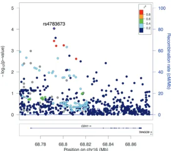
Figure 1 displays the single variant associations of CDH1
with ER-negative breast cancer risk. The best signal locus was an intronic SNP, rs4783673. The T allele was associated with 19% reduced risk of ER-negative breast cancer (OR = 0.81, 95% CI 0.73, 0.90, P = 9.2E−5), which remained significant after correction for multiple testing at the gene level (corrected P = 0.02) (Table 2). When this SNP was removed, CDH1 remained significant at the gene level (P = 0.005) but the Hippo pathway was no longer sig-nificant (P = 0.13). Although CDH1 was also associated with over-all breast cancer risk at the gene level, no individual variants in the gene were significantly associated with overall breast cancer after correction for multiple testing (data not shown). The most significant SNP in CDH1 for overall breast cancer was rs4783673, the T allele of which was associated with a 12% reduced risk at a borderline significance level (OR = 0.88, 95% CI, 0.82, 0.94,
P = 2E−4, corrected P = 0.06). This was the same variant identified above with ER-negative breast cancer, and thus the association with overall breast cancer risk was likely driven by this subtype.
No gene in the Hippo pathway was associated with ER-positive breast cancer at the gene level (Table 1). However, an intronic SNP rs142697907 in PTPN14 was significant at the single marker-level after within gene adjustment for multiple testing. The A allele was associated with a 75% increased risk of
Table 1. P values of pathway- and gene-level test with breast cancer risk (P values lower than 0.05 is in bold)
Gene # Total marker # Effective marker Overall ER+ ER− ER− versus ER+
Hippo pathway 7017 2244 0.36 0.83 0.02 0.24
AREG 666 186 0.45 0.89 0.29 0.41
CDH1 666 269 0.02 0.08 0.004 0.50
CDK1 195 74 0.73 0.65 0.09 0.01
CTGF 26 12 0.76 0.58 0.91 0.80
CTNNA1 939 149 0.72 0.99 0.97 0.96
CTNNB1 138 56 0.32 0.69 0.59 0.53
DLG5 714 166 0.63 0.64 0.64 0.75
FAT1 976 455 0.83 0.35 0.97 0.48
PTPN14 1151 384 0.29 0.21 0.09 0.20
RASSF1 53 32 0.76 0.57 0.99 0.41
SIAH1 10 7 0.35 0.22 0.14 0.21
STK4 586 113 0.96 0.96 0.79 0.22
TEAD4 430 188 0.27 0.52 0.41 0.69
ER-positive cancer (OR = 1.75, 95% CI 1.33, 2.30, P = 7.4E−5, cor-rected P = 0.03).
A 3′ UTR SNP, rs2456773 in CDK1 was likely the variant driving the association of CDK with ER status in case-only analysis. The C allele was associated with 25% increased odds of ER-negative versus ER-positive cancer (OR = 1.25, 95% CI 1.11, 1.42, P = 3.2E−4, corrected P = 0.02) (Table 2). A second nearby 3′ UTR SNP, rs10711 in perfect LD with rs2456773 (r2 = 1.0), was also associated with ER status. In case-control analyses performed separately by ER status, rs2456773 was only associated with ER-negative cancer (OR = 1.20, 95% CI 1.06, 1.34, P = 0.003), but not with ER-positive cancer (OR = 0.96, 95% CI 0.88, 1.06, P = 0.42).
Discussion
Here, we report findings of a comprehensive analysis of germline variations in the Hippo signaling pathway with breast cancer risk and by tumor ER status. The unique strengths of the study include a large population of AA women with breast cancer and controls and a systematic interrogation of common genetic vari-ations in all available genes in this pathway. We found evidence of the overall Hippo pathway being associated with ER-negative breast cancer risk, which may be attributed to CDH1. Our find-ing corroborates that from laboratory studies linkfind-ing the Hippo pathway with ER-negative and triple-negative breast cancer.
CDH1 encodes a classical member of the cadherin fam-ily, which plays an important role in maintaining the epithe-lial integrity. Down regulation of CDH1 has been considered as one of the main molecular alterations for tumor invasion and metastasis (45). Complete E-cadherin loss has been reported in 86% to 100% of invasive lobular breast cancer (46). Interestingly, E-cadherin reduction has been found in triple negative breast cancer patients with lymph node metastasis (47,48). Given the well established role of somatic changes in CDH1 in cancer invasion and metastasis, a number of studies have investigated the associations of CDH1 germline variants with risk of vari-ous human cancers. A meta-analysis concluded that one SNP in the promoter region, rs16260 (-160 C>A), was associated with increased risk of all cancers, but not with breast cancer in strati-fied analyses by cancer type (49). In our AA population, we did not find any association of rs16260 with breast cancer risk.
We also found a low frequency variant in PTPN14 associated with 75% increased risk of ER-positive breast cancer risk. PTPN14
encodes a member of the protein tyrosine phosphatase, which has been shown to mediate the dephosphorylation of tyrosine residues in some adherens junction proteins such as β-catenin (50). In addition, it was reported that PTPN14 suppressed metas-tasis by reducing the intracellular protein trafficking through the secretory pathway (51). We and other have previously dem-onstrated that PTPN14 negatively regulated YAP oncogenic func-tion through direct interacfunc-tion with YAP (52) and activation of LATS1/2 proteins (53). Interestingly, PTPN14 loss-of-function and deleterious missense mutations were found in skin cancer (54). To our knowledge, there is no published study of germline variants in PTPN14 with cancer risk. It should be noted, however, that the variant we identified with ER-positive breast cancer had a low frequency of 0.02 and was imputed with a moderate info score of 0.83. Thus, the result should be interpreted with caution because of possible imputation inaccuracy. Nevertheless, given the growing research interest in PTPN14 in cancer, our data may provide support for further study of variants in this gene in breast cancer.
To explore whether the significant gene we identified in AA women were also associated with breast cancer in EA women, we queried all available variants in CDH1 using publicly avail-able data from the GAME-ON GWAS look up tool. The T allele of rs4783673 in CDH1 was associated with slightly decreased risk of ER-negative breast cancer in an EA population (P = 0.07), which is consistent with our finding of this SNP in AA women. However, none of the variants in this gene was associated with overall or ER-negative breast cancer risk after correcting for multiple com-parisons. The low replication rate of significant genetic variants from EA to AA populations and vice versa is not unexpected, as observed in previous studies from us and others (35,55,56). This low replication rate may be due to distinct differences in genetic architecture between the two populations.
Several limitations should be noted in our study. Although we included a large number of genes and variants in the analysis,
Table 2. Top variants associated with breast cancer risk after gene-wide correction for multiple test (P ≤ 0.05)
SNP Gene A1/A2 Function A1 frequency Info score OR (95% CI) P Corrected P
ER-positive breast cancer
rs142697907 PTPN14 A/G Intronic 0.02 0.83 1.75 (1.33–2.30) 7.42E−05 0.03
ER-negative breast cancer
rs4783673 CDH1 T/C Intronic 0.65 0.99 0.81 (0.73–0.90) 9.21E−05 0.02
ER-negative versus ER-positive breast cancer
rs2456773 CDK1 C/G 3′ UTR 0.25 0.98 1.25 (1.11–1.42) 3.22E−04 0.02
several genes in the core Hippo signaling pathway, such as LAST1/2 and YAP1, were not included as candidates for tagSNP selection in development of the chip. Although SNPs in exonic regions of these genes were typed as the standard content in the exome chip array, variants in other regions of these genes were not typed. As a result, the marker density of these genes was much lower and was biased to exons, making imputed data from non-exonic regions more error-prone compared to genes selected as candidates in the genotyping process. Thus, we did not included Hippo pathway genes with only exonic SNPs typed in the analysis, and future studies with better coverage of the pathway are warranted. Another limitation of our study came from the lack of complete information on all immunohisto-chemical markers needed to classify triple-negative or basal-like breast cancer subtype. Given the emerging evidence from laboratory studies linking the Hippo pathway with triple nega-tive breast cancer, it would be interesting to analyze genetic variants in this pathway with this subtype. In the AMBER con-sortium, central staining and defining of breast cancer subtypes is ongoing, and follow up analysis will be conducted when such data become available. Lastly, the lack of functionality of the identified SNPs is a typical limitation of SNP association stud-ies, including ours. However, the identified associations provide clues for future experimental studies to characterize the func-tional impact of those genetic variations.
To conclude, in the first large study of common genetic vari-ants in the Hippo signaling pathway with breast cancer risk in AA women, we found that this pathway was specifically associ-ated with ER-negative breast cancer risk. Considering that AA women are at higher risk of ER-negative cancer than European American women, further studies are needed to assess whether the Hippo pathway may be a part of the biological differences underlying breast cancer disparities.
Supplementary material
Supplementary Table 1 can be found at http://carcin. oxfordjournals.org/
Funding
National Cancer Institute (NCI) (R21CA179693 to J. Z, P01CA151135 to J.R.P., C.B.A. and A.F.O., R01CA058420 to L.R., UM1CA164974 to L.R., R01CA098663 to J.R.P., R01CA100598 to C.B.A., P50CA58223 to M.A.T. and A.F.O.); the University Cancer Research Fund of North Carolina (M.A.T. and A.F.O.); the Breast Cancer Research Foundation (C.B.A.); the Roswell Park Alliance Foundation; and the American Cancer Society Research Scholar RSG-14-214-01-TBE (to J.Z.).
Conflict of Interest Statement: None declared.
References
1. Pan, D. (2010) The hippo signaling pathway in development and cancer. Dev. Cell, 19, 491–505.
2. Harvey, K. et al. (2007) The Salvador-Warts-Hippo pathway - an emerg-ing tumour-suppressor network. Nat. Rev. Cancer, 7, 182–191. 3. Moroishi, T. et al. (2015) The emerging roles of YAP and TAZ in cancer.
Nat. Rev. Cancer, 15, 73–79.
4. Harvey, K.F. et al. (2013) The Hippo pathway and human cancer. Nat. Rev. Cancer, 13, 246–257.
5. Johnson, R. et al. (2014) The two faces of Hippo: targeting the Hippo pathway for regenerative medicine and cancer treatment. Nat. Rev. Drug Discov., 13, 63–79.
6. Bossuyt, W. et al. (2014) An evolutionary shift in the regulation of the Hippo pathway between mice and flies. Oncogene, 33, 1218–1228. 7. Yu, F.X. et al. (2012) Regulation of the Hippo-YAP pathway by
G-protein-coupled receptor signaling. Cell, 150, 780–791.
8. Chen, D. et al. (2012) LIFR is a breast cancer metastasis suppressor upstream of the Hippo-YAP pathway and a prognostic marker. Nat. Med., 18, 1511–1517.
9. Calvo, F. et al. (2013) Mechanotransduction and YAP-dependent matrix remodelling is required for the generation and maintenance of cancer-associated fibroblasts. Nat. Cell Biol., 15, 637–646.
10. Aragona, M. et al. (2013) A mechanical checkpoint controls multicel-lular growth through YAP/TAZ regulation by actin-processing factors. Cell, 154, 1047–1059.
11. Dupont, S. et al. (2011) Role of YAP/TAZ in mechanotransduction. Nature, 474, 179–183.
12. Zhao, B. et al. (2012) Cell detachment activates the Hippo pathway via cytoskeleton reorganization to induce anoikis. Genes Dev., 26, 54–68. 13. Chan, S.W. et al. (2008) A role for TAZ in migration, invasion, and
tumo-rigenesis of breast cancer cells. Cancer Res., 68, 2592–2598.
14. Li, Y.W. et al. (2015) Characterization of TAZ domains important for the induction of breast cancer stem cell properties and tumorigenesis. Cell Cycle, 14, 146–156.
15. Skibinski, A. et al. (2014) The Hippo transducer TAZ interacts with the SWI/SNF complex to regulate breast epithelial lineage commitment. Cell Rep., 6, 1059–1072.
16. Cordenonsi, M. et al. (2011) The Hippo transducer TAZ confers cancer stem cell-related traits on breast cancer cells. Cell, 147, 759–772. 17. Bartucci, M. et al. (2015) TAZ is required for metastatic activity and
chemoresistance of breast cancer stem cells. Oncogene, 34, 681–690. 18. Matteucci, E. et al. (2013) Bone metastatic process of breast cancer
involves methylation state affecting E-cadherin expression through TAZ and WWOX nuclear effectors. Eur. J. Cancer, 49, 231–244.
19. Overholtzer, M. et al. (2006) Transforming properties of YAP, a candidate oncogene on the chromosome 11q22 amplicon. Proc. Natl. Acad. Sci. USA, 103, 12405–12410.
20. Lamar, J.M. et al. (2012) The Hippo pathway target, YAP, promotes metastasis through its TEAD-interaction domain. Proc Natl Acad Sci USA, 109, E2441–50.
21. Frangou, C. et al. (2014) Molecular profiling and computational network analysis of TAZ-mediated mammary tumorigenesis identifies action-able therapeutic targets. Oncotarget, 5, 12166–12176.
22. Kim, T. et al. (2015) A basal-like breast cancer-specific role for SRF-IL6 in YAP-induced cancer stemness. Nat. Commun., 6, 10186.
23. Lai, D. et al. (2011) Taxol resistance in breast cancer cells is mediated by the hippo pathway component TAZ and its downstream transcrip-tional targets Cyr61 and CTGF. Cancer Res., 71, 2728–2738.
24. Takahashi, Y. et al. (2005) Down-regulation of LATS1 and LATS2 mRNA expression by promoter hypermethylation and its association with biologically aggressive phenotype in human breast cancers. Clin. Can-cer Res., 11, 1380–1385.
25. Amend, K. et al. (2006) Breast cancer in African-American women: dif-ferences in tumor biology from European-American women. Cancer Res., 66, 8327–8330.
26. Brawley, O.W. (2013) Health disparities in breast cancer. Obstet. Gynecol. Clin. North Am., 40, 513–523.
27. DeSantis, C. et al. (2014) Breast cancer statistics, 2013. CA. Cancer J. Clin., 64, 52–62.
28. Dietze, E.C. et al. (2015) Triple-negative breast cancer in African-Amer-ican women: disparities versus biology. Nat. Rev. Cancer, 15, 248–254. 29. Palmer, J.R. et al. (2014) A collaborative study of the etiology of breast
cancer subtypes in African American women: the AMBER consortium. Cancer Causes Control, 25, 309–319.
30. Newman, B. et al. (1995) The Carolina Breast Cancer Study: integrating population-based epidemiology and molecular biology. Breast Cancer Res. Treat., 35, 51–60.
32. Bandera, E.V. et al. (2013) Rethinking sources of representative controls for the conduct of case-control studies in minority populations. BMC Med. Res. Methodol., 13, 71.
33. Rosenberg, L. et al. (1995) The Black Women’s Health Study: a follow-up study for causes and preventions of illness. J. Am. Med. Womens. Assoc., 50, 56–58.
34. Kolonel, L.N. et al. (2000) A multiethnic cohort in Hawaii and Los Ange-les: baseline characteristics. Am. J. Epidemiol., 151, 346–357.
35. Yao, S. et al. (2016) Genetic variations in vitamin D-related pathways and breast cancer risk in African American women in the AMBER con-sortium. Int. J. Cancer, 138, 2118–2126.
36. Subramanian, A. et al. (2005) Gene set enrichment analysis: a knowl-edge-based approach for interpreting genome-wide expression pro-files. Proc. Natl. Acad. Sci. USA, 102, 15545–15550.
37. Abecasis, G.R. et al. (2012) An integrated map of genetic variation from 1,092 human genomes. Nature, 491, 56–65.
38. Laurie, C.C. et al. (2010) Quality control and quality assurance in genotypic data for genome-wide association studies. Genet. Epidemiol., 34, 591–602. 39. Howie, B.N. et al. (2009) A flexible and accurate genotype imputation
method for the next generation of genome-wide association studies. PLoS Genet., 5, e1000529.
40. Price, A.L. et al. (2006) Principal components analysis corrects for strati-fication in genome-wide association studies. Nat. Genet., 38, 904–909. 41. Purcell, S. et al. (2007) PLINK: a tool set for whole-genome association
and population-based linkage analyses. Am. J. Hum. Genet., 81, 559–575. 42. Yu, K. et al. (2009) Pathway analysis by adaptive combination of
P-val-ues. Genet. Epidemiol., 33, 700–709.
43. Gao, X. (2011) Multiple testing corrections for imputed SNPs. Genet. Epidemiol., 35, 154–158.
44. Pruim, R.J. et al. (2010) LocusZoom: regional visualization of genome-wide association scan results. Bioinformatics, 26, 2336–2337.
45. Celebiler Cavusoglu, A. et al. (2009) Predicting invasive phenotype with CDH1, CDH13, CD44, and TIMP3 gene expression in primary breast can-cer. Cancer Sci., 100, 2341–2345.
46. Acs, G. et al. (2001) Differential expression of E-cadherin in lobular and ductal neoplasms of the breast and its biologic and diagnostic implica-tions. Am. J. Clin. Pathol., 115, 85–98.
47. Tang, D. et al. (2012) The expression and clinical significance of the androgen receptor and E-cadherin in triple-negative breast cancer. Med. Oncol., 29, 526–533.
48. Kashiwagi, S. et al. (2010) Significance of E-cadherin expression in tri-ple-negative breast cancer. Br. J. Cancer, 103, 249–255.
49. Deng, Q.W. et al. (2014) Roles of E-cadherin (CDH1) genetic variations in cancer risk: a meta-analysis. Asian Pac. J. Cancer Prev., 15, 3705–3713. 50. Wadham, C. et al. (2003) The protein tyrosine phosphatase Pez is a
major phosphatase of adherens junctions and dephosphorylates beta-catenin. Mol. Biol. Cell, 14, 2520–2529.
51. Belle, L. et al. (2015) The tyrosine phosphatase PTPN14 (Pez) inhibits metastasis by altering protein trafficking. Sci. Signal., 8, ra18. 52. Lin, J.I. et al. (2013) The Hippo size control pathway–ever expanding.
Sci. Signal., 6, pe4.
53. Wilson, K.E. et al. (2014) PTPN14 forms a complex with Kibra and LATS1 proteins and negatively regulates the YAP oncogenic function. J. Biol. Chem., 289, 23693–23700.
54. Bonilla, X. et al. (2016) Genomic analysis identifies new drivers and pro-gression pathways in skin basal cell carcinoma. Nat. Genet, 48, 398–406. 55. Yao, S. et al. (2013) Genetic variants in microRNAs and breast cancer risk in African American and European American women. Breast Can-cer Res. Treat., 141, 447–459.