R E S E A R C H
Open Access
Low-flow CO
2
removal integrated into a
renal-replacement circuit can reduce acidosis
and decrease vasopressor requirements
Christian Forster, Jens Schriewer, Stefan John, Kai-Uwe Eckardt and Carsten Willam
*Abstract
Introduction:Lung-protective ventilation in patients with ARDS and multiorgan failure, including renal failure, is often paralleled with a combined respiratory and metabolic acidosis. We assessed the effectiveness of a hollow-fiber gas exchanger integrated into a conventional renal-replacement circuit on CO2removal, acidosis, and
hemodynamics.
Methods:In ten ventilated critically ill patients with ARDS and AKI undergoing renal- and respiratory-replacement therapy, effects of low-flow CO2removal on respiratory acidosis compensation were tested by using a hollow-fiber
gas exchanger added to the renal-replacement circuit. This was an observational study on safety, CO2-removal
capacity, effects on pH, ventilator settings, and hemodynamics.
Results:CO2elimination in the low-flow circuit was safe and was well tolerated by all patients. After 4 hours of
treatment, a mean reduction of 17.3 mm Hg (−28.1%) pCO2was observed, in line with an increase in pH. In
hemodynamically instable patients, low-flow CO2elimination was paralleled by hemodynamic improvement, with
an average reduction of vasopressors of 65% in five of six catecholamine-dependent patients during the first 24 hours.
Conclusions:Because no further catheters are needed, besides those for renal replacement, the implementation of a hollow-fiber gas exchanger in a renal circuit could be an attractive therapeutic tool with only a little additional trauma for patients with mild to moderate ARDS undergoing invasive ventilation with concomitant respiratory acidosis, as long as no severe oxygenation defects indicate ECMO therapy.
Keywords:Lung-protective ventilation, Low-flow CO2removal, ARDS, AKI, Renal-replacement therapy
Introduction
In modern intensive care medicine, patients with multiorgan failure frequently need respiratory ventilation and renal-replacement therapy to bridge organ dysfunc-tion. Ventilation itself, in particular with the use of high tidal volumes and high airway pressures, has been shown to be deleterious for patient outcomes [1,2], and thus protective ventilation strategies, including lower tidal volumes, have been implemented into clinical practice [1,3]. Although these ventilation specifications often lead to respiratory acidosis, the concept of permissive hyper-capnia and concomitant acidosis is presently widely
accepted, albeit still controversially discussed in the scientific community. Whereas evidence has been pro-vided for immunologic, redox, and vasoactive protective effects, acidosis has also been associated with higher hemodynamic instability [4-7]. Extracorporeal
mem-brane oxygenation (ECMO) [2,8-10] and pumpless CO2
-removal systems (PECLA) [11,12] are increasingly used to support lung-protective ventilation strategies and to improve CO2 removal and respiratory acidosis. Import-antly, in the recent Xtravent study by Bein et al. [13], pumpless CO2removal enabled efficient low-tidal venti-lation (about 3 ml/kg PBW) without severe acidosis, which was also associated with more ventilator-free days for patients having a severe oxygenation deficit (PaO2/ FIO2<150). However, in all of these therapies, a higher * Correspondence:[email protected]
Department of Nephrology and Hypertension, Friedrich-Alexander-University Erlangen-Nuremberg, Ulmenweg 18, 91054 Erlangen, Germany
extracorporeal blood-flow circuit with additional risk and trauma by using special cannulas with big diameters to the patient is mandatory.
About 35% to 60% of the patients undergoing re-spiratory therapies in multiorgan failure also need renal-replacement therapies (RRTs) [14,15]. Because, in these patients with concomitant renal failure, extra-corporeal blood circuits have necessarily already been established for renal-replacement therapy, we won-dered whether addition of a hollow-fiber gas exchanger to the low-flow blood circuit could support lung-protective strategies by improving respiratory acidosis. Although renal-replacement circuits allow only a blood flow of 300 to 500 ml/min, partial elimination of CO2 appears to be feasible [16]. Arterial blood with a pCO2 of 40 mm Hg (5.3 kPa) contains approximately 500 ml CO2/L (pH 7.4). In sheep experiments, Younget al. [17] achieved 130 to 180 ml CO2elimination (500 ml/L pCO2 in blood at 40 mm Hg (5.3 kPa) calculated) by using blood-flow rates of about 500 ml/min, combining a hollow-fiber gas exchanger and a hemofiltration device. Livigni et al. [18] also tested effects of low-flow CO2 elimination in sheep by using a veno-venous pump-driven bypass. They found an average CO2reduction of hypercapnic ventilated sheep of 17% to 22%. Batchinsky et al.[19] were able to achieve normocapnia in hypercap-nic ventilated pigs by using a veno-venous pump-driven system (400 to 600 ml/min blood flow), including a gas exchanger. Altogether, experimental evidence suggests, therefore, that a significant amount of basal CO2 produc-tion can be eliminated in low-flow veno-venous systems. First experiences in critically ill patients applying CO2 re-moval with low-flow veno-venous systems were gained by using a specialized device with a hollow-fiber gas exchan-ger adapted to low blood flows (about 350 ml/min; “DecapSmart”). First, case reports described successful ap-plication of this system in single patients [20-23]. Eventu-ally, a clinical study using the DecapSmart system was able to demonstrate effects of low-flow CO2removal in critically ill patients. Here in 10 patients being ventilated with a plateau pressure between 28 and 30 cm H2O and having respiratory acidosis, additional low-flow CO2
re-moval reduced pCO2 from 73.6 to 50.4 mm Hg and
in-creased pH from 7.20 to 7.32 in 60 to 90 minutes [24]. However, the DecapSmart system still needs a specialized device and cannulation, which needs vascular access side to side with cannulas needed for renal-replacement ther-apy in severe AKI. We thus wondered whether implemen-tation of a hollow-fiber gas exchanger into the renal circuit could combine low-flow CO2 removal and renal-replacement therapy by using one system and blood ac-cess and tested for ventilatory and hemodynamic effects in 10 severely ill patients with combined respiratory and renal failure.
Materials and methods
CRRT circuit
We used a CVVHD device (bm11/14; Edwards-Lifescience, Irvine, CA, USA) with a standard setup and adjustment for continuous venovenous hemodialysis. A high-flux polysulfone capillary hemofilter with a membrane surface area of 1.4 m2 (Polyflux 140 H; Gambro, Hechingen, Germany ) was used.
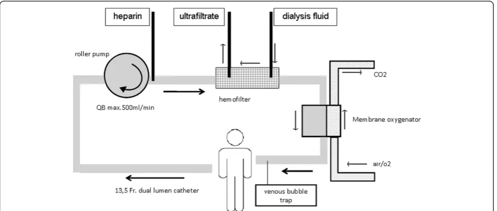
For decarboxylation, a small standard hollow-fiber gas exchanger (D902 Liliput 2 ECMO; Sorin Group Milan, Italy) was applied. This gas exchanger has a surface area of 0.67 m2 and is intended for extracorporeal cir-cuits with a maximum blood flow of 2,300 ml/min. According to the manufacturer’s description, the filter is coated with phosphorylcholine, which should form a phospholipid-like structure and reduce coagulation. The gas exchanger was integrated into the continuous hemodialysis system after the dialysis filter. For the connection, we used standard tubes with a Luer-lock sys-tem. The gas exchanger was attached to the hemodialysis machine with a conventional clamp (Figure 1). Venous and arterial pressures were monitored continuously. Gas flow through the gas exchanger was set to 4 L/min (blood flow, <300 ml/min) or 4 to 6 L/min (blood flow, >300 ml/ min) with a FiO2of 0.21. Only in cases in which oxygen-ation by the ventilator was critical, the FiO2 was varied (0.5 to 1.0 FiO2), in the hope of counteracting systemic hypoxia. For reasons of simplicity, we called this setting “lung-assisting renal replacement system”or“LARRS.”
The LARRS system was tested beforehand in“dummy”
experiments, yielding no detectable changes in venous, arterial, and transmembrane pressures and confirming the full function of alarms, bubble catcher, and emer-gency shutter. In particular, flushing the system with air by artificial occlusion of all three ventilation tubes of the oxygenator led to prompt emergency standby of the CVVHD circuit, according to security standards in renal-replacement therapy devices. Also when the sys-tem was subsequently applied to patients, the pressure control of the hemodialysis did not show any differences in the presence of the gas exchanger in comparison to conventional renal-replacement therapy.
Blood flow was primarily adjusted to 400 ml/min, but could be adapted to individual needs and circumstances (300 to 500 ml/min). A 13.5-French double-lumen cath-eter was placed into the jugular vein. In two patients, we used a second 13.5-French double-lumen catheter in the femoral vein to allow higher blood flows. In this case, the two lumina of one catheter were linked by a y-adapter to form a single blood line. This allowed in-creasing the blood flow about 30% to 40% in these two patients. The dialysate contained Na, 140 mM; K, 4.0M;
Ca, 1.5 mM; Mg, 0.5 mM; Cl, 113 mM; HCO3, 35.0 mM;
and D-Glucose, 9.0 g/L. Anticoagulation was performed with systemic heparin, and doses were prescribed
Forsteret al. Critical Care2013,17:R154 Page 2 of 11
targeting a PTT of 60 seconds or an activated clotting time (ACT), which was measured in bedside assays of 160 to 200 seconds. To prevent cooling of the patient, we used a tube heating system (Fresenius, Germany), which was set to 37°C.
Patient inclusion
Patients were treated with the hollow-fiber gas exchanger in the renal circuit according to the individual decision of the treating physician, based on the patient’s characte-ristics and needs. Inclusion criteria were primarily the need for renal-replacement and mechanical-ventilation therapy with concomitant hypercapnic respiratory acid-osis (pH <7.25). The treatment protocol was approved by the local Ethics Committee of the University of Erlangen-Nuremberg, Germany. In all cases, written in-formed consent was obtained from a legal guardian of the patient before application of the gas-exchange filter was started.
All patients had a central venous catheter in addition to the double-lumen catheters used for RRT, a urinary catheter (unless they were anuric), and an invasive blood-pressure measurement. Heart rate, blood pressure, SaO2, and temperature were monitored continuously; di-uresis was measured hourly, and arterial blood gases were measured in variable intervals during the whole stay at the intensive care unit. Norepinephrine infusions were applied in parallel to fluid administration to main-tain a mean arterial pressure of 65 mm Hg.
Statistics
Because of the small number of patients, most results were presented for each case in absolute values or by
calculating differences between baseline and 24 hours after commencing treatment. Means are expressed as mean ± SD of the mean.
Results
Patient characteristics
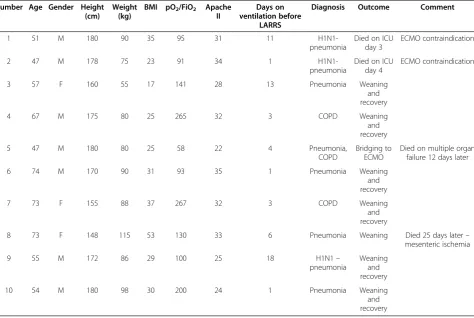
We treated 10 patients with the hollow-filter gas exchan-ger (LARRS) between November 2009 and January 2011. All patients were already undergoing CVVHD because of acute renal failure and oliguria when the CO2hollow filter was applied. At inclusion, the arterial pH varied from 7.07 to 7.24; patient 7 was included to prevent pro-gressive respiratory failure and exhaustion due to a still-compensated respiratory acidosis (HCO3, 31 mM; pCO2, 55 mm Hg; pH 7.37). The patients’ baseline characteris-tics are summarized in Table 1. Eight patients had community-acquired pneumonia; the other two patients had acute infectious exacerbation of COPD. Two pa-tients (1 and 2) fulfilled ARDS criteria, but had contrain-dications against ECMO therapy. One patient (5) with severe ARDS was treated briefly for bridging until ECMO therapy was installed. Although this patient had preexisting high bicarbonate values (37 mm Hg) because of long-term adaptation to his COPD, this patient deve-loped progressive respiratory acidosis (pH 7.34; pCO2, 88 mm Hg) owing to his severe respiratory failure and increasing difficulties in ventilation support. The mean APACHE II score was 29.6 (APACHE II, 22 to 35), with a mean predicted mortality of 72% (40% to 82%).
CO2elimination in the CRRT circuit
[image:3.595.57.542.89.295.2]was well tolerated in all cases, and no change of function of the CVVHD device and the renal-replacement filter was detectable or necessary. Blood-flow rates from 250 ml to 500 ml could be achieved in all patients (mean, 378 ml/min at 24 hours). In particular, intraluminal
[image:4.595.62.537.111.429.2]pressures in the system were not altered. All patients tolerated the intervention well, and no complications occurred during the therapy. Mean heparin doses were 815 (t= 0 hours) and 1,116 IU/h (t= 24 hours), resulting in a mean PTT of 43 (t= 0 hours) and 72 (t= 24 hours)
Table 1 Baseline patients’characteristics and outcomes of the 10 patients treated with the hollow-fiber gas-exchange device included in the renal-replacement circuit
Number Age Gender Height (cm)
Weight (kg)
BMI pO2/FiO2 Apache
II
Days on ventilation before
LARRS
Diagnosis Outcome Comment
1 51 M 180 90 35 95 31 11
H1N1-pneumonia
Died on ICU day 3
ECMO contraindication
2 47 M 178 75 23 91 34 1
H1N1-pneumonia
Died on ICU day 4
ECMO contraindication
3 57 F 160 55 17 141 28 13 Pneumonia Weaning
and recovery
4 67 M 175 80 25 265 32 3 COPD Weaning
and recovery
5 47 M 180 80 25 58 22 4 Pneumonia,
COPD
Bridging to ECMO
Died on multiple organ failure 12 days later
6 74 M 170 90 31 93 35 1 Pneumonia Weaning
and recovery
7 73 F 155 88 37 267 32 3 COPD Weaning
and recovery
8 73 F 148 115 53 130 33 6 Pneumonia Weaning Died 25 days later–
mesenteric ischemia
9 55 M 172 86 29 100 25 18 H1N1–
pneumonia
Weaning and recovery
10 54 M 180 98 30 200 24 1 Pneumonia Weaning
and recovery
Table 2 Treatment modalities for low-flow CO2removal integrated into the RRT circuit
No. Double-lumen catheter
Blood flow (ml/ min)
Dialysate flow (L/h)
Ultrafiltration (ml/h)
Volume (ml)
Gas flow (L/min)
Oxy FiO2
(vol. %)
Treatment time (hours)
t= 4 h t= 24 h t= 4 h t= 24 h t= 4 h t= 24 h t= 24 h
1 1 250 300 4.0 4.0 100 200 −645 6 1.00 30
2 1 330 400 2.0 2.0 200 0 −705 4 1.00 84
3 1 400 400 2.0 2.0 0 150 605 4 0.50 45
4 1 400 400 2.0 2.0 0 100 1,715 6 0.40 215
5 2 300a n.a. 2.0a n.a. 100a n.a. n.a. 6 0.21 2.5
6 1 500 450 3.0 3.0 0 100 1,340 6 0.21 191
7 1 320 250 2.0 2.0 100 150 −3,345 4 0.21 83
8 2 400 450 2.0 1.5 40 100 −2,055 6 0.21 71
9 1 400 500 2.0 2.0 0 0 180 6 0.21 92
10 1 250 250 2.0 2.0 50 0 2,945 4 0.21 134
Patient 5 was switched to ECMO after 2.5 hours of treatment and was therefore not applicable att= 4 hours andt= 24 hours.at = 2.5 hours. n.a., not applicable.
Forsteret al. Critical Care2013,17:R154 Page 4 of 11
[image:4.595.58.536.549.725.2]seconds. Mean activated clotting time was 177 (t= 0 hours) and 213 seconds (t= 24 hours) in bedside tests (see Additional file 1: Table S1). Two episodes of clotting were observed. In one case, the renal filter clotted after 23 hours of use and had to be exchanged. In a second case, the hollow-fiber gas exchanger clotted, which led to a rapid decrease of the arterial pH and increase of pCO2in the extracorporeal system and activated pressure alarms of the dialysis machine. In this case, the hollow-fiber gas exchanger was exchanged immediately, which quickly re-stored CO2removal and pH compensation.
Overall, in-device blood gas analysis directly after the hollow-fiber gas exchanger showed a mean reduction of
extracorporeal pCO2 of 39 mm Hg (63 mm Hg before
gas-exchanger to 24 mm Hg after gas-exchanger filter; see Additional file 2: Figure S1), resulting in a mean in-crease of pH of 0.31 (pH 7.28 before filter and 7.59 after filter, mean blood flow of 377 ml/min). The pO2in the extracorporeal system increased from 41 mm Hg (before filter) to 122 mm Hg (after filter), but no change oc-curred in the patients’systemic pO2. (Additional file 2: Figure S1).
Blood-gas changes during the course of therapy
Implementation of the hollow-fiber gas exchanger
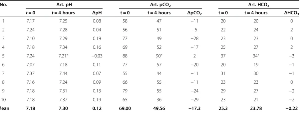
re-duced the average systemic arterial pCO2 by 17.3 mm
Hg or 28.1% in about 4 hours (Table 3). The average
pCO2 could then be kept constant during the next 24
hours. The pH concomitantly increased by 0.12 (0.04 to 0.19) in the first 4 hours, remaining approximately con-stant over the next 24 hours. No change was seen in ar-terial bicarbonate concentrations, presumably due to continuous dialysis against bicarbonate-containing di-alysate. Despite an FiO2of 1.0 in the gas flow to the
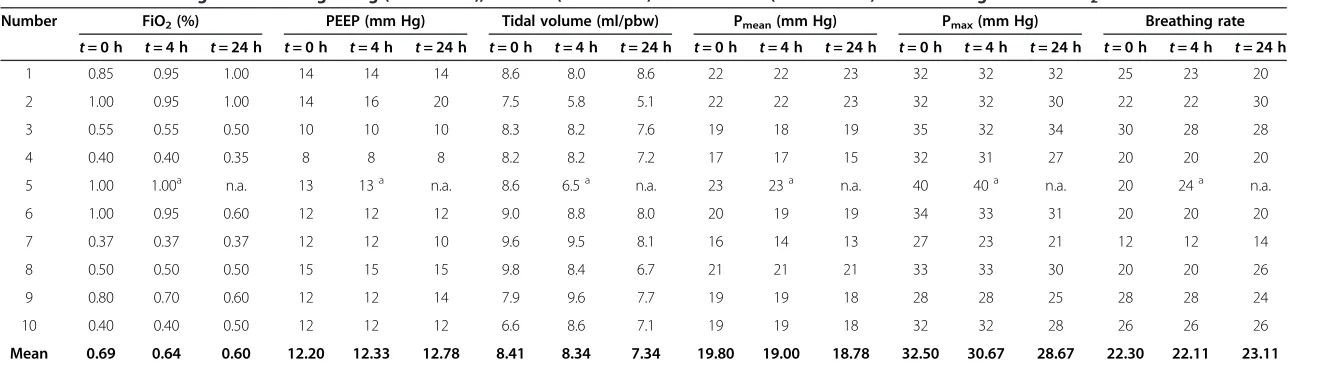
gas-exchange device and a concomitant increase in the extracorporeal pO2 in the first two patients, no change occurred in their arterial pO2, which is consistent with the dominant role of pulmonary gas exchange for arter-ial pO2. The absence of a measurable effect on oxygen-ation can most likely be attributed to the low blood flow and thus to the low amount of oxygen provided for sys-temic circulation. We therefore subsequently reduced the FiO2at the membrane oxygenator to 0.21 to avoid potential side effects or counterregulatory vascular ef-fects in the pulmonary circulation arising from possibly increased oxygen tensions inside the pulmonary artery and pulmonary capillary bed. Despite the improvements in CO2 elimination, ventilator settings remained un-changed or were slightly deescalated. In particular, att=
24 hours, mean Pmax could be reduced from 32.50 to
28.67 mm Hg att= 24 hours. Mean tidal volumes were
slightly decreased from 8.41 to 7.34 ml PBW, eventually still not achieving 6 ml/kg PBW (Table 4).
Hemodynamic stability
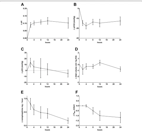
[image:5.595.61.540.535.716.2]In parallel to pH correction, a marked stabilization of hemodynamics was observed (Figure 2). When the hollow-fiber gas-exchanger CVVHD system was started, nine of 10 patients received norepinephrine therapy, five of these in doses between 0.6 and 5.0 mg/h (0.13 to 0.93 μg/kg/min). In four of these five patients with higher doses, a marked hemodynamic stabilization with de-creased norepinephrine doses (average 65% reduction) could be achieved during the therapy (Table 5). In the remaining hemodynamically unstable patient (number 1), acidosis could not be corrected, and no hemodynamic stabilization occurred. In terms of volume control, patients were either ultrafiltrated because of volume
Table 3 Changes in systemic arterial pH, pCO2, bicarbonate (HCO3) from the beginning (t= 0 hours), 4 hours (t= 4 hours), and 24 hours (t= 24 hours) after starting low-flow CO2removal
No. Art. pH Art. pCO2 Art. HCO3
t= 0 t= 4 hours ΔpH t = 0 t = 4 hours ΔpCO2 t = 0 t = 4 hours ΔHCO3
1 7.17 7.25 0.08 58 47 −11 20 20 0
2 7.24 7.28 0.04 56 51 −5 22 24 2
3 7.10 7.29 0.19 77 49 −28 23 23 0
4 7.18 7.34 0.16 69 52 −17 25 27 2
5 7.24 7.21a −0.03 88 90a 2 37 34a −3
6 7.07 7.18 0.11 77 57 −20 20 19 −1
7 7.37 7.44 0.07 55 44 −11 31 30 −1
8 7.16 7.24 0.09 66 55 −11 23 23 0
9 7.18 7.31 0.13 79 55 −24 29 27 −2
10 7.18 7.37 0.19 65 36 −29 23 21 −2
Mean 7.18 7.30 0.12 69.00 49.56 −17.3 25.3 23.78 −0.22
Table 4 Ventilator settings from the beginning (t= 0 hours), 4 hours (t= 4 hours) and 24 hours (t= 24 hours) after starting low-flow CO2removal
Number FiO2(%) PEEP (mm Hg) Tidal volume (ml/pbw) Pmean(mm Hg) Pmax(mm Hg) Breathing rate
t= 0 h t= 4 h t= 24 h t= 0 h t= 4 h t= 24 h t= 0 h t= 4 h t= 24 h t= 0 h t= 4 h t= 24 h t= 0 h t= 4 h t= 24 h t= 0 h t= 4 h t= 24 h
1 0.85 0.95 1.00 14 14 14 8.6 8.0 8.6 22 22 23 32 32 32 25 23 20
2 1.00 0.95 1.00 14 16 20 7.5 5.8 5.1 22 22 23 32 32 30 22 22 30
3 0.55 0.55 0.50 10 10 10 8.3 8.2 7.6 19 18 19 35 32 34 30 28 28
4 0.40 0.40 0.35 8 8 8 8.2 8.2 7.2 17 17 15 32 31 27 20 20 20
5 1.00 1.00a n.a. 13 13a n.a. 8.6 6.5a n.a. 23 23a n.a. 40 40a n.a. 20 24a n.a.
6 1.00 0.95 0.60 12 12 12 9.0 8.8 8.0 20 19 19 34 33 31 20 20 20
7 0.37 0.37 0.37 12 12 10 9.6 9.5 8.1 16 14 13 27 23 21 12 12 14
8 0.50 0.50 0.50 15 15 15 9.8 8.4 6.7 21 21 21 33 33 30 20 20 26
9 0.80 0.70 0.60 12 12 14 7.9 9.6 7.7 19 19 18 28 28 25 28 28 24
10 0.40 0.40 0.50 12 12 12 6.6 8.6 7.1 19 19 18 32 32 28 26 26 26
Mean 0.69 0.64 0.60 12.20 12.33 12.78 8.41 8.34 7.34 19.80 19.00 18.78 32.50 30.67 28.67 22.30 22.11 23.11
n.a., not applicable. Patient 5 was switched to ECMO after 2.5 hours of treatment and was therefore excluded from calculation of means att= 4 hours and t= 24 hours.a
t = 2.5 hours.
Forster
et
al.
Critical
Care
2013,
17
:R154
Page
6
o
f
1
1
http://ccfo
rum.com/con
overload in lung failure in the first 24 hours (four pa-tients, -645 to−3,345 ml/24 hours) or received volume replacement as part of hypertensive shock therapy (six patients, 180 to 2,945 ml/24 hours). No obvious
differ-ence was seen in the response to CO2elimination
be-tween patients in whom either negative or positive fluid balance was achieved. Exemplary time courses of
pH, pCO2, and norepinephrine dose in two patients
who were successfully treated with the low-flow RRT system are shown in Figure 3.
Patients’outcome
No serious adverse events could be attributed to the hollow-fiber gas exchanger or the RRT circuit during
treatment of the patients. Seven of 10 patients were suc-cessfully weaned from the low-flow CO2removal system, improved their pulmonary function, and recovered from critical illness (Table 1).
In the two patients (patients 1 and 2), who had
contra-indications for ECMO therapy, low-flow CO2 removal
[image:7.595.58.537.87.528.2]was applied into the preexisting CVVHD devices as an additional limited therapy option in very severe multiple organ failure: both patients had H1N1 infection with pul-monary ARDS and very low oxygenation indices (95/91). Patient 1 had also a severe liver failure secondary to preexisting alcoholic liver cirrhosis Child C. He died at day 3 after study inclusion. Patient 2 had Hodgkin disease and was undergoing chemotherapy. He had acquired Figure 2Clinical effects of low-flow CO2removal 4 and 24 hours after commencement of treatment.Graphs illustrate mean changes of
H1N1 and concomitantAspergillus fumigatuspneumonia. We eventually limited intensive care support because of the overall poor prognosis and in line with the patient’s provision. He died at day 4 after commencing intensive care therapy. Patient 5, with ARDS due to pneumonia, was bridged to ECMO therapy for only 2.5 hours, before ECMO cannulas were applied and an ECMO device was available. He died 12 days later of multiorgan failure des-pite continued ECMO therapy.
Discussion
For the first time we here report the use of a low-flow hollow-fiber gas exchanger implemented in a CRRT cir-cuit in a small series of critically ill patients. The simple device proved to be efficient in terms of CO2elimination, was well tolerated, and did not lead to adverse events. Concomitant renal-replacement therapy was in no way compromised, and alarm functions of the CRRT system ensured safety control for the gas-exchange device.
The concept of permissive hypercapnia has been de-veloped to reduce baro- and volutrauma during
ventila-tion. Increased CO2 levels have been shown to be
associated with some potentially beneficial (for example, antiinflammatory effects), but the resulting acidosis also induces hemodynamic instability and potential cellular adverse effects [4-7]. In our case series in eight of 10 pa-tients, application of the gas-exchange filter led to a rapid, partial, or complete correction of the pH and a significant reduction of the pCO2within 4 hours. Ranges
of CO2 reduction and pH correction were overall
com-parable to results obtained in the previous study of Terragni et al. [24] using a standalone low-flow CO2 -removal system. In five of six hemodynamically unstable patients requiring higher doses of norepinephrine
(>0.5 mg/h/>0.1 μg/kg/min), pH correction was in line with a marked reduction of vasopressor needs and an improved hemodynamic stability. Norepinephrine doses could be reduced to approximately one half after 6 to 8 hours on average; in three patients, vasopressors could even be stopped. Hemodynamic stability correlated with pH correction by enhanced CO2elimination.
In the CRRT circuit used, blood flow was limited to 500 ml/min at maximum, provided that catheter flow was optimal. We here used a high ratio of blood to gas
flow to achieve maximum CO2 elimination, although
lower blood/gas-flow ratios would be potentially effect-ive as well. Achieved CO2removal allowed correction of acidosis, but we did not find a significant contribution to the systemic oxygenation of the patients even when higher FiO2 together with high gas/blood-flow ratios were applied. Thus, additional systemic oxygenation can-not be achieved with this low-flow device and is reserved for extracorporeal lung replacement therapies with higher flow rates, as in ECMO or PECLA therapy.
[image:8.595.59.539.111.291.2]As it was a pilot, nonrandomized proof-of-concept study, our study has several inherent limitations. First, implementation of the device was one of multiple inter-ventions in the severely ill patients under investigation. Treatment included volume administration, antibiotic treatment, and further co-medication to treat the under-lying disease, which was sepsis in eight of 10 cases. Treatment of acidosis was not confined to CO2 elimin-ation by the respirator and the hollow-fiber gas exchan-ger, but also included bicarbonate filtrate substitution and proton dialysis during the course of renal-replacement therapy. However, the time course of blood pH and pCO2in individual patients with immediate re-sponses after the implementation of the hollow-fiber gas
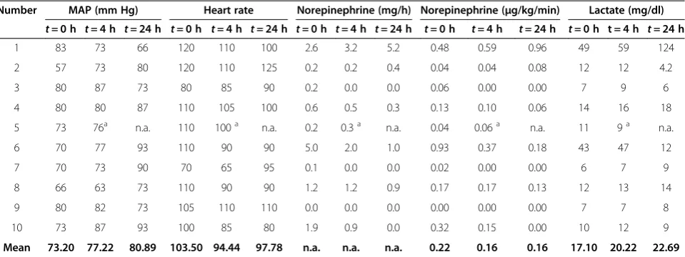
Table 5 Hemodynamic parameters from the beginning (t= 0 h), 4 hours (t= 4 h), and 24 hours (t= 24 h) after starting low-flow CO2removal
Number MAP (mm Hg) Heart rate Norepinephrine (mg/h) Norepinephrine (μg/kg/min) Lactate (mg/dl)
t= 0 h t= 4 h t= 24 h t= 0 h t= 4 h t= 24 h t= 0 h t= 4 h t= 24 h t= 0 h t= 4 h t= 24 h t= 0 h t = 4 h t= 24 h
1 83 73 66 120 110 100 2.6 3.2 5.2 0.48 0.59 0.96 49 59 124
2 57 73 80 120 110 125 0.2 0.2 0.4 0.04 0.04 0.08 12 12 4.2
3 80 87 73 80 85 90 0.2 0.0 0.0 0.06 0.00 0.00 7 9 6
4 80 80 87 110 105 100 0.6 0.5 0.3 0.13 0.10 0.06 14 16 18
5 73 76a n.a. 110 100a n.a. 0.2 0.3a n.a. 0.04 0.06a n.a. 11 9a n.a.
6 70 77 93 110 90 90 5.0 2.0 1.0 0.93 0.37 0.18 43 47 12
7 70 73 90 70 65 95 0.1 0.0 0.0 0.02 0.00 0.00 6 7 9
8 66 63 73 110 90 90 1.2 1.2 0.9 0.17 0.17 0.13 12 13 14
9 80 82 73 105 110 110 0.0 0.0 0.0 0.00 0.00 0.00 7 7 8
10 73 87 93 100 85 80 1.9 0.9 0.0 0.32 0.15 0.00 10 12 9
Mean 73.20 77.22 80.89 103.50 94.44 97.78 n.a. n.a. n.a. 0.22 0.16 0.16 17.10 20.22 22.69
n.a., not applicable. Patient 5 was switched to ECMO after 2.5 hours of treatment and was therefore excluded from calculation of means att= 4 hours and t = 24 hours. a
t = 2.5 hours.
Forsteret al. Critical Care2013,17:R154 Page 8 of 11
exchanger clearly argue for a causal impact of the device. Moreover, metabolic acidosis was already balanced by
bi-carbonate dialysis, when low-flow CO2 removal was
started, and bicarbonate levels remained more or less stable during renal-replacement therapy and therefore most likely did not contribute to the observed effects. Second, we did not apply a standardized ventilation protocol, which would have allowed testing for effects of the intervention on ventilatory requirements or achiev-ing low tidal volumes of 6 ml/kg PBW or less. However, in our clinical setting, the hollow-fiber gas exchanger in
a low-flow circuit CO2at least compensated for respira-tory acidosis and avoided further increases in ventilation settings or even led to slight reductions of peak pressure despite ongoing respiratory failure. Statistically, the over-all survival in this smover-all series was much higher than predicted by the calculated APACHE II scores of the pa-tients, but a sample size of 10 patients certainly does not allow drawing any conclusions in respect to outcomes.
[image:9.595.59.536.90.558.2]Despite this principal limitation of the hollow-fiber gas exchanger inserted into an RRT circuit, the system may have significant advantages in settings where the control Figure 3Exemplary time courses for clinical effects of low-flow CO2removal in two patients.Patient 10 had acute pneumonia(A,B,C);
of hypercapnia is desirable, including less-severe ARDS, when ECMO therapy is either still not indicated or simply not available. The advantages of applying a hollow-fiber gas exchanger in a CRRT circuit include its simplicity and its potential applicability in nonspecialized centers, which are experienced in renal-replacement therapy, as well as the fact that no additional catheter placements are needed. In contrast to many ECMO devices, available RRT ma-chines are more secure and are equipped with distinct alarm systems, which allow emergency shut-off due to air or clots in the blood circuits. Regional anticoagulation with citrate was not performed in conjunction with the described setting, because of the necessity for increased blood flows, but regional anticoagulation protocols could possibly be developed, because a hemofilter allowing clearance of calcium-citrate complexes is included in the system, which is not the case in systems with isolated hollow-fiber gas exchangers.
Conclusions
Implementation of a holfiber gas exchanger in a low-flow CRRT circuit was feasible and safe and let to a signifi-cant CO2 removal and rapid correction of arterial pH in critically ill patients with acute renal and respiratory failure, with a positive impact on hemodynamic stability. Integra-tion of a hollow-fiber gas exchanger could thus be poten-tially an additive tool in the armamentarium of treatment modalities in patients with multiorgan failure. To this end, additional, larger, and controlled studies are certainly needed to assess the impact of low-flow CO2removal on ventilator settings and patient prognosis.
Key messages
Implementation of a hollow-fiber gas exchanger in a
low-flow CRRT circuit was feasible and safe
Low-flow CO2removal in a CRRT circuit
significantly removed CO2and allowed rapid
correction of arterial pH in critically ill patients with acute renal and respiratory failure
Correction of arterial pH contributed to
hemodynamic stabilization of the patient
Low-flow CO2did not significantly contribute to
systemic oxygenation
Low-flow CO2removal complements but did not
substitute ECMO or PECLA therapy, when ECMO/ PECLA was indicated
Additional files
Additional file 1: Table S1.Anticoagulation therapy and clotting values after starting low-flow CO2removal. n.a., not applicable.
Additional file 2: Figure S1.In device pre- and postfilter pH, pCO2and
pO2blood-gas measurements values.
Abbreviations
ACT:Activated clotting time; AKI: Acute kidney injury; ALI: Acute lung injury; ARDS: Acute respiratory distress syndrome; CRRT: Continuous renal-replacement therapy; CVVHD: Continuous veno-venous hemodialysis; ECMO: Extracorporeal membrane oxygenation; LARRS: Lung-assisting renal-replacement system; PBW: Predicted body weight; PECLA: Pumpless extracorporeal lung assist; RRT: Renal-replacement therapy.
Competing interests
Sorin (Milan, Italy) provided the hollow-fiber gas exchangers without cost. The authors declare that they have no competing interests.
Authors’contributions
CF participated in the design of the study, carried out the data acquisition and analysis, and participated in drafting the manuscript. JS primarily established the LARRS system and controlled the technical application. SJ and KUE participated in the design of the study and participated in drafting the manuscript. CW conceived of and designed the study, carried out the coordination, and drafted the manuscript. All authors read and approved the final manuscript.
Received: 22 March 2013 Accepted: 9 July 2013 Published: 24 July 2013
References
1. Malhotra A:Low-tidal-volume ventilation in the acute respiratory distress syndrome.N Engl J Med2007,357:1113–1120.
2. Walkey AJ, Summer R, Ho V, Alkana P:Acute respiratory distress syndrome: epidemiology and management approaches.J Clin Epidemiol
2012,4:159–169.
3. Amato MB, Barbas CS, Medeiros DM, Magaldi RB, Schettino GP, Lorenzi-Filho G, Kairalla RA, Deheinzelin D, Munoz C, Oliveira R, Takagaki TY, Carvalho CR:Effect of a protective-ventilation strategy on mortality in the acute respiratory distress syndrome.N Engl J Med1998,338(6):347–354.
4. O’Croinin D, Ni Chonghaile M, Higgins B, Laffey JG:Bench-to-bedside review: permissive hypercapnia.Crit Care2005,9:51–59.
5. Curley G, Contreras MM, Nichol AD, Higgins BD, Laffey JG:Hypercapnia and acidosis in sepsis: a double-edged sword?Anesthesiology2010,
112:462–472.
6. Ijland MM, Heunks LM, van der Hoeven JG:Bench-to-bedside review: hypercapnic acidosis in lung injury–from“permissive”to”therapeutic.
Crit Care2010,14:237.
7. Ismaiel NM, Henzler D:Effects of hypercapnia and hypercapnic acidosis on attenuation of ventilator-associated lung injury.Minerva Anestesiol
2011,77:723–733.
8. Sidebotham D, McGeorge A, McGuinness S, Edwards M, Willcox T, Beca J: Extracorporeal membrane oxygenation for treating severe cardiac and respiratory failure in adults: part 2, technical considerations.
J Cardiothorac Vasc Anesth2010,24:164–172.
9. Allen S, Holena D, McCunn M, Kohl B, Sarani B:A review of the fundamental principles and evidence base in the use of extracorporeal membrane oxygenation (ECMO) in critically ill adult patients.J Intensive Care Med2011,26:13–26.
10. Peek GJ, Mugford M, Tiruvoipati R, Wilson A, Allen E, Thalanany MM, Hibbert CL, Truesdale A, Clemens F, Cooper N,et al:Efficacy and economic assessment of conventional ventilatory support versus extracorporeal membrane oxygenation for severe adult respiratory failure (CESAR):
a multicentre randomised controlled trial.Lancet2009,374:1351–1363. 11. Bein T, Zimmermann M, Hergeth K, Ramming M, Rupprecht L, Schlitt HJ,
Slutsky AS:Pumpless extracorporeal removal of carbon dioxide combined with ventilation using low tidal volume and high positive end-expiratory pressure in a patient with severe acute respiratory distress syndrome.Anaesthesia2009,64:195–198.
12. Zimmermann M, Bein T, Arlt M, Philipp A, Rupprecht L, Mueller T, Lubnow M, Graf BM, Schlitt HJ:Pumpless extracorporeal interventional lung assist in patients with acute respiratory distress syndrome: a prospective pilot study.Crit Care2009,13:R10.
13. Bein T, Weber-Carstens S, Goldmann A, Muller T, Staudinger T, Brederlau J, Muellenbach R, Dembinski R, Graf BM, Wewalka M,et al:Lower tidal volume strategy (approximately 3 ml/kg) combined with extracorporeal CO2removal versus”conventional”protective ventilation (6 ml/kg) in
Forsteret al. Critical Care2013,17:R154 Page 10 of 11
severe ARDS: the prospective randomized Xtravent study.Intensive Care Med2013,39:847–856.
14. Esteban A, Alia I, Gordo F, de Pablo R, Suarez J, Gonzalez G, Blanco J: Prospective randomized trial comparing pressure-controlled ventilation and volume-controlled ventilation in ARDS, for the Spanish Lung Failure Collaborative Group.Chest2000,117:1690–1696.
15. Liu KD, Matthay MA:Advances in critical care for the nephrologist: acute lung injury/ARDS.Clin J Am Soc Nephrol2008,3:578–586.
16. Gattinoni L, Kolobow T, Tomlinson T, Iapichino G, Samaja M, White D, Pierce J:Low-frequency positive pressure ventilation with extracorporeal carbon dioxide removal (LFPPV-ECCO2R): an experimental study.Anesth Analg1978,57:470–477.
17. Young JD, Dorrington KL, Blake GJ, Ryder WA:Femoral arteriovenous extracorporeal carbon dioxide elimination using low blood flow.Crit Care Med1992,20:805–809.
18. Livigni S, Maio M, Ferretti E, Longobardo A, Potenza R, Rivalta L, Selvaggi P, Vergano M, Bertolini G:Efficacy and safety of a low-flow veno-venous carbon dioxide removal device: results of an experimental study in adult sheep.Crit Care2006,10:R151.
19. Batchinsky AI, Jordan BS, Regn D, Necsoiu C, Federspiel WJ, Morris MJ, Cancio LC:Respiratory dialysis: reduction in dependence on mechanical ventilation by venovenous extracorporeal CO2removal.Crit Care Med 2011,39:1382–1387.
20. Ruberto F, Pugliese F, D’Alio A, Perrella S, D’Auria B, Ianni S, Anile M, Venuta F, Coloni GF, Pietropaoli P:Extracorporeal removal CO2using a
venovenous, low-flow system (Decapsmart) in a lung transplanted patient: a case report.Transplant Proc2009,41:1412–1414. 21. Iacovazzi M, Oreste N, Sardelli P, Barrettara B, Grasso S:Extracorporeal
carbon dioxide removal for additional pulmonary resection after pneumonectomy.Minerva Anestesiol2012,78:381–384.
22. Moscatelli A, Ottonello G, Nahum L, Lampugnani E, Puncuh F, Simonini A, Tumolo M, Tuo P:Noninvasive ventilation and low-flow veno-venous extracorporeal carbon dioxide removal as a bridge to lung
transplantation in a child with refractory hypercapnic respiratory failure due to bronchiolitis obliterans.Pediatr Crit Care Med2010,11:e8–e12. 23. Gramaticopolo S, Chronopoulos A, Piccinni P, Nalesso F, Brendolan A,
Zanella M, Cruz DN, Ronco C:Extracorporeal CO2removal: a way to
achieve ultraprotective mechanical ventilation and lung support: the missing piece of multiple organ support therapy.Contrib Nephrol2010, 165:174–184.
24. Terragni PP, Del Sorbo L, Mascia L, Urbino R, Martin EL, Birocco A, Faggiano C, Quintel M, Gattinoni L, Ranieri VM:Tidal volume lower than 6 ml/kg enhances lung protection: role of extracorporeal carbon dioxide removal.Anesthesiology2009,111:826–835.
doi:10.1186/cc12833
Cite this article as:Forsteret al.:Low-flow CO2removal integrated into a
renal-replacement circuit can reduce acidosis and decrease vasopressor requirements.Critical Care201317:R154.
Submit your next manuscript to BioMed Central and take full advantage of:
• Convenient online submission
• Thorough peer review
• No space constraints or color figure charges
• Immediate publication on acceptance
• Inclusion in PubMed, CAS, Scopus and Google Scholar
• Research which is freely available for redistribution