http://www.scirp.org/journal/jbm ISSN Online: 2327-509X ISSN Print: 2327-5081
Point-of-Care Testing Using Three Dimensional
Optical Biosensor Based on Microfluidic
Technology
Chunxiu Liu
*, Haoyuan Cai, Jian Jia, Tianyang Cao, Tong Li, Tianjun Ma, Chang Liu
*State Key Laboratory of Transducer Technology, Institute of Electronics, Chinese Academy of Sciences, Beijing, China
Abstract
We have presented a three dimensional optical protein chip that fulfills the demand-ing for point-of-care diagnostics in terms of ease-of-use (one step assay), miniaturi-zation (5 μl). The artful combination of magnetic nanoparticles on chip and total in-ternal reflection imaging (TIRI) technology permits the sensitive and rapid detection of hs-CRP (high-sensitivity C-reactive protein). The whole test was complete within 10 min using “all in one step” assay with a limit of detection of 0.1 ng/mL hs-CRP. The measuring range for hs-CRP could be extended to 10 ng/mL. The chip can also be used to detect more parameters in blood samples.
Keywords
Point-of-Care Testing (POCT), Three Dimensional Optical Chip, hs-CRP
1. Introduction
This POCT (point-of-care testing) has the advantages of simple equipment operation, accurate and reliable results, those novel technologies and devices to all visitors with a new concept, namely “A number of measurement results can be obtained in place close to the patients in a very short period of time”. POCT is a new field of medical develop-ment, achieving the rapid progress with the development of high technology and com-prehensive application, conforming to the current, fast-paced work of the people [1]- [5]. The patient can obtain timely diagnosis and treatment.
The development of microfluidic technology provides a new idea and platform for the rapid and high-throughput detection of protein, and greatly reduces the cost of de-tection. Compared with the traditional analysis method, the microfluidic detection technology has the advantages of high efficiency, fast, less reagent consumption, auto-How to cite this paper: Liu, C.X., Cai,
H.Y., Jia, J., Cao, T.Y., Li, T., Ma, T.J. and Liu, C. (2016) Point-of-Care Testing Using Three Dimensional Optical Biosensor Based on Microfluidic Technology. Journal of Bio- sciences and Medicines, 4, 56-61.
http://dx.doi.org/10.4236/jbm.2016.412009
mation and so on.
Study on POCT microfluidic chip challenged the detection of minimal volume, com-plex pretreatment samples. The precision and efficiency of the real-time diagnostic test and its device is an important change in the field of modern life and health. The micro-fluidic detection and analysis chip is the main technology to make real-time diagnosis accurate and effective [6] [7] [8] [9].
Total internal reflection imaging (TIRI) technology as a branch of optical detection combined with the microfluidic chip can be used for the detection and analysis of a va-riety of substances [10] [11] [12]. The detection is based on COMS camera capture of the reflected light of the chip reaction interface. This work developed the fast POCT detection of low concentration hs-CRP protein by three dimensional microfluidic chip and total internal reflection imaging (TIRI).
2. Materials and Methods
2.1. Experimental Materials
A pair of commercially available sandwich anti-CRP monoclonal antibodies (mAb) was purchased from Abzymo Biosciences Co. (Beijing, China). The tracer monoclonal an-tibody was functionalized with biotin. The Carboxyl Streptavidin Magnetic Particles (500 nm diameter, 0.5% w/v) were purchased from ACME microspheres Co. (Indian-apolis, IN, USA). Polylysine (PLL, P4832, Sigma), BSA and other reagents were pur-chased from Sigma-Aldrich Inc. (St. Louis, MO, USA). The buffer solutions were pre-pared with ultra-pure water. Concentration values stated in this study are used by serial dilutions in PBS buffer.
2.2. Experimental Equipment
The Poly-Pico System (PolyPico., Ireland) device was used to precisely and uniformly dispense pL-sized arrayed capture monoclonal antibodies particles. The high precision laser engraving machine DPSS (Suzhou, China) can be used to cut double sided adhe-sive tape to form the microfluidic channel, and reaction chamber of the chip. The TIRI home made system containing the optics module and the magnetic module was used to take image and collect data.
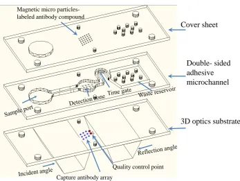
2.3. Fabrication of the 3D Optical Biosensor
The optical chip was composed of a 3D glass bottom board, double-sided adhesive on the micro channel, and cover sheet (Figure 1). The basic structure of the micro channel was cut by the laser cutting machine on the double-sided adhesive, and the double-sided adhesive was pasted on the surface of the 3D glass base plate. The trace mAb and capture mAb were immobilized on the top and bottom of the reaction chamber. The chip can be divided into the sample port, reaction and detection zone, time gate and waste reservoir.
Figure 1. The schematic illustration and assembly of 3D optical TIRI chip.
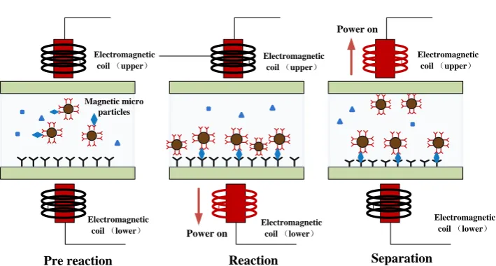
noassay method shown in Figure 2. When the samples were added, the sample rehy-drated the trace mAb and magnetic particle in the detection area and then the antigen action with the composites. The bidirectional magnetic force was open and the capture antibody immobilization in the bottom acted with the Mag-mAb-Ag complex and form magnetic spots at the bottom of the reaction area. The total internal optical signals are subject to magnetic speckle interference, and the signal was inverse correlation with the antigen concentrations. Matlab software was used to assay the obtained magnetic spot imaging.
3. Results
3.1. Optimization on the Reflection Angle of the 3D Chip
One major aim of the assay development was the capture of the clear images that oc-curred during one-step sandwich immunoassay. The incidence angle and reflection an-gle of the 3D optical chip was symmetric in the center of the reaction zone. The inci-dence angle should meet the conditions of total internal reflection imaging according to the refractive index of the material of the 3D chip. The angular range of the reflection angle has important influence on imaging quality. When the reflection angle continues to increase, the image sharpness decreased rapidly with the increasing angle of reflec-tion. The image changed from circles to ellipse when the angle gradually increased and the image distortion aggravated when the angle reached 70 degree angle (Figure 3). It was too small to form a total internal reflection if the reflection angle is less than 45 de-gree.
Cover sheet
Double- sided
adhesive
microchannel
3D optics substrate
Magnetic microparticles-labeled antibody compound
Capture antibody array
Figure 2. Schematic diagram of detection principle of sandwich immunoassay method.
Figure 3. The TIRI image of 3D chip at different reflection angle.
3.2. Detection of hs-CRP
The standard curves with a dilution series of standard CRP samples by PBS buffer were created. The assay achieved an LOD value of 0.1 ng/mL in standard CRP samples. The linear fits to the curves are shown in Figure 4. The sensitivity of the assay is sufficient to distinguish hs-CRP levels during 0.1 - 10 ng/mL (Figure 4). To enable the measure-ment of lower and higher concentrations of CRP, further detection and improvemeasure-ments to the chip system are necessary. The assay required no sample preparation and is therefore easy to handle, which is suitable for POC measurement at the patient.
4. Conclusion
A new, highly sensitive assay to quantify CRP via a POCT device was developed. The device fulfills all the important characteristics of a POCT application. Our assay has a
Electromagnetic coil (upper)
Electromagnetic
coil (lower) Power on
Magnetic micro particles
Reaction Separation Pre reaction
Electromagnetic coil (upper)
Electromagnetic coil (lower)
Power on
Electromagnetic coil (upper)
Electromagnetic coil (lower)
Figure 4. Standard curves of CRP with the sandwich assay using paired monoclonal antibody, number of concentra-tions: 3, number of repeats: 3.
volume (20 µL). There is no need for sample pretreatment because the device uses ready to-use 3D optical chip. Furthermore, it is simple and safe to handle. Furthermore, the assay operates in the necessary sensitivity range (0.1 - 10 ng/mL) with standard samples.
Acknowledgements
This work is supported by the NSFC (No.60801032, 61271147, 61372052), the major national scientific research plan (2011CB933202), the project of CAS (KFJ-EW-STS- 046) and the high resolution major projects (06-Y30B04-9002-13/15).
References
[1] Natalia, L.B., Jorge, D.G. and Johann, F.O. (2016) The Future Point-of-Care Detection of Disease and Its Data Capture and Handling. Anal Bioanal Chem, 408, 2827-2837.
http://dx.doi.org/10.1007/s00216-015-9249-2
[2] Jung, W., Han, J., Choi, J.W. and Ahn, C.H. (2015) Point-of-Care Testing (POCT) Diagnos-tic Systems Using Microfluidic Lab-on-a-Chip Technologies. Microelectronic Engineering, 132, 46-57. http://dx.doi.org/10.1016/j.mee.2014.09.024
[3] Ouellette, A.L., Li, J.J., Cooper, D.E., Ricco, A. and Kovacs, G.T.A. (2009) Evolving Point- of-Care Diagnostics Using Up-Converting Phosphor Bioanalytical Systems. Analytical Chemistry, 81, 3216-3221. http://dx.doi.org/10.1021/ac900475u
[4] Luppa, P.B., Müller, C., Schlichtiger, A. and Schlebusch, H. (2011) Point-of-Care Testing (POCT): Current Techniques and Future Perspectives. Trac Trends in Analytical Chemi-stry, 30, 887-898. http://dx.doi.org/10.1016/j.trac.2011.01.019
[5] Yang, Z. and Zhou, D.M. (2006) Cardiac Markers and Their Point-of-Care Testing for Di-agnosis of Acute Myocardial Infarction. Clinical Biochemistry, 39, 771-780.
http://dx.doi.org/10.1016/j.clinbiochem.2006.05.011 100
110 120 130 140
0.1 1 10 100
L
um
ina
nce V
a
lues
[6] Pfäfflin, A. and Schleicher, E. (2009) Inflammation Markers in Point-of-Care Testing (POCT). Analytical & Bioanalytical Chemistry, 393, 1473-1480.
http://dx.doi.org/10.1007/s00216-008-2561-3
[7] Kiechle, L.F. (2015) Three POCT Molecular Applications: A View into Future Develop-ment, Point of Care. The Journal of Near-Patient Testing & Technology, 14, 95-98.
http://dx.doi.org/10.1097/poc.0000000000000052
[8] Baldini, F., Bolzoni, L., Giannetti, A., et al. (2009) A New Procalcitonin Optical Immuno-sensor for POCT Applications. Analytical and Bioanalytical Chemistry, 393, 1183-1190.
http://dx.doi.org/10.1007/s00216-008-2547-1
[9] Rascher, D., Geerlof, A., Kremmer, E., Krämer, P. and Schmid, M. (2014) Total Internal Reflection (TIRF)-Based Quantification of Procalcitonin for Sepsis Diagnosis—A Point- of-Care Testing Application. Biosensors & Bioelectronics, 59C, 251-258.
http://dx.doi.org/10.1016/j.bios.2014.03.052
[10]Mejri, M.B., Baccar, H., Ktari, T., Aouni, M. and Abdelghani, A. (2011) Detection of E. coli Bacteria Using Impedance Spectroscopy and Surface Plasmon Resonance Imaging Based Biosensor. Sensor Letters, 9, 1-3. http://dx.doi.org/10.1166/sl.2011.1747
[11]Michael, J.O., Victor, H.P., Brueck, S.R.J. and Gabriel, P.L. (2001) A Surface Plasmon Re-sonance Array Biosensor Based on Spectroscopic Imaging. Biosensors & Bioelectronics, 16, 97-108. http://dx.doi.org/10.1016/S0956-5663(00)00137-8
[12]Tan, H.M., Pechprasarn, S., Zhang, J., Pitter, M.C. and Somekh, M.G. (2016) High Resolu-tion Quantitative Angle-Scanning Widefield Surface Plasmon Microscopy. Scientific Re-ports, 6, Article Number: 20195. http://dx.doi.org/10.1038/srep20195
Submit or recommend next manuscript to SCIRP and we will provide best service for you:
Accepting pre-submission inquiries through Email, Facebook, LinkedIn, Twitter, etc. A wide selection of journals (inclusive of 9 subjects, more than 200 journals)
Providing 24-hour high-quality service User-friendly online submission system Fair and swift peer-review system
Efficient typesetting and proofreading procedure
Display of the result of downloads and visits, as well as the number of cited articles Maximum dissemination of your research work