© 2018, IRJET | Impact Factor value: 7.211 | ISO 9001:2008 Certified Journal | Page 2262
RETINAL DISEASE SCREENING THROUGH LOCAL BINARY PATTERNS
1
Abhilash M,
2Sachin Kumar
1Student, M.tech, Dept of ECE ,LDN Branch, Navodaya Institute of Technology, Raichur - 584101.
2Asst.Prof. Dept. of ECE, Navodaya Institute of Technology, Raichur - 584101.
---***---Abstract:
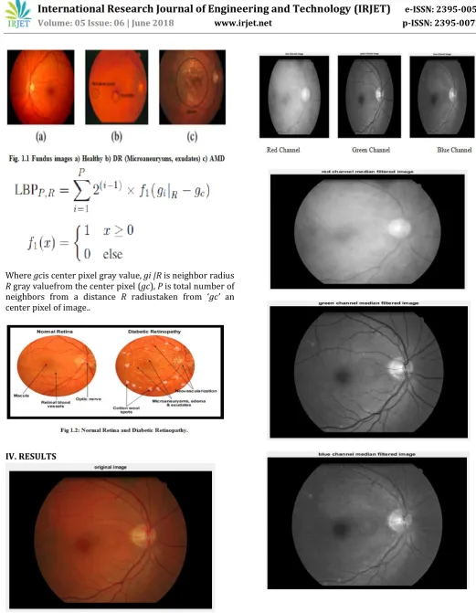
When sugar level (glucose) in the blood fails to regulate the insulin properly in human body, diabetic is occurred. The effect of diabetic on eye causes diabetic retinopathy. Diabetic Retinopathy is one of the complicated diabetes which can cause blindness. It is metabolic and the disordered patients perceive no symptoms until the disease is at late stage. So early detection and proper treatment has to be ensured. To serve this purpose, various automated systems have been designed). A key feature to recognize Diabetic Retinopathy is to detect Microaneurysm in the fundus of the eye. This work investigates discrimination capabilities in the texture of fundus images to differentiate between pathological and healthy images. For this purpose, the performance of Local Binary Patterns (LBP) as a texture descriptor for retinal images has been explored. The goal is to distinguish between diabetic retinopathy (DR) and normal fundus images analyzing the texture of the retina background and avoiding a previous lesion segmentation stage. We propose preprocessing technique such as Contrast Limited Adaptive Histogram Equalization (CLAHE) to enhance the contrast of the input image and we use candidate extractors such as Circular Hough Transform to improve the red lesion detection. Finally the output image was classified as Normal and Diabetic retinopathy (DR). These results suggest that the method presented in this paper is a robust algorithm for describing retina texture and can be useful in a diagnosis aid system for retinal disease screening.
I. INTRODUCTION
The development of a telemedicine system for screening of retinal disease depends on identification of retinal lesions in images which includes fundus. Diabetic retinopathy (DR) is occurred mainly due to reduction in the sugar level. The present survey behind the cause of blindness says that it is due to DR in aging population. DR may be managed using available methods of treatment, which are effective if diagnosed early. DR is metabolic and disordered patients will not find any symptoms till the disease occurs at severe phase. Thus early revealing and better treatment has to be taken care.
M. Pietikinenet.all proposed local binary pattern as a texture operator- Multiresolution grayscale with classification of texture using rotation invariant method [3]. They gave a presentation in a general aspect which involves the realization of gray-scale with LBP feature using rotation-invariant method including angular space quantization, resolution and this method combines different operators for analysis of multi-resolution.This approach is found to be very use full for variations which includes gray scale. The grayscale transformations are also realized using look up table. Better results were obtained from the experiments using different scenarios such as classifier is used at one-rotation angle for training and tested with different rotation angle
II. BACKGROUND
© 2018, IRJET | Impact Factor value: 7.211 | ISO 9001:2008 Certified Journal | Page 2263
along with thresholding and mathematical morphology operation. After masking the optic disc and blood vessel, the LBP and VAR are applied to the image which extracts the texture surface of the image. Different statistical information is extracted from these histograms to use it as features in the classification stage. The calculated statistical values are mean, standard deviation, median, entropy, skewness and kurtosis. Finally the output image was classified as Normal and Diabetic retinopathy (DR)..
III. DESIGN&ALGORITHMS
Software requirements
MATLAB 8.3 Version R2014a Image processing Toolbox
mage Processing Toolbox
Image processing device box permits carrying out image improvement, deblurring of image, characteristic identification, decreasing of noise, image segmentation, feature extraction, as well as classification of image.
Features of Matlab
Interactive background meant for aim investigation as well as resolving the difficulty. MATLAB is a sophisticated language intended for creating, calculating as well as building up a purpose. It contains numerical tasks such as figures, calculus, sorting out, developments, mathematical integration, as well as working out equations. Graphics integrated intended for visualization. Intended for generating traditional plot integrated equipments is accessible. Troubles as well as way outs are given in well-known numerical symbol
The noise present in the image is removed by median filterBetter Detection of Blood vessels and Optic diskThe patient retinal images can be classified as normal or diseasedThe texture surface of the image is extracted.
ALGORITHM.
Cubic Convolution Interpolation (CCI)
CCI finds the grey level value for average of 16 closest pixels to the specific input coordinates, then gives that value to output coordinates. At first 4 one dimension convolutions are done in horizontal direction and one more in vertical direction. For one dimension CCI, the grid points required to assess the interpolation function is 4. For Bi cubic Interpolation (CCI in two dimensions), the grid points are required for evaluation of inter polation function is sixteen. The grid points required for one-dimension and two-one-dimension CCI are shown in below figure
© 2018, IRJET | Impact Factor value: 7.211 | ISO 9001:2008 Certified Journal | Page 2264
Where gcis center pixel gray value, gi |R is neighbor radius
R gray valuefrom the center pixel (gc), P is total number of neighbors from a distance R radiustaken from ‘gc’ an center pixel of image..
[image:3.595.30.552.46.718.2]IV. RESULTS
[image:3.595.39.277.402.505.2]© 2018, IRJET | Impact Factor value: 7.211 | ISO 9001:2008 Certified Journal | Page 2265
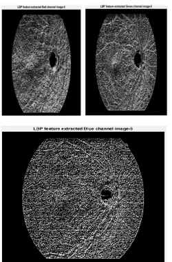
Fig : LBP feature for R, G and B Components
Fig : VAR feature for R, G, B components
Results for classification
[image:4.595.36.287.92.473.2]Depending on the feature set values using SVM classifier the image is classified as DR or Healthy. A pop up is displayed as fallows.
[image:4.595.37.295.516.715.2]© 2018, IRJET | Impact Factor value: 7.211 | ISO 9001:2008 Certified Journal | Page 2266
V. CONCLUSION
An approach is made for DR diagnosis based on texture features on fundus images were introduced for differentiate healthy and pathological images. Compared with the other texture description the routine accuracy of the LBP is better in the screening of a retinal disease.
The classification of different stages of a patient such as severe, moderate and mild helps the patients to the reduction of cost in diagnosis and results in a prevention of better health
In the future work experiments with more images were carried out and tested by considering various phenomenon’s such as exclusion of tessellated images and including the calculation of some more statistical parameters.
VI. REFERENCES
[1] World Health Organization (WHO), “Universal eye health: a global action plan 2014-2019,” 2013.
[2] World Health Organization (WHO), “Action plan for the prevention of avoidable blindness and visual impairment 2009-2013,” 2010.
[3] T. Ojala, M. Pietikinen, and T. Menp, “A generalized local binary pattern operator for multiresolution gray scale and rotation invariant texture classification,” in Advances in Pattern Recognition, 2nd International Conference on, 2001, pp. 397–406..
[4] T. Ojala, M. Pietikainen, and T. Maenpaa, “Multiresolution gray-scale and rotation invariant texture classification with local binary patterns,” Pattern Analysis and Machine Intelligence, IEEE Transactions on, vol. 24, no. 7, pp. 971–987, 2002..
[5] T. Ahonen, A. Hadid, and M. Pietikainen, “Face description with local binary patterns: Application to face recognition,” Pattern Analysis and Machine Intelligence, IEEE Transactions on, vol. 28, no. 12, pp. 2037– 2041, 2006.
[6] M. Heikkil, M. Pietikinen, and C. Schmid, “Description of interest regions with local binary patterns,” Pattern Recognition, vol. 42, no. 3, pp. 425 – 436, 2009.
[7] Z. Yang and H. Ai, “Demographic classification with local binary patterns,” in Advances in Biometrics, ser. Lecture Notes in Computer Science, S.-W. Lee and S. Li, Eds., 2007, vol. 4642, pp. 464–473.
[8] L. Kotu, K. Engan, T. Eftestol, L. Woie, S. Orn, and A. Katsaggelos, “Local binary patterns used on cardiac MRI to
classify high and low risk patient groups,” in Signal Processing Conference (EUSIPCO), Proceedings of the 20th European, 2012, pp. 2586–2590.
[9] K. Oppedal, K. Engan, D. Aarsland, M. Beyer, O. B. Tysnes, and T. Eftestol, “Using local binary pattern to classify dementia in MRI,” in Biomedical Imaging (ISBI), 9th IEEE International Symposium on, May 2012, pp. 594– 597.
[10] L. Nanni, A. Lumini, and S. Brahnam, “Local binary patterns variants as texture descriptors for medical image analysis,” Artificial Intelligence in Medicine, vol. 49, no. 2, pp. 117 – 125, 2010.
[11] S. Zabihi, M. Delgir, and H.-R.Pourreza, “Retinal vessel segmentation using color image morphology and local binary patterns,” in Machine Vision and Image Processing (MVIP), 6th Iranian, 2010, pp. 1–5.
[12] S. Dhanushkodi and M. Vasuki, “Diagnosis system for diabetic retinopathy to prevent vision loss,” Applied Medical Informatics, vol. 33, no. 3, pp. 1–11, 2013.
[13] M. Mookiah, U. R. Acharya, R. J. Martis, C. K. Chua, C. Lim, E. Ng, and A. Laude, “Evolutionary algorithm based classifier parameter tuning for automatic diabetic retinopathy grading: A hybrid feature extraction approach,” Knowledge-Based Systems, vol. 39, no. 0, pp. 9 – 22, 2013.
[14] M. M. R. Krishnan and A. Laude, “An integrated diabetic retinopathy index for the diagnosis of retinopathy using digital fundus image features,” Journal of Medical Imaging and Health Informatics, vol. 3, no. 2, pp. 306–313, 2013.
[15] M. Garnier, T. Hurtut, H. Ben Tahar, and F. Cheriet, “Automatic multiresolution age-related macular degeneration detection from fundus images,” in SPIE, Proceedings, vol. 9035, 2014, pp. 903532–903532–7
[16] T. M¨aenp¨ a¨a and M. Pietik¨ainen, “Multi-scale binary patterns for texture analysis,” in Image Analysis, ser. Lecture Notes in Computer Science, J. Bigun and T. Gustavsson, Eds., 2003, vol. 2749, pp. 885–892.
[17] V. Ojansivu and J. Heikkil, “Blur insensitive texture classification using local phase quantization,” in Image and Signal Processing, ser. Lecture Notes in Computer Science, A. Elmoataz, O. Lezoray, F. Nouboud, and D. Mammass, Eds., 2008, vol. 5099, pp. 236–243.
© 2018, IRJET | Impact Factor value: 7.211 | ISO 9001:2008 Certified Journal | Page 2267
for age-related macular degeneration characterization and classification,” Computers in Biology and Medicine, vol. 63, pp. 208 – 218, 2015.
[19] S. Liao, M. Law, and A. Chung, “Dominant local binary patterns for texture classification,” Image Processing, IEEE Transactions on, vol. 18, no. 5, pp. 1107–1118, May 2009.
[20] Y. Zheng, M. H. A. Hijazi, and F. Coenen, “Automated disease / no disease grading of age-related macular degeneration by an image mining approach,” Investigative Ophthalmology & Visual Science, vol. 53, no. 13, pp. 8310– 8, 2012.
BIOGRAPHIES
Mr. Sachin kumar M.Tech (P.hd) Asst prof
Novadaya engineering college , ECE branch
Raichur-584101
Mr. Abhilash M
Pursuing Masters in DCN(Digital communication and networking) Novadaya engineering college , ECE branch