Two different functions for CD44 proteins in
human myelopoiesis.
J Moll, … , H Ponta, P Herrlich
J Clin Invest.
1998;
102(5)
:1024-1034.
https://doi.org/10.1172/JCI2494
.
CD44 is important during myelopoiesis, although the contributions of variant CD44 proteins
are unclear. We show here that in human long-term bone marrow culture antibodies
recognizing a CD44 NH2-terminal epitope (mab 25-32) or a CD44v6 epitope (mab VFF18)
inhibit myelopoiesis. However, mab 25-32 but not mab VFF18 affects myeloid colony
formation. These data suggest that an early precursor cell compartment is the target for the
25-32 antibody, whereas the mab VFF18 targets later stages in myelopoiesis. Since the
bulk of hemopoietic precursor cells are negative for the v6 epitope and only a minor subset
of myeloid cells express the v6 epitope, we have used several human myeloid progenitor
cell lines to unravel the function of different CD44 proteins. These cell lines produce variant
CD44 proteins, predominantly a new variant CD44v4-v10, when stimulated towards
myeloid differentiation. Features that can be acquired by the expression of CD44v4-v10 are
an increased hyaluronate (HA) and a de novo chondroitin sulphate A (CS-A) binding.
Although, the expression of CD44v4-v10 per se is necessary for HA and CS-A binding, the
protein backbone seems to require appropriate glycosylation. HA binding results in
CD44-mediated cellular self-aggregation and adhesion to the stromal cell line MS-5. In summary,
our data suggest that different CD44 proteins are important for at least two different steps in
myelopoiesis.
Research Article
Find the latest version:
J. Clin. Invest.
© The American Society for Clinical Investigation, Inc. 0021-9738/98/09/1024/11 $2.00
Volume 102, Number 5, September 1998, 1024–1034 http://www.jci.org
Two Different Functions for CD44 Proteins in Human Myelopoiesis
J. Moll,* S. Khaldoyanidi,* J.P. Sleeman,* M. Achtnich,‡ I. Preuss,* H. Ponta,* and P. Herrlich*
*Forschungszentrum Karlsruhe, Institut für Genetik, P.O. Box 3640, D-76021 Karlsruhe, Germany; and ‡Klinikum Mannheim,
Wiesbadenerstr. 7-11, D-68305 Mannheim, Germany
Abstract
CD44 is important during myelopoiesis, although the con-tributions of variant CD44 proteins are unclear. We show here that in human long-term bone marrow culture antibod-ies recognizing a CD44 NH2-terminal epitope (mab 25–32)
or a CD44v6 epitope (mab VFF18) inhibit myelopoiesis. However, mab 25–32 but not mab VFF18 affects myeloid colony formation. These data suggest that an early precur-sor cell compartment is the target for the 25–32 antibody, whereas the mab VFF18 targets later stages in myelopoiesis. Since the bulk of hemopoietic precursor cells are negative for the v6 epitope and only a minor subset of myeloid cells express the v6 epitope, we have used several human myeloid progenitor cell lines to unravel the function of different CD44 proteins. These cell lines produce variant CD44 pro-teins, predominantly a new variant CD44v4–v10, when stimulated towards myeloid differentiation. Features that can be acquired by the expression of CD44v4–v10 are an in-creased hyaluronate (HA) and a de novo chondroitin sul-phate A (CS-A) binding. Although, the expression of CD44v4–v10 per se is necessary for HA and CS-A binding, the protein backbone seems to require appropriate glycosy-lation. HA binding results in CD44-mediated cellular self-aggregation and adhesion to the stromal cell line MS-5. In summary, our data suggest that different CD44 proteins are important for at least two different steps in myelopoiesis. (J. Clin. Invest. 1998. 102:1024–1034.) Key words: hyaluronate •
chondroitin sulphate A • CD44 • myelopoiesis • long-term
bone marrow culture
Introduction
The CD44 transmembrane glycoproteins are structurally di-verse in their extracellular domains due to extensive alterna-tive splicing of variant exons (nine in humans [1, 2]). Further diversity is created by differential glycosylation (3, 4). The ex-tracellular domains of CD44 proteins interact with several components in the extracellular matrix including hyaluronate (HA;1 5, 6) and with growth factors such as osteopontin (7) or
fibroblast growth factors (8). Diversity and domain structure
suggest that the CD44 proteins are involved in several func-tions. While the smallest protein containing no variant exon encoded sequences (CD44s; “hemopoietic” or standard iso-form) is expressed in many different cell types including cells of the hemopoietic system, the occurrence of larger splice vari-ants is restricted in time and/or location, suggesting that they exert more specialized functions. Upon activation of lympho-cytes, CD44 levels are increased, and larger splice variants are synthesized transiently (9, 10, 11). In agreement with a role in proliferation control, a Thy-1 promoter-regulated CD44v4–v7 transgene accelerates and enhances lymphocyte activation (12) and bone marrow repopulation (13). This latter result trig-gered our interest in a possible involvement of CD44 proteins in normal hemopoiesis.
The development of mature blood cells is a highly regu-lated process during which undifferentiated pluripotent stem cells develop into different hemopoietic lineages. It is the stro-mal microenvironment consisting of strostro-mal cells and extracel-lular matrix that is thought to regulate the future fate of stem cells and committed progenitors along specific lineages. The interaction between progenitor cells and the microenviron-ment is likely mediated by growth factors/growth factor recep-tors, by cell adhesion molecules and their cell-bound ligands, as well as by components of the extracellular matrix.
In bone marrow, HA and CS-A account for the majority of extracellular matrix constituents (14–16), and a CD44-depen-dent HA binding activity of CD341 hematopoietic progenitors has been shown (17). We explore here the role of different CD44 isoforms during hemopoiesis in humans. Antibody in-terference experiments in long-term bone marrow culture (LTBMC) indicate a role for several CD44 proteins, including variant proteins in the generation of myeloid cells. Leukemic precursor cell lines express CD44 variant proteins at least when they are stimulated to differentiate towards the myeloid lineage. The major CD44 protein appears to be CD44v4–10. We have characterized properties of CD44 proteins on these cell lines. These proteins mediate efficient binding to HA, which in time results in binding to the stromal cell line MS-5. CD44 variants but not standard proteins bind specifically to CS-A, but in contrast to HA, this binding does not mediate cell adhesion.
Methods
Cell culture.The cell lines KG-1 (ATCC CCL 246), K562 (ATCC CCL 243), TF-1 (18), and HL-60 (ATCC CCL240) were grown in Is-cove’s Modified Dulbecco’s Medium (IMDM, GIBCO BRL, Gaithers-burg, MD) supplemented with 10% FCS and 1 mM glutamine. MS-5 cells (19) were kept in a-MEM supplemented with 15% FCS and Glutamax I (GIBCO). To induce myeloid differentiation, cells were The first three authors shared equal contributions.
Address correspondence to Dr. J. Moll, Forschungszentrum Karlsruhe, Institut für Genetik, P.O. Box 3640, D-76021 Karlsruhe, Germany. Phone: 149 7247 82 2634; FAX: 149 7247 82 3354; E-mail: [email protected]
Received for publication 12 December 1997 and accepted in re-vised form 2 July 1998.
treated with 10 ng/ml PMA for various periods of time as stated in the figure legends. HL-60 cells were induced to differentiate towards other lineages by adding to the medium either retinoic acid (1 mM; Sigma Chemical Co., St. Louis, MO) or DMSO (1.25%; Fluka Chemie AG, Buchs, Switzerland). Glycosylation was inhibited by the addition of 5 mg/ml tunicamycin (Sigma). Differentiation of cells was monitored by morphological criteria such as adhesive properties, by their capacity to reduce nitroblue tetrazolium (20) or by the appear-ance of surface antigens such as CD41 as a marker for the megakaryo-cytic lineage.
Isolation of mononuclear cells (MNCs). Human bone marrow was collected from the crista illiaca posterior superior by puncture. MNCs were isolated by density gradient centrifugation. In brief, bone mar-row cells were diluted in PBS, and 5 ml of the cell suspension was lay-ered gently onto 5 ml of 1.077 g/ml Ficoll. Preparations were centri-fuged at room temperature and 400 g for 30 min. MNCs were harvested at the interface and washed twice in IMDM supplemented with 10% FCS.
LTBMC. MNC were diluted to a final concentration of 1.5–2 3 106 cells/ml in LTBMC medium (10% horse serum [Linaris], 10%
FCS, 5 3 1027 M hydrocortisone [Sigma] in IMDM). The cell
suspen-sion was inoculated into 6-well plates and incubated at 338C and 5% CO2. At intervals of 7 d, half of the growth medium containing
non-adherent cells was removed, and fresh medium was added to keep the volume constant. The cells harvested at the time of feeding were counted, and the number of clonogenic cells was determined. To study the effect of antibodies LTBMC assays were performed in the presence of 10 mg/ml of dialyzed mAbs VFF18 or 25–32 (21) as indi-cated in the figure legends.
CFU assay. Cells collected from LTBMC were counted and plated into 24-well plates at a concentration of 2 3 104/ml in methyl
cellulose containing 30% FCS, 1% BSA, 1024 M 2-mercaptoethanol,
2 mM L-glutamine (StemCell Technologies, Vancouver, Canada), and 15% conditioned medium from 5,637 (ATCC HTBg) cells as a source of cytokines. The plates were incubated at 378C in a humidified atmo-sphere of 5% CO2 for 10 d before scoring.
Isolation of polyA1 mRNA, RT-PCR, and exon run-on analysis. PolyA1 mRNA was isolated from cells according to standard proce-dures (22). RT-PCR and CD44 exon run-on analysis were performed as described (23). This method allows the identification of the exon composition of expressed CD44 isoforms. Different exon specific 59 primers, together with a constant 39 primer are used to identify the presence of variant exons. In brief, cDNA was synthesized from 0.5
mg polyA1 RNA and a PCR preamplification step was used to obtain all possible CD44 PCR products using primers (59 primer C12A and 39 primer C13; 23) in the constant region. PCR conditions: 25 cycles of 60 s at 948C, 60 s at 568C, and 90 s at 728C in a volume of 50 ml. Exon-specific amplifications were performed using the same condi-tions but 1 ml of the preamplification as a template and for each CD44v exon a specific 59 primer (C13, pv2–pv10) and a constant 39 primer (C2A; 23). The PCR amplification products were resolved on a 1% agarose gel.
Flow cytometry. Antibodies mAb IM7.8.1 (ATCC TIB235), mAb Hermes-3, and mAb Hermes-1 (24) recognize N-terminal epitopes of CD44; mAb VFF18 (Bender, Vienna, Austria) recognizes an epitope encoded by variant exon v6 of human CD44 (25), respec-tively. Source of other antibodies are as follows: mAb CD41, mAb CD54-PE (Biozol, Munich, Germany), mAb CD34-PE (HPCA-2; Dako, Glostrup, Denmark), polyclonal rabbit serum, which is sup-posed to recognize RHAMM (kindly provided by Dr. M. Hofmann, Karlsruhe), Cy-Chrome™ labeled mAb IM7.8.1, goat F(ab9)2 anti–
mouse FITC IgG (Southern Biotechnology Association, Birming-ham, AL), goat anti–mouse IgG-PE, mouse anti–rat IgG-PE, strep-tavidin-PE, isotype controls (PharMingen, San Diego, CA), and biotinylated F(ab9)2 swine anti–rabbit Ig (Dako). Rooster comb HA (Sigma) and chondroitin sulphate A (CS-A; Fluka) were biotinylated or FITC labeled as previously described (26). For flow cytometry, 106
cells were stained using either first antibody, biotinylated HA, or
biotinylated CS-A at a concentration of 0.1 mg/ml on ice for 30 min. Phycoerythrin labeled secondary antibodies or streptavidin-PE were added after three washes in FACS buffer (PBS, 3% FCS, 0.01% NaN3), and cells were kept on ice for an additional 30 min. After
washing, cells were analyzed on a Becton Dickinson (San Jose, CA) FACStar Plus cytometer according to standard procedures.
Cell adhesion assays. MS-5 cells (19) were grown to subconfluent density in 24-well plates (Costar, Cambridge, MA) and washed three times in PBS. One million myeloid cells, treated as stated in the figure legends, were added and suspended in medium or PBS, 5 mM EDTA. 20 min before addition onto the MS-5 cell layer, myeloid cells were treated in medium containing 5 mg/ml of the dye bisbenzimide (Höchst 33342; Calbiochem, Lucerne, Switzerland). The plates were agitated on a shaker at 30 rpm and room temperature for 30 min. Nonadherent cells were washed off, and adherent cells were analyzed by microscopy. Where indicated, CS-A or HA were added at 0.5 mg/ ml to the adhesion assay or MS-5 cells were pretreated with hyal-uronidase (5 U/ml; Sigma) at 378C for 30 min, before addition of my-eloid cells. In cases where cells formed self-aggregates, the aggregates were allowed to adhere at a reduced shaking rate (10 rpm) for an ad-ditional 15 min to compensate for the increased shearing forces, be-fore washing off the nonadherent cells.
CPC precipitations. Confluent cells on 10-cm plates were washed three times in PBS. The cells were then lysed in 1 ml 0.5% Triton X-100, PBS, 1 mM PMSF and incubated on ice for 15 min. The lysate was then centrifuged at 10,000 g for 5 min to remove insoluble mate-rial. Aliquots of lysate (100 ml) were subjected to CPC precipitations (27) by using 50 ml of 1 mg/ml aqueous solutions of HA and CS-A (Sigma). As a positive control, 10 ml of nontreated lysate was run out alongside the CPC precipitations on SDS PAGE. Gels were blotted and probed with 5 mg/ml Hermes 3 antibody (a kind gift of Dr. S. Jal-kanen, Turku) as described (24).
Results
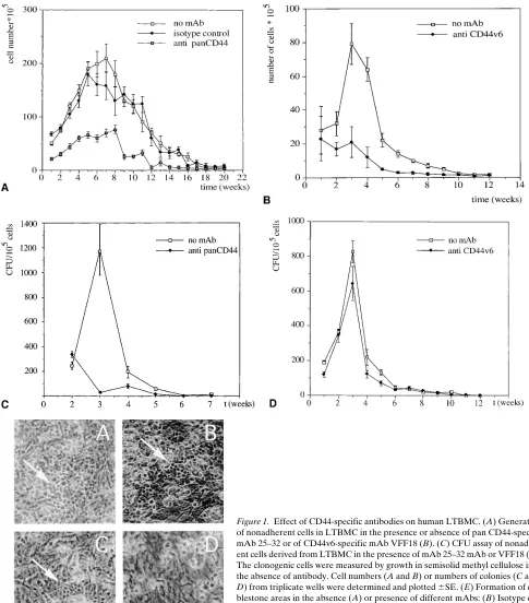
Essential CD44 functions in the maturation of myeloid progeni-tors in LTBMC. To investigate whether CD44 proteins are es-sential during human hemopoiesis, we decided to exploit hu-man LTBMC, which allows the study of several subsequent steps during myelopoiesis, including the formation of the he-mopoietic stromal cell microenvironment, the development of “cobblestone” areas, which are centers of hemopoietic stem cells, and the production of differentiated cells in the superna-tant. We compared the development of myelopoietic cells in LTBMC in the absence or presence of specific antibodies. At each weekly feeding, half of the culture medium containing nonadherent cells was collected, and the number of cells was counted. The mAb 25–32 which recognizes an NH2-terminal
epitope in all known CD44 proteins, inhibited the yield of non-adherent myeloid cells over the whole period of incubation, while an isotype control did not (Fig. 1 A). In a separate series of experiments, the mAb VFF18, which recognizes an epitope encoded by CD44 exon v6, also inhibited the yield of nonad-herent cells (Fig. 1 B).
Figure 1. Effect of CD44-specific antibodies on human LTBMC. (A) Generation of nonadherent cells in LTBMC in the presence or absence of pan CD44-specific mAb 25–32 or of CD44v6-specific mAb VFF18 (B). (C) CFU assay of nonadher-ent cells derived from LTBMC in the presence of mAb 25–32 mAb or VFF18 (D). The clonogenic cells were measured by growth in semisolid methyl cellulose in the absence of antibody. Cell numbers (A and B) or numbers of colonies (C and D) from triplicate wells were determined and plotted 6SE. (E) Formation of cob-blestone areas in the absence (A) or presence of different mAbs: (B) Isotype con-trol IgG2a, (C) mAb VFF18, (D) mAb 25–32. For easier identification cobble-stone areas are marked by an arrow. All experiments were performed at least three times.
that of the other. This suggests that there are two steps involv-ing CD44 action: before (inhibited by 25–32) and after the cob-blestone stage (the second one involving a variant carrying the v6 epitope).
An analysis of the types of cells inhibited in LTBMC sup-ported the idea of two steps of CD44 action. The nonadherent cells in the LTBMC are composed of mature myeloid cells,
VFF18 interferes with a later stage of myelopoiesis, e.g., mye-loid maturation but not with stem cell proliferation, while 25– 32 may act on an earlier step alone or on both steps.
Lineage-specific induction of CD44v4–v10 in leukemic pro-genitor cell lines. Within the CD341/CD441 hemopoietic pre-cursors, only a very small number of cells express the v6 epitope (28). However, CD44v6 epitopes are detected on the
surface of myeloid cells upon maturation and activation (9). We also detected CD44v61 cells of myeloid origin in periph-eral blood (data not shown); however, the numbers of these cells in blood are very low (, 0.1%). We therefore decided to use the corresponding immature myeloid cell lines to elucidate a functional role for CD44v in the myeloid lineage. One of them, the myeloid cell line K562 expresses no CD44 proteins.
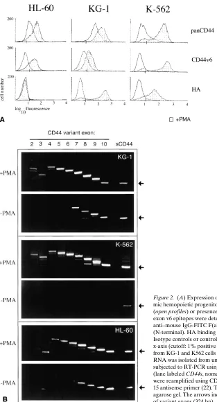
Figure 2. (A) Expression of CD44 isoforms and HA binding capacity of leuke-mic hemopoietic progenitor cell lines. The cells were cultured in the absence (open profiles) or presence (shaded profiles) of PMA for 36 h. N-terminal and exon v6 epitopes were detected by using mAb VFF18 (v6) followed by goat anti–mouse IgG-FITC F(ab9)2 or directly Cy-Chrome™-labeled mAb IM.7.8.1
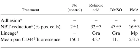
[image:5.612.58.371.55.635.2]K562 cells react neither with a pan CD44 antibody nor with the CD44 exon v6 specific antibody (Fig. 2 A). HL-60 and the myeloid precursor-like cell line KG-1 express CD44s protein, as these cells react with the pan-specific antibody, but no exon v6 epitope can be detected (Fig. 2 A). Although these cell lines are apparently “frozen” in a precursor cell compartment, they can be induced to enter differentiation pathways. We asked whether the expression pattern of CD44 epitopes changes with differentiation. HL-60 cells are particularly useful because they can be induced to differentiate along several myeloid pathways (20, 29), namely, macrophage-like cells, neutrophils, and eosinophils. PMA-induced differentiation of HL-60 cells along the monocyte/macrophage lineage (as judged by adhe-sion to plastic, Table I) is matched with the upregulation of CD44, as determined by staining with an antibody recognizing an N-terminal epitope (24; Fig. 2 A, top, left histogram). Flow cytometric analysis also indicates the appearance of a v6 epitope (Fig. 2 A, middle histogram). In contrast, both retinoic acid and DMSO induced differentiation towards neutrophils (monitored by the ability of cells to reduce nitroblue tetrazo-lium) and led to loss of CD44 from the surface of cells (Table I), compatible with a similar reduction in CD44 expression during normal granulopoiesis (30).
The notion from the HL-60 data that CD44 epitopes en-coded by exon v6 appear during myeloid differentiation is con-firmed by studies with KG-1 cells. PMA-induced myeloid dif-ferentiation of KG-1 cells also induces v6 epitope expression (Fig. 2 A). KG-1 cells spontaneously carry some N-terminal CD44 epitope, which is also upregulated upon treatment with PMA. Another differentiation pathway, namely, differentia-tion along the megakaryocytic lineage can be studied using K562 cells cultured in the presence of PMA (31, 32). Concomi-tantly with the appearance of the megakaryocytic marker CD41 (data not shown), N-terminal and CD44v6 epitopes ap-pear on the cell surface (Fig. 2 A).
To evaluate exactly which CD44 isoforms are upregulated during differentiation, RT-PCR followed by a subsequent CD44 exon run-on analysis was performed (23). For the RT-PCR, primers complementary to sequences in exons 5 and 15 (constant exons outside the v exon region [33]) were used. The result of the first amplification is shown in the outer right lane of Fig. 2 B. With RNA from K562 cells, bands of 324 bp (calcu-lated size, if no v exon is inserted) and of z 720, 820, and 1,050 bp were most prominent. In KG-1 and HL-60 cells, the yield
was too low to allow size calculations. Since the efficiency of amplification decreases with distance between the primers, the abundance of larger bands is underestimated. K562 cells ex-press only low levels of CD44s whereas KG-1 and HL-60 cells express CD44s and low levels of CD44v7–v10 (34; Fig. 2 B). After phorbol ester treatment large amounts of high molecular weight isoforms are detected in all cell lines (see Fig. 2, A and
B). Run-on analysis of the products generated by RT-PCR with primers from all variant exons allows the definition of which variant exon sequence is present in the amplification product and how they are aligned. In all investigated cell lines the major isoform induced by PMA treatment is CD44v4–v10. In addition, minor amounts of cDNAs encoding other, shorter isoforms that bear CD44v2, v3, v8, or v9 are detectable in K562, and CD44v2,v3 in KG-1 or v2 in HL-60 cells (Fig. 2 B). Thus, upon myeloid differentiation, leukemic progenitor cells upregulate CD44 promoter activity and acquire a splice pattern that predominately results in surface expression of CD44v4–v10.
Gain of CD44 variant-dependent HA binding. The occur-rence of CD44 variants on progenitor cell lines after PMA treatment permit the exploration of molecular properties asso-ciated with CD44v4–v10, which could be relevant for the inter-action of precursor cells with stromal cells and/or matrix components. HA is a major component of bone marrow extra-cellular matrix, and it has been shown previously that CD44 carries HA-binding motifs (35). We therefore tested the cells for HA binding.
KG-1 and K562 cells do not bind soluble HA spontane-ously (Fig. 2 A, bottom panel). Whereas, in the case of K562 cells, this might be due to the absence of CD44, KG-1 cells ap-pear to bear CD44 on their surface, which is apparently unable to bind to soluble HA. Strong binding to soluble HA is, how-ever, achieved upon phorbol ester–induced differentiation of K562 and KG-1 cells (36; Fig. 2 A, bottom panel). Differences in HA-binding capacity might be due to differences in expres-sion levels of CD44 between the two cell lines. Interestingly, HL-60 cells, which after phorbol ester treatment carry levels of CD44 v6 epitope comparable with KG-1 cells, do not bind to soluble HA. This lack of HA binding is not due to preoccupa-tion of HA receptors with HA, since hyaluronidase pretreat-ment of cells did not restore HA binding (data not shown). It has been shown in other cell types that the presence of CD44 on the surface of cells does not necessarily lead to HA binding (1, 37, 38). The CD44 variant isoform expressed on PMA-treated HL60 cells is obviously also not in an active HA-bind-ing conformation, although the exon run-on analysis revealed that all PMA-treated cell lines express the same major isoform CD44v4–v10. Hence, additional postranslational modifications such as glycosylation might define HA-binding capacity in the myeloid cell lines.
[image:6.612.56.299.600.685.2]Since CD44 is not the only protein that has potential affin-ity for HA, the surface levels of other known HA receptors on the progenitor cell lines, namely RHAMM (39) and ICAM-1 (40), were determined. A RHAMM-specific polyclonal antise-rum did not bind to the surface of HL-60, KG-1, or K562 cells before and after PMA treatment (data not shown). ICAM-1 is not expressed on untreated HL-60 cells but upregulated upon PMA stimulation (data not shown), although these cells do not bind HA (Fig. 2 A). K562 and KG-1 cells express ICAM-1 spontaneously, and PMA treatment increases ICAM-1 expres-sion in KG-1 (data not shown). To determine the contribution
Table I. CD44 Expression upon Differentiation of HL-60 Cells into Different Lineages
Treatment
No (control)
Retinoic
acid DMSO PMA
Adhesion* 2 2 2 1
NBT-reduction‡ (% pos. cells) 261 3263 4765 1663
Lineage§ 2 Gra Gra Mp
Mean pan CD44i fluorescence 150.1 45.7 11.1 551.7
* 1, . 95% adherent cells; 2, , 5% adherent cells. ‡Values were
calcu-lated from triplicate wells 6SD. §Gra, neutrophilic granulocytes; Mp,
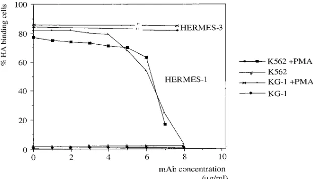
of the CD44 isoforms to HA binding, the HA-binding domain of CD44 was specifically blocked by using the mAb Hermes-1. Hermes-1 efficiently blocks the induced binding to HA of both KG-1 and K562 cells (Fig. 3). The mAb Hermes-3 recognizes an epitope outside the HA-binding motif of CD44 and is un-able to block HA binding (Fig. 3). mAbs recognizing ICAM-1, RHAMM, or other epitopes of CD44 including those recog-nizing variant exons v5, v6, v7, and v10 also do not influence HA binding (data not shown).
In summary, the induced HA affinity of K562 and KG-1 cells is exclusively mediated by CD44, and, as will be shown below, HA binding is the function predominantly of CD44 variants. The presence of ICAM-1 does not confer HA-bind-ing capacity. Although the bindHA-bind-ing to soluble HA is strongly enhanced by the presence of CD44 splice variants (41) and also is strongly upregulated here with the appearance of CD44v4–v10, we note that an anti-v6 antibody does not affect HA binding. Furthermore, cells must require a property in ad-dition to the protein backbone structure of CD44v4–v10, e.g., an accessory protein or a modification of CD44, to bind HA.
CS-A is a specific ligand for CD44v4–v10. We have
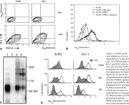
previ-ously shown using a rat adenocarcinoma cell line that expres-sion of larger CD44 variants containing exon v6 and v7 encoded epitopes expand their repertoire of binding to GAGs such as CS, heparan, and heparansulfate (42). Since CS-A is a highly abundant matrix component of bone marrow, we exam-ined binding of myeloid precursor cell lines to CS-A as this may be of significance in the process or regulation of progeni-tor differentiation. Untreated K562 and KG-1 cells, which do not express CD44 variant epitopes, do not bind CS-A. Upreg-ulation of CD44v4–v10 isoform expression, monitored by the appearance of exon v6 epitope on the cell surface, is accompa-nied by gain of CS-A binding ability (Fig. 4 A). Similar to the situation with HA, the binding motif for CS-A is in the N-ter-minal domain of CD44, and antibodies recognizing the v6 epitope are unable to block this binding (42), whereas Her-mes-1 the antibody that blocks HA binding by CD44 also blocks CS-A binding in PMA-treated K562 cells (Fig. 4 B) and KG-1 cells (data not shown).
To determine whether CS-A binding was specific for CD44v isoforms, GAG-associated proteins from phorbol es-ter–induced KG-1 cells were purified by CPC precipitation (see Methods), resolved by SDS-PAGE and analyzed by
Western blotting using the mAb Hermes-3. The relative abun-dance of CD44 proteins staining with Hermes-3 can be judged from Fig. 4 C (lane 1), which shows the separation of the cell lysate before precipitation (1/10 of volume subjected to CPC precipitation, Fig. 4 C). Four protein bands are visible in cell lysates of KG-1 with apparent molecular weights of: 85,000 (the CD44s form), 120,000, 160,000, and 180,000. The larger protein bands may represent different variants or differentially modified proteins of CD44. Incubation of both HA or CS-A with cell lysates leads to CPC-precipitable material. HA asso-ciates with CD44s and with the high molecular weight variant CD44 isoforms in this assay (Fig. 4 C, lane 3), although more high molecular weight isoforms seem to be precipitated. CPC precipitation is specific for GAG-associated proteins since, without the addition of GAG, no CD44 proteins are precipi-tated (Fig. 4 C, lane 2). This finding suggests that the larger variants exhibit higher affinity to HA, especially when the ra-tio of CD44s and CD44v detected in a Western blot is com-pared with the ratio that binds to HA. On the other hand, CPC precipitation of protein after CS-A loading leads to almost ex-clusive precipitation of all large CD44 bands but hardly any CD44s (Fig. 4 C, lane 4). Interestingly, the largest band barely visible in lane 1 is enriched in lane 4, obviously binding to CS-A particularly well.
We conclude that CD44v isoforms strongly enhance HA-binding capacity and almost exclusively confer CS-A HA-binding ability on hemopoietic progenitor cell lines during differentia-tion. The presence of CD44 high molecular weight isoforms per se is not sufficient for binding to either GAG, since HL-60 cells induced to express CD44v levels equivalent to those of KG-1 or K562 cells do not bind GAGs.
CS-A and HA binding are modulated by N-glycosylation.
HL-60 carry abundant levels of CD44 variants, but this is not sufficient to mediate affinity for GAGs, whereas this is the case for activated K562 and KG-1 cells. In previous experi-ments, we have been unable to find accessory proteins, which may facilitate GAG binding by CD44 (data not shown). We therefore examined whether modifications of CD44 proteins could be responsible for the affinity to GAGs. CD44 proteins are highly glycosylated (3, 4) and can be phosphorylated at cy-toplasmic tail serine residues (43, 44). Both modifications have been implicated in HA binding. We could not find any correla-tion of HA binding and phosphorylacorrela-tion of CD44 in the
[image:7.612.58.368.555.733.2]loid cell lines, which we have used (data not shown); however, inhibition of N-linked glycosylation by tunicamycin reduced HA binding in differentiating K562 and KG-1 cells (Fig. 4 D). CD44 surface levels and splicing were hardly changed in the case of K562 cells, but CD44 expression was reduced in case of KG-1 cells as judged by flow cytometry (Fig. 4 D) and exon run-on analysis (data not shown). CS-A binding was totally abolished in tunicamycin-treated cultures (Fig. 4 D). In the case of K562 cells, the HA-, and CS-A–binding functions of CD44 are regulated by or dependent on N-glycosylation. This is supported by the finding that tunicamycin did not affect CD44 expression levels on K562 cells but inhibits GAG bind-ing. Therefore, a qualitative change on CD44 molecules such as glycosylation is probably responsible for the changed bind-ing behavior, although we cannot exclude an indirect mecha-nism of inhibition of CS-A or HA binding by tunicamycin.
HA-mediated adhesion of K562 and KG-1 cells to the bone marrow stromal cell line MS-5. The acquired affinity to GAGs
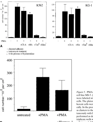
ubiquitously present in the bone marrow may help cells to ad-here to a specific microenvironment. The specific component of the microenvironment is likely to be stromal cells, and we asked whether differentiating hemopoietic cells gain a CD44-dependent affinity for stromal cells. To assay for this property, we chose the bone marrow stromal cell line MS-5, which has been shown to support the growth of growth factor–dependent hemopoietic cell lines (45). KG-1 and K562 cells do not adhere to MS-5 cells unless stimulated by phorbol ester. Both cell types then adhere, and K562 cells adhere particularly strongly (Fig. 5 A) as compared with KG-1 cells. This may indicate that CD44 variant isoforms are responsible for the interaction, as they are particularly abundant in K562 cells.
We investigated the molecular mechanism of the interac-tion of stromal cells with PMA-treated leukemic progenitor cells. Since MS-5 cells also express CD44s, one could envisage that the adhesion could be mediated by HA molecules bridg-ing CD44 proteins on the stromal cells and on the progenitor
[image:8.612.57.549.58.459.2]cells. This has been shown to occur between keratinocytes and CD44 carrying tumor cells (46). mAb Hermes-1, which blocks binding of HA to the PMA-treated K562 and KG-1 cells, was able to reduce the number of adherent cells (Fig. 5 B; data not shown), although, no total block could be achieved suggesting that other adhesion molecules participate in MS-5 and K562 interaction.
The presence of soluble HA or CS-A (0.5 mg/ml) during the adhesion assay did not abrogate binding of differentiated K562 or KG-1 cells to MS-5 cells (Fig. 5 A), although HA but not CS-A induced self-aggregation of K562 or KG-1 cells lead-ing to a clustered pattern of adhesion to MS-5 cells (not shown). Nevertheless, the presence of hyaluronidase during the adhesion assay abrogated binding dramatically (Fig. 5 A), suggesting HA-dependent adhesion. We interpret these data to suggest that MS-5 cells present sufficient HA themselves, and therefore additional exogenous HA does not enhance ad-hesion, while K562 and KG-1 cells do not appear to synthesize HA as HA addition triggers self-aggregation.
The presence of 0.5 mM EDTA did not influence the adhe-sive properties of the cells (not shown). This excludes a contri-bution from integrins which mediate Ca21-ion-dependent
ad-hesion. Tunicamycin treatment of induced KG-1 or K562 cells destroys adhesion to MS-5 cells drastically (Fig. 5 A), which matches with the finding that tunicamycin inhibits binding of soluble HA to CD44v and CD44s. We conclude that CD44 contributes to the HA-dependent adhesion of the myeloid pre-cursor cell lines KG-1 and K562 to the bone marrow stromal cell line MS-5, and that binding is regulated by N-glycosylation. The CD44v-mediated affinities for GAGs and stromal cells by differentiating progenitor cells may be of significance in regulating hemopoiesis. However, other CD44v-dependent in-teractions must also occur in the bone marrow to regulate my-elopoiesis as the VFF18 antibody inhibits myeloid differentia-tion but does not inhibit CD44v-mediated GAG binding or stromal cell interaction (data not shown).
Discussion
[image:9.612.57.364.331.734.2]CD44 has been shown to play a functional role during he-mopoiesis. In mouse monoclonal antibodies to Pgp-1/CD44 block lymphopoiesis or myelopoiesis in LTBMC (47, 48) and deletion of CD44 by homologous recombination leads to a hematological impairment (49). On the other hand, a LTBMC
enhancing antibody recognizing canine CD44 (50) has been described. Other mAbs recognizing CD44 have diverse effects on hemopoiesis in humans (51). These data show a role for CD44 proteins in hemopoiesis, although, it is not clear at which stages CD44 acts and which CD44 protein family mem-bers are involved.
The antibody blocking experiments presented in this paper suggest that CD44 proteins are required in at least two steps in human myelopoiesis. First, the mAb 25–32, which recognizes an N-terminal epitope on all CD44 proteins, inhibits an early step in the generation of progenitor cells. Second, the CD44 exon v6 specific antibody VFF18 blocks a later step in myeloid maturation but not the production of early progenitors. CD341 cells that express CD44 could be the target for inhibi-tion by the mAb 25–32. Since the majority of CD341/CD441 cells show no CD44v6 expression, it is likely that CD44s are the target for inhibition by CD44 pan mAb. Furthermore, the absence of the VFF18 epitope on the surface of the bulk of CD34 stem cells suggests that the function inhibited by VFF18 may not be required in the production of progenitors but rather in the transition towards the myeloid lineage. This view is supported by the finding that there exists a small subpopula-tion of myeloid lineage committed cells in human bone mar-row that show expression of exon v10–containing CD44 iso-forms (52).
Leukemic hemopoietic progenitor cell lines mimic part of the process of normal myeloid maturation (20, 29). We there-fore studied cellular properties of such cell lines with respect to CD44 expression and function. If our findings with these pro-genitor cell lines are extrapolated to normal myelopoiesis, CD44 variant expression seems to function after entry into myeloid maturation. Myeloid differentiation of KG-1 and K562 cells leads to a CD44v-dependent increase in affinity to HA and CS-A. The strong or even exclusive binding of GAGs to CD44 variant isoforms is likely due to their ability to homo-oligomerize and to cluster in the plasma membrane (53).
The same CD44 proteins expressed in different cells can be high affinity receptors for GAGs or nonbinders. In addition to the binding motif, covalent modifications are required to cre-ate high GAG affinity. In K562 cells, for example, a specific
N-glycosylation seems to be necessary for GAG binding as
suggested by tunicamycin inhibition experiments, consistent with observations in rat pancreatic carcinoma cells. This does not always seem to be the case. An increase in CD44-depen-dent HA binding by tunicamycin has recently been described (8, 54, 55) and also in our hands a murine T cell lymphoma (56) shows enhanced HA binding upon tunicamycin treat-ment, which might be due to the lack of negatively charged end-standing sugar residues (data not shown).
What could be the function in bone marrow of induced af-finity to GAGs? There are a number of options. 1.) GAGs may serve to present growth factors or growth inhibitors. The CD44 variant proteins could bind GAGs together with their associated factors. This binding may allow presentation of the associated factors either to other cells or to high-affinity recep-tors on the cells carrying the variant. For instance, a growth factor relevant for hemopoiesis, GM-CSF, has been found in association with HA (57). 2.) The affinity of CD44v proteins for CS-A may allow binding of chondroitin sulphate-modified proteins carried by other cells or located in soluble form in the extracellular space. Candidates for such proteins are the CS-modified M-CSF (58), osteopontin (7), serglycin (59, 60), and
the invariant chain (61). For osteopontin, serglycin, and the in-variant chain, interactions with CD44 have been described. We propose that CD44v might interact in two ways with such proteins. The N-terminal part binds to the GAG portion, and the variant exon part interacts with the protein backbone deter-mining the specificity. While the CD44v-mediated binding of CS-A is not inhibited by the v6-specific antibody, this latter in-teraction might well be blocked. 3.) GAGs could form the glue in cell–cell interactions through CD44. In the PMA-treated K562 and KG-1 cells, HA indeed causes cell–cell aggregation. These cells also acquire the ability to bind to the stromal bone marrow cell line MS-5 in a CD44- and HA-dependent manner. This phenomenon could also be of relevance for primary he-mopoietic precursor cells in the bone marrow, since a subpopu-lation of CD341/CD441 cells do bind HA (17). Such a HA- and CD44-dependent cell–cell contact between progenitor cells and stromal cells might therefore be important for hemopoiesis.
Cell–cell contact through CD44 without the participation of GAGs is another interesting possibility that might be rele-vant in the bone marrow. CD44-mediated association of cells, for instance, has been reported between cells carrying CD44s and other cells expressing a CD44 variant isoform (62). A hy-bridoma cell line was shown to bind to HA-coated wells as well as to a stromal cell layer (48). Also, this effect could be blocked by hyaluronidase treatment or by mAbs recognizing CD44. The only difference is that in our case exogeneously added HA did not block adhesion but rather induced cluster-ing of PMA-activated cells. Therefore, a mechanism of direct CD44 interaction as has been suggested (62) might participate in cell adhesion in our case. Irrespective of the mechanism of cell–cell interaction, as a result, conditions favorable to differ-entiation may be created, perhaps by signalling to the stromal cells such that they release factors.
In conclusion, we have been able to define two discrete roles for CD44 during hemopoiesis. The first pathway involves CD44s and influences the production of progenitor cells. The second involves CD44 variant proteins bearing v6 and influ-ences myeloid differentiation. We postulate that part of the role of CD44 in myeloid differentiation is to mediate binding to different GAGs. However, other as yet undefined roles for CD44 in myeloid differentiation must also exist.
Note added in proof: During the preparation of this work, doubt came up on the relevance of ICAM-1 and RHAMM as cell surface HA-receptors (63–65).
Acknowledgments
We thank Anne Denninger and Tanja Schüler for excellent technical assistance and Dr. K.H. Heider and Dr. G. Adolf (Bender, Vienna) for providing us with the human CD44v-specific mAbs.
This work was supported by a grant from the Deutsche Forsch-ungsgemeinschaft (He 551/8-2).
References
1. Lesley, J., R. Hyman, and P.W. Kincade. 1993. CD44 and its interaction with the extracellular matrix. Adv. Immunol. 54:271–335.
2. Herrlich, P., M. Zöller, S.T. Pals, and H. Ponta. 1993. CD44 splice vari-ants: metastases meet lymphocytes. Immunol. Today. 14:395–399.
3. Rudy, W., M. Hofmann, R. Schwartz-Albiez, M. Zöller, K.H. Heider, H. Ponta, and P. Herrlich. 1993. The two major CD44 proteins expressed on a met-astatic rat tumor cell line are derived from different splice variants: each one in-dividually suffices to confer metastatic behavior. Cancer Res. 53:1262–1268.
Whit-tle. 1995. Proteoglycan forms on the lymphocyte homing receptor CD44 are al-ternatively spliced variants containing the v3 exon. J. Cell Biol. 128:673–685.
5. Aruffo, A., I. Stamenkovic, M. Melnick, C.B. Underhill, and B. Seed. 1990. CD44 is the principal cell surface receptor for hyaluronate. Cell. 61:1303–1313.
6. Miyake, K., C.B. Underhill, J. Lesley, and P.W. Kincade. 1990. Hyal-uronate can function as a cell adhesion molecule and CD44 participates in hyaluronate recognition. J. Exp. Med. 172:69–75.
7. Weber, G.F., S. Ashkar, M.J. Glimcher, and H. Cantor. 1996. Receptor-ligand interaction between CD44 and osteopontin (Eta-1). Science. 271:509– 512.
8. Bennett, K.L., D.G. Jackson, J.C. Simon, E. Tanczos, R. Peach, B. Modrell, I. Stamenkovic, G. Plowman, and A. Aruffo. 1995. CD44 isoforms containing exon v3 are responsible for the presentation of heparin-binding growth factor. J. Cell Biol. 128:687–698.
9. Arch, R., K. Wirth, M. Hofmann, P. Ponta, S. Matzku, P. Herrlich, and M. Zöller. 1992. Participation in normal immune responses of a splice variant of CD44 that encodes a metastasis-inducing domain. Science. 257:682–685.
10. Koopman, G., K.H. Heider, E. Horst, G.R. Adolf, F. van den Berg, H. Ponta, P. Herrlich, and S.T. Pals. 1993. Activated human lymphocytes and ag-gressive non-Hodgkin lymphomas express a homologue of the rat metastasis-associated variant of CD44. J. Exp. Med. 177:897–904.
11. Screaton, G.R., J.F. Caceres, A. Mayeda, M.V. Bell, M. Plebanski, D.G. Jackson, J.I. Bell, and A.R. Krainer. 1995. Identification and characterization of three members of the human SR family of pre-mRNA splicing factors. EMBO J. 14:4336–4349.
12. Moll, J., A. Schmidt, H. van der Putten, R. Plug, H. Ponta, P. Herrlich, and M. Zöller. 1996. Accelerated immune response in transgenic mice express-ing rat CD44v4-v7 on T cells. J. Immunol. 156:2085–2094.
13. Zöller, M., A. Schmidt, A. Wenzel, and J. Moll. 1997. Constitutive ex-pression of a CD44 variant isoform on T cells facilitates regaining of immuno-competence in allogeneic bone marrow transplantation. Blood. 90:873–885.
14. Gallagher, J.T., E. Spooncer, and T.M. Dexter. 1983. Role of the extra-cellular matrix in hemopoiesis. I. Synthesis of glycosaminoglycans by mouse bone marrow cell cultures. J. Cell. Sci. 63:155–161.
15. Wight, T.N., M.G. Kinsella, A. Keating, and J.W. Singer. 1986. Pro-teoglycans in human long-term bone marrow cultures: biochemical and ultra-structural analysis. Blood. 67:1333–1343.
16. Siczkowski, M., A.S. Amos, and M.Y. Gordon. 1993. Hyaluronic acid regulates the function and distribution of sulfated glycosaminoglycans in bone marrow stromal cultures. J. Exp. Hematol. 21:126–130.
17. Legras, S., J.P. Levesque, R. Charrad, K. Morimoto, C. Le Bousse, D. Clay, C. Jasmin, and F. Smadja-Joffe. 1997. CD44-mediated adhesiveness of hu-man hematopoietic progenitors to hyaluronan is modulated by cytokines. Blood. 89:1905–1914.
18. Kitamura, T., T. Tange, T. Terasawa, T. Chiba, T. Kuwaki, K. Miya-gawa, Y.F. Piao, K. Miyazono, A. Urabe, and F. Takaku. 1989. Establishment and characterization of a unique human line that proliferates dependently on GM-CSF, IL-3, or erythropoietin. J. Cell. Physiol. 140:323–334.
19. Itoh, K., H. Tezuka, H. Sakoda, M. Konno, K. Nagata, T. Uchiyama, H. Uchino, and K.J. Mori. 1989. Reproducible establishment of hematopoietic supportive stromal cell lines from murine bone marrow. Exp. Hematol. 17:145– 153.
20. Collins, S.J. 1987. The HL-60 promyelocytic leukemia cell line: prolifer-ation, differentiprolifer-ation, and cellular oncogene expression. Blood. 70:1233–1244.
21. Zuckermann, F.A., R.M. Binns, R. Husmann, H. Yang, M.M. Carr, Y.B. Kim, W.C. Davis, M. Mistfeldt, and J.K. Lunney. 1993. Analysis of monoclonal antibodies reactive with porcine CD44 and CD45. Vet. Immunol. Immuno-pathol. 43:293–305.
22. Sambrook, J., E.F. Fritsch, and T. Maniatis. 1989. Molecular Cloning: A Laboratory Manual. Cold Spring Harbor Laboratory Press. Cold Spring Har-bor, NY.
23. van Weering, D.H.J., P.D. Baas, and J.L. Bos. 1993. A PCR-based method for the analysis of human CD44 splice products. PCR Meth. Appl. 3: 100–106.
24. Jalkanen, S.T., R.F. Bargatze, L.R. Herron, and E.C. Butcher. 1986. A lymphoid cell surface glycoprotein involved in endothelial cell recognition and lymphocyte homing in man. Eur. J. Immunol. 16:1195–1202.
25. Heider, K.H., M. Hofmann, E. Horst, F. van den Berg, H. Ponta, P. Herr-lich, and S.T. Pals. 1993. A human homologue of the rat metastasis-associated variant of CD44 is expressed in colorectal carcinomas and adenomatous polyps. J. Cell Biol. 120:227–233.
26. de Belder, A.N., and K.O. Wik. 1975. Preparation and properties of flu-orescein-labeled hyaluronate. Carbohydr. Res. 44:251–257.
27. Lee, T.H., H.G. Wisniewski, and J. Vilcek. 1992. A novel secretory tu-mor necrosis factor-inducible protein (TSG-6) is a member of the family of hy-aluronate binding proteins, closely related to the adhesion receptor CD44. J. Cell Biol. 116:545–557.
28. Neu, S., A. Geiselhart, M. Sproll, D. Hahn, S. Kuci, D. Niethammer, and R. Handgretinger. 1997. Expression of CD44 isoforms by highly enriched CD34-positive cells in cord blood, bone marrow and leukaphereses. Bone Mar-row Transplant. 20:593–598.
29. Fischkoff, S., A. Pollak, A. Gleich, J. Testa, S. Misawa, and T. Reber.
1984. Eosinophilic differentiation of the human promyelocytic leukemia cell line HL-60. J. Exp. Med. 160:179–196.
30. Kansas, G.S., M.J. Muirhead, and M.O. Daily. 1990. Expression of the CD11/CD18 leukocyte adhesion molecule 1, and CD44 adhesion molecules during normal myeloid and erythroid differentiation. Blood. 76:2483–2492.
31. Tettero, P.A.T., F. Massaro, A. Mulder, R. Schrender-von Gelder, and A.E.G. van den Borne. 1984. Megakaryoblastic differentiation of proerythro-blastic K562 cell line cells. Leuk. Res. 8:197–206.
32. Murate, T., S. Saga, T. Hotta, H. Asano, T. Ito, K. Kato, K. Tsushita, T. Kinoshita, A. Ichikawa, S. Yoshida, et al. 1993. The close relationship between DNA replication and the selection of differentiation lineages of human erythro-leukemia cell lines K562, HEL, and TF1 into erythroid or megakaryocytic lin-eages. Exp. Cell. Res. 208:35–43.
33. Screaton, G.R., M.V. Bell, D.G. Jackson, F.B. Cornelis, U. Gerth, and J.I. Bell. 1992. Genomic structure of DNA encoding the lymphocyte homing re-ceptor CD44 reveals at least 12 alternatively spliced exons. Proc. Natl. Acad. Sci. USA. 89:12160–12164.
34. Dougherty, G.J., P.M. Landorp, D.L. Cooper, and R.K. Humphries. 1991. Molecular cloning of CD44R1 and CD44R2, two novel isoforms of the human CD44 “homing” receptor expressed by hemopoietic cells. J. Exp. Med. 174:1–5.
35. Peach, R.J., D. Hollenbaugh, I. Stamenkovic, and A. Aruffo. 1993. Iden-tification of hyaluronic acid binding sites in the extracellular domain of CD44. J. Cell Biol. 122:257–264.
36. Morimoto, K., E. Robin, M.C. LeBousse-Kerdiles, Y. Ly, D. Clay, C. Jasmin, and F. Smadja-Joffe. 1994. CD44 mediates hyaluronate binding by hu-man myeloid KG1A and KG1 cells. Blood. 83:657–662.
37. Moll, J., J.P. Sleeman, K. Kondo, A. Hekele, R. Plug, L. Sherman, A. Schmidt, M. Zöller, E. Horst, H. Ponta, et al. 1994. Analysis of molecular func-tions of the tumor metastasis promoting surface molecule CD44v4-v7 using transgenic mice. In Molecular and Cellular Basis for Cell to Cell Interaction: Its Significance in Cancer. S. Hirohashi, H.L. Moses, E. Ruoslahti, T. Sugimura, M. Takeichi, and M. Terada, editors. Princeton Scientific Publ. Co., Princeton, NJ. 142–151.
38. Sherman, L., J.P. Sleeman, P. Herrlich, and H. Ponta. 1994. Hyaluronate receptors: key players in growth, differentiation, migration and tumor progres-sion. Curr. Opin. Cell. Biol. 6:726–733.
39. Hardwick, C., K. Hoare, R. Owens, H.P. Hohn, M. Hook, D. Moore, V. Cripps, L. Austen, D.M. Nance, and E.A. Turley. 1992. Molecular cloning of a novel hyaluronan receptor that mediates tumor cell motility. J. Cell Biol. 117: 1343–1350.
40. McCourt, P.A., B. Ek, N. Forsberg, and S. Gustafson. 1994. Intercellular adhesion molecule-1 is a cell surface receptor for hyaluronan. J. Biol. Chem. 269:30081–30084.
41. Sleeman, J.P., S. Arming, J. Moll, H. Hekele, W. Rudy, L. Sherman, G. Kreil, H. Ponta, and P. Herrlich. 1996. Hyaluronate-independent metastatic be-havior of CD44 variant-expressing pancreatic carcinoma cells. Cancer Res. 56: 3134–3141.
42. Sleeman, J.P., K. Kondo, J. Moll, H. Ponta, and P. Herrlich. Variant ex-ons v6 and v7 together expand the repertoire of glycosaminoglycans bound by CD44. J. Biol. Chem. 272:31837–31844.
43. Uff, C.R., S.J. Neame, and C.M. Isacke. 1995. Hyaluronan binding by CD44 is regulated by a phosphorylation-dependent mechanism. Eur. J. Immu-nol. 25:1883–1887.
44. Puré, E., R.L. Camp, D. Perrit, R.A. Panettieri, A.L. Lazaar, and S. Nayak. 1995. Defective phosphorylation and hyaluronate binding of CD44 with point mutations in the cytoplasmatic domain. J. Exp. Med. 181:55–62.
45. Sugimoto, K., Y. Tsurumaki, H. Hoshi, S. Kadowaki, M.C. LeBousse-Kerdiles, F. Smadja-Joffe, and K.J. Mori. 1994. Effects of anti-CD44 mono-clonal antibody on adhesion of erythroid leukemic cells (ELM-I-1) to hemo-poietic supportive cells (MS-5): CD44, but not hyaluronate-mediated, cell-cell adhesion. Exp. Hematol. 22:488–494.
46. Hudson, D.L., J. Sleeman, and F.M. Watt. 1995. CD44 is the major pea-nut lectin-binding glycoprotein of human epidermal keratinocytes and plays a role in cellular adhesion. J. Cell. Sci. 108:1959–1970.
47. Miyake, K., K.L. Medina, S.I. Hayashi, S. Ono, T. Hamaoka, and P.W. Kincade. 1990. Monoclonal antibodies to Pgp-1/CD44 block lympho-hemopoie-sis in long-term bone marrow cultures. J. Exp. Med. 171:477–488.
48. Miyake K., C.B. Underhill, J. Lesley, and P.W. Kincade. 1990. Hyal-uronate can function as a cell adhesion molecule and CD44 participates in hyaluronate recognition. J. Exp. Med. 172:69–75.
49. Schmits, R., J. Filmus, N. Gerwin, G. Senaldi, F. Kiefer, T. Kundig, A. Wakeham, A. Shahinian, C. Catzavelos, J. Rak, et al. 1997. CD44 regulates he-mopoietic progenitor distribution, granuloma formation, and tumorigenicity. Blood. 90:2217–2233.
50. Rossbach, H.C., L. Krizanac-Bengez, E.B. Santos, T.A. Gooley, and B.M. Sandmaier. 1996. An antibody to CD44 enhances hematopoiesis in long-term marrow cultures. Exp. Hematol. 24:221–227.
51. Ghaffari, S., G.J. Dougherty, A.C. Eaves, and C.J. Eaves. 1997. Diverse effects of anti-44 antibodies on the stromal cell-mediated support of normal but not leukaemic (CML) haemopoiesis in vitro. Br. J. Haematol. 97:22–28.
Eaves. 1995. Differentiation-associated changes in CD44 isoform expression during normal hematopoiesis and their alteration in chronic myeloid leukemia. Blood. 15:2976–2985.
53. Sleeman J., W. Rudy, M. Hofmann, J. Moll, P. Herrlich, and H. Ponta. 1996. Regulated clustering of variant CD44 proteins increase their hyaluronate binding capacity. J. Cell Biol. 135:1139–1150.
54. Lesley, J., N. English, A. Perschl, J. Gregoroff, and R. Hyman. 1995. Variant cell lines selected for alterations in the function of the hyaluronan re-ceptor CD44 show differences in glycosylation. J. Exp. Med. 182:431–437.
55. Katoh, S., Z. Zheng, K. Oritani, T. Shimozato, and P.W. Kincade. 1995. Glycosylation of CD44 negatively regulates its recognition of hyaluronan. J. Exp. Med. 182:419–429.
56. Lugasi, H., S. Hajos, J.R. Murphy, T.B. Strom, J. Nichols, C. Penarroja, and D. Naor. 1990. Murine spontaneous T-cell leukemia constitutively express-ing IL-2 receptor: a model for human T-cell malignancies expressexpress-ing IL-2 re-ceptor. Int. J. Cancer. 45:163–167.
57. Gordon, M.Y., G.P. Riley, S.M. Watt, and M.F. Greaves. 1987. Com-partmentalization of a hematopoietic growth factor (GM-CSF) by glycosami-noglycans in the bone marrow microenvironment. Nature. 326:403–405.
58. Suzu, S., T. Inaba, N. Yanai, T. Kawashima, N. Yamada, T. Oka, R. Machinami, T. Ohtsuki, F. Kimura, and S. Kondo. 1994. Proteoglycan form of macrophage colony-stimulating factor binds low density lipoprotein. J. Clin. In-vest. 94:1637–1641.
59. Toyama-Sorimachi, N., and M. Miyasaka. 1994. A novel ligand for CD44 is sulfated proteoglycan. Int. Immunol. 6:655–660.
60. Toyama-Sorimachi, N., H. Sorimachi, Y. Tobita, F. Kitamura, H. Yagita, K. Suzuki, and M. Miyasaka. 1995. A novel ligand for CD44 is serglycin, a hematopoietic cell lineage-specific proteoglycan. J. Biol. Chem. 270:7437– 7444.
61. Naujokas, M., M. Morin, M.S. Anderson, M. Peterson, and J. Miller. 1993. The chondroitin sulfate form of invariant chain can enhance stimulation of T cell responses through interaction with CD44. Cell. 74:257–268.
62. Droll, A., S.T. Dougherty, R.K. Chiu, J.F. Dirks, W.H. McBride, D.L. Cooper, and G.J. Dougherty. 1995. Adhesive interactions between alternatively spliced CD44 isoforms. J. Biol. Chem. 270:11567–11571.
63. Peter, A., G. McCourt, and S. Gustafson. 1997. On the adsorption of hy-aluronan and ICAM-1 to modified hydrophobic resins. Int. J. Biochem. Cell. Biol. 29:1179–1183.
64. Assmann, V., J.F. Marshall, C. Fieber, M. Hofmann, and I.R. Hart. 1998. The human hyaluronan receptor RHAMM is expressed as an intracellu-lar protein in breast cancer cells. J. Cell. Sci. 111:1685–1694.