0095-1137/08/$08.00⫹0 doi:10.1128/JCM.00051-08
Copyright © 2008, American Society for Microbiology. All Rights Reserved.
Rapid Identification of Penicillin and Macrolide Resistance Genes and
Simultaneous Quantification of
Streptococcus pneumoniae
in Purulent
Sputum Samples by Use of a Novel Real-Time
Multiplex PCR Assay
䌤
†
Kazuko Y. Fukushima,
1Katsunori Yanagihara,
1* Yoichi Hirakata,
2Kazuyuki Sugahara,
1Yoshitomo Morinaga,
1,2Shigeru Kohno,
2and Shimeru Kamihira
1Department of Laboratory Medicine1and Second Department of Internal Medicine,2Nagasaki University Graduate School of
Biomedical Sciences, 1-7-1 Sakamoto, Nagasaki City 852-8501, Japan
Received 10 January 2008/Returned for modification 3 March 2008/Accepted 29 April 2008
We evaluated a real-time quantitative PCR combined with a multiplex PCR assay for the quantification of
Streptococcus pneumoniae and the simultaneous detection of drug-resistant genes by gel-based PCR, using
purulent sputum samples. This assay correctly quantifiedS. pneumoniaeand identified their penicillin and erythromycin susceptibilities directly from samples within 3 h.
Streptococcus pneumoniaeis a crucial pathogen that causes community-acquired pneumonia (CAP). CAP in adults is often treated with a combination of-lactam antibiotics and macro-lides (18, 22). However, alarmingly high frequencies of peni-cillin- and macrolide-resistant pneumococci have been re-ported, especially in several Asian countries, including Japan (4, 24, 30). The resistance ofS. pneumoniaeto penicillin has been shown to be closely associated with mosaic mutations in thepbp1a, pbp2b, andpbp2xgenes (12, 35). Macrolide resis-tance is generally mediated by two mechanisms: 23S rRNA methylation encoded by theerm(B) gene or macrolide efflux via themef(A) gene (23, 32). Detection of drug-resistant S. pneumoniaeby classical techniques usually takes several days (17, 27). Recently, the utility of the real-time PCR method in the detection and quantification of S. pneumoniae has been examined using lower respiratory tract samples (2, 5, 15, 20, 36). The results of these studies suggest that clinical infection correlates with increased pneumococcal load, and these studies also mentioned the capacity of quantitative real-time PCR to distinguish between colonization and true infection (15, 36). Many investigators have evaluated the accuracy of multiplex PCR methods, which are used in the screening of S. pneu-moniaestrains demonstrating penicillin and macrolide resis-tance (13, 14, 21, 33, 34). Most of these assays, however, re-quire separate tubes, are only able to detect two or three gene fragments, and have not been evaluated for clinical respiratory tract samples. In the present study, we developed a simulta-neous single-tube real-time quantitative PCR combined with a multiplex PCR (RQ-mPCR) assay that rapidly quantifies S. pneumoniae; identifies alterations inpbp1a, pbp2b, andpbp2x
genes; and detects the presence oferm(B) andmef(A) genes.
We first verified this method by using clinicalS. pneumoniae
strains and then evaluated the effectiveness of the method using purulent sputum samples.
We used 200 clinical isolates ofS. pneumoniaescreened by optochin susceptibility (susceptible) and bile solubility (solu-ble) that were collected from April 2004 to March 2006 by a laboratory at the Nagasaki University Hospital. Strains were propagated on 5% sheep blood agar (Nissui Co., Ltd., Tokyo, Japan) at 37°C with 5% CO2. A mixture of 24 bacterial species from the American Type Culture Collection (ATCC; see Table S1 in the supplemental material) were selected from species commonly isolated from respiratory tract and from species that are genetically related toS. pneumoniae (3). In addition, 17 clinical strains ofStreptococcus mitisand 12 clinical strains of
Streptococcus oralis, which had been isolated from respiratory tract samples, were collected for cross-reactivity studies.
The MICs were determined by using broth microdilution techniques as described by the Clinical and Laboratory Stan-dards Institute (CLSI) guidelines (7, 8).S. pneumoniaeATCC 49619 was used for quality control.
A total of 200 purulent sputum samples, which were col-lected from April 2004 to March 2005 and from June 2007 to August 2007 by a laboratory at the Nagasaki University Hos-pital, were used. Only good-quality sputum samples (P2 and P3 according to the classification of Miller and Jones (19) were used. Sputum samples were diluted 1:100 and 1:10,000 with 0.45% sodium chloride and treated with Sputazyme solution (Kyokuto Pharmaceutical Industries Co., Ltd., Tokyo, Japan). The diluted samples were spread on 5% sheep blood agar plates with a DS500 spiral plater (InterScience, Inc., Markham, Ontario, Canada) and then incubated at 37°C in 5% CO2. Optochin sensitivity and bile solubility were used to identifyS. pneumoniae. Nucleic acids were isolated from clinical strains and sputum samples by using a QIAamp DNA blood minikit (Qiagen, Hilden, Germany).
ThelytA,pbp1a,pbp2b,pbp2x,ermB, andmefAgenes were amplified by PCR. Primers LytA-F and LytA-R and probes
LytA-DCR and LytA-ACR were designed to target a 173-bp * Corresponding author. Mailing address: Department of
Labora-tory Medicine, Nagasaki University Graduate School of Biomedical Sciences, 1-7-1 Sakamoto, Nagasaki City 852-8501, Japan. Phone: 81-95-849-7418. Fax: 81-95-849-7257. E-mail: [email protected].
† Supplemental material for this article may be found at http://jcm .asm.org/.
䌤Published ahead of print on 7 May 2008.
2384
on May 16, 2020 by guest
http://jcm.asm.org/
fragment of the single copy autolysin (lytA) gene ofS. pneu-moniaeand were gleaned from published sequence (26). The primers for amplification of thepbp1a,pbp2b, andpbp2xgenes were newly designed as follows:pbp1a(353 bp), 5⬘-1709AGTA TATCAAGAACACTGGCTACG1732 and 5⬘-2061GCTTGGA GTGGTTGAGCTA2079-3⬘;pbp2b(442 bp), 5⬘-1291AAATTG GCATATGGATCTTTTC1312-3⬘and 5⬘-1732TATTCATCTCT GTCGGTTGC1751-3⬘; andpbp2x(339 bp), 5⬘-990AAGTAAC TATGAACCAGGATCAG1012-3⬘ and 5⬘-1388CGAAGCATT TGTGTTTGTGT1407-3⬘. The resistance pbp1a primers were designed to target four amino acid substitutions (Thr-5743Asn, Ser-5753Thr, Gln-5763Gly, and Phe-5773Thr) that are common to all penicillin G (PCG)-intermediate and -resistant isolates (29). The resistancepbp2xprimers were de-signed to target amino acid substitutions in the 337STMK motif, and the resistancepbp2bprimers were designed to tar-get amino acid substitutions close to the 448SSN motif (25, 28). The primers for amplification of theermB(224-bp) andmefA
(294-bp) genes were gleaned from a published sequence (21). All of the primers used for RQ-mPCR had almost identical annealing temperatures (range, from 59.0 to 62.5°C), which reduces the occurrence of unwanted bands originating from nonspecific amplification. The PCR product amplified fromS. pneumoniaeATCC 49619 using theLytA-F andLytA-R primer set was ligated into the pTAC-1 plasmid vector (BioDynamics,
Tokyo, Japan) by using the TA PCR cloning technique. Plas-mid standards containing 2.9⫻106to 2.9⫻100copies/l were prepared by diluting the plasmid extracts in water. The stan-dard curve was generated and exported by using LightCycler software (v3.5). PCR was performed on a LightCycler instru-ment. The final 20-l single-tube reaction mixture contained 2⫻LightCycler FastStart DNA Master HybProbe (Roche Di-agnostics, Basal, Switzerland), 5 mM MgCl2, 0.5M concen-trations of each primer (LytA-F, LytA-R, pbp1a-F, pbp1a-R,
pbp2b-F,pbp2b-R,pbp2x-F,pbp2x-R,mef-F,mef-R,erm-F, and
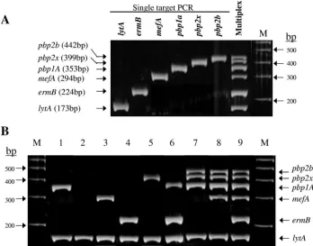
[image:2.585.117.465.69.342.2]erm-R), 0.2 M concentrations of each hybridization probe (LytA-DCR andLytA-ACR), and 2l of DNA template. The cycling conditions were as follows: 95°C for 10 min, followed by 40 cycles of 95°C for 5 s, 60°C for 10 s, and 72°C for 15 s. All runs included a negative control of water and a calibrator/ positive control of 2.9⫻106copies of the plasmid/l used for the standard curve. The data were analyzed by using Light-Cycler Software (v3.5) in the F2/F1 mode with a fit point calculation method. After amplification, 10 l of the PCR product was separated by electrophoresis on a 3% agarose gel (Cambrex BioScience/Rockland, Inc., Rockland, ME) for 30 min at 100 V. The positions of DNA fragments are shown in Fig. 1A, lane M (100-bp ladder; Amersham Biosciences). Fig-ure 1B shows the presence of the amplified products after agarose gel electrophoresis when DNA was extracted from FIG. 1. New multiplex PCR assay for simultaneous detection oflytA, penicillin resistance genes (alteredpbp1a,pbp2x, andpbp2b), and macrolide resistance genes [erm(B) andmef(A)]. (A) Comparison of single-target versus multiplex PCRs using “resistant” control strain 1824/F. Lane M, 100-bp ladder (Amersham Biosciences). (B) Agarose gel electrophoresis of PCR products amplified with RQ-mPCR using control strains. Lane 1, ATCC 49619 (MICs of 0.25g/ml for PCG and⬍0.5g/ml for EM); lane 2, clinical strain 4808/S (MICs of⬍0.015g/ml for PCG and
⬍0.5g/ml for EM); lane 3, clinical strain 1512/F (MICs of 0.03g/ml for PCG and 2g/ml for EM); lane 4, clinical strain 1565/F (MICs of 0.03
g/ml for PCG and 32g/ml for EM); lane 5, clinical strain 8315/F (MICs of 0.12g/ml for PCG and 0.5g/ml for EM); lane 6, clinical strain 4827/F (MICs of 0.12g/ml for PCG and 32g/ml for EM); lane 7, clinical strain 8605/Z (MICs of 8g/ml for PCG and⬍0.5g/ml for EM); lane 8, clinical strain 8729/Z (MICs of 4g/ml for PCG and 8g/ml for EM); lane 9, clinical strain 1824/F (MICs of 2g/ml for PCG and 16g/ml
for EM); lane M, 100-bp ladder (Amersham Biosciences).
on May 16, 2020 by guest
http://jcm.asm.org/
representative PCG- and erythromycin (EM)-resistantS. pneu-moniaestrains (lane 1, ATCC 49619 (MICs of PCG⫽ 0.25
g/ml and EM ⬍ 0.5 g/ml); lane 2, clinical strain 4808/S (MICs of PCG⬍0.015g/ml and EM⬍0.5g/ml); lane 3, clinical strain 1512/F (MICs of PCG⫽0.03g/ml and EM⫽ 2g/ml); lane 4, clinical strain 1565/F (MICs of PCG⫽0.03
g/ml and EM ⫽ 32 g/ml); lane 5, clinical strain 8315/F (MICs of PCG⫽0.12g/ml and EM ⫽ 0.5g/ml); lane 6, clinical strain 4827/F (MIC of PCG⫽0.12g/ml and EM⫽32
g/ml); lane 7, clinical strain 8605/Z (MICs of PCG⫽8g/ml and EM⬍0.5g/ml); lane 8, clinical strain 8729/Z (MICs of PCG⫽ 4g/ml and EM ⫽ 8g/ml); lane 9, clinical strain 1824/F (MICs of PCG⫽2g/ml and EM⫽16g/ml); lane M, 100-bp ladder).
Six, S. pneumoniae ATCC strains were all positive for the
lytA gene. None of the DNA extracts (ⱖ10 ng/l) from 47 nonpneumococcal organisms (including 17 clinical isolates of
S. mitisand 12 clinical isolates ofS. oralis) cross-react with the primer-probe set, showing that thelytAprimer-probe set was 100% specific for detectingS. pneumoniaestrains. We usedS. pneumoniae ATCC 49619 (positive for lytA and pbp1a), S. pneumoniaeclinical strain 4808/S (positive forlytAonly) and
S. pneumoniaeclinical strain 1824/F [positive forlytA,erm(B),
mef(A), pbp1a, pbp2x, and pbp2b] to examine the analytical sensitivity ofS. pneumoniaequantification by RQ-mPCR (see Fig. S1A in the supplemental material). The detection limit of
lytA quantification was 20 copies/assay (10 copies/l), which corresponds to 5⫻102CFU/ml. To verify the analytical sen-sitivities of drug resistance genes in RQ-mPCR, we used S. pneumoniaeclinical strain 1824/F and assessed the appearance of PCR products by gel electrophoresis (see Fig. S1B in the supplemental material). The detection limits of the five drug-resistant genes were between 5.8⫻101and 5.8⫻102copies/ assay (between 103 and 104 CFU/ml). To validate the RQ-mPCR technique, all of the 200 S. pneumoniae strains that were tested by RQ-mPCR were also screened for the presence of individual resistance genes by a single PCR using the PCR
conditions described above. The results of the two methods were in full agreement (data not shown), suggesting that the multiplex PCR primer sets are reliable. The RQ-mPCR results of 200S. pneumoniaestrains and the MIC distribution of PCG and EM are shown in Tables 1 and 2. All of the 200 strains were positive for thelytAgene. The multiplex PCR correctly identified penicillin susceptibility (MICⱕ0.06g/ml) or non-susceptibility (MICⱖ 0.12g/ml) in 189 (94.5%) of 200 iso-lates evaluated. The sensitivity, specificity, positive predictive values, and negative predictive values of our assay were 98.1% (103/105), 90.5% (86/95), 91.9% (103/112), and 97.7% (86/88), respectively. Our assay also correctly identified EM suscepti-bility (MICⱕ0.5g/ml) or resistant (MICⱖ1g/ml) in 200 (100%) out of 200 isolates evaluated. All of these isolates yielded 100% sensitivity, 100% specificity, 100% positive pre-dictive values, and 100% negative prepre-dictive values.
Of 200 purulent sputum samples, 56 samples wereS. pneu-moniaepositive, and the remaining 144 samples wereS. pneu-moniae negative as determined by the conventional culture method. All 56S. pneumoniae-positive samples were also pos-itive for the lytA gene, and 143 of the 144 S. pneumoniae -negative samples were -negative for thelytAgene. Therefore, the sensitivity and specificity for the identification ofS. pneu-moniae using purulent sputum samples in RQ-mPCR com-pared to the conventional culture method were 100% (56/56) and 99.3% (143/144), respectively. The correlation between the conventional culture counts and the level of lytA gene expression by RQ-mPCR using the 56 pneumococcal culture-positive sputum samples is shown in Fig. S2 in the supplemen-tal material.
The penicillin- and macrolide-resistant genes detected by RQ-mPCR in purulent sputum samples are shown in Table 3. We compared these results to those in isolatedS. pneumoniae
from the same sputum samples. Of 56 pneumococcal culture-positive sputum samples, all were culture-positive forlytA gene, and for 51 samples the detected genes were in complete agreement with the isolatedS. pneumoniae. For the remaining five sam-TABLE 1. RQ-mPCR results and PCG MICs in 200 pneumococcal isolatesa
RQ-mPCR result
PCG MIC distribution (no. of isolates)
ⱕ0.015g/ml 0.03g/ml 0.06g/ml 0.12g/ml 0.25g/ml 0.5g/ml 1g/ml 2g/ml 4g/ml 8g/ml
None 50 26 10 2
pbp2xonly 1 6 9 4
pbp1aonly 2 4 7 4
pbp1a⫹pbp2x 4 1 3 2
pbp1a⫹pbp2x⫹pbp2b 4 6 16 20 18 1
[image:3.585.41.541.80.162.2]aThe CLSI PCG MIC breakpoints forS. pneumoniaeare as follows: susceptible,ⱕ0.06g/ml; intermediate, 0.12 to 1g/ml; and resistant,ⱖ2g/ml (7).
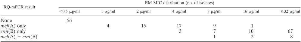
TABLE 2. RQ-mPCR results and EM MICs in 200 pneumococcal isolatesa
RQ-mPCR result
EM MIC distribution (no. of isolates)
⬍0.5g/ml 1g/ml 2g/ml 4g/ml 8g/ml 16g/ml ⱖ32g/ml
None 56
mef(A) only 4 15 17 9 1
erm(B) only 3 7 10 67
mef(A)⫹erm(B) 1 2 8
a
The CLSI EM MIC breakpoints forS. pneumoniaeare as follows: susceptible,ⱕ0.25g/ml; intermediate, 0.5g/ml; and resistant,ⱖ1g/ml (7).
on May 16, 2020 by guest
http://jcm.asm.org/
[image:3.585.42.542.646.716.2]ples, the genes were not in complete agreement with the iso-lated S. pneumoniae: two samples were false positives for
erm(B); one sample was false positive for mef(A), pbp1a,
pbp2x, andpbp2b; one sample was false positive forpbp2xand
pbp2b; and one sample was false positive formef(A) and false negative for pbp1a. The sensitivities and specificities of this assay for detecting genes directly from sputum samples relative to isolatedS. pneumoniaewere 100 and 93.9% forerm(B), 100 and 94.8% for mef(A), 94.4 and 97.5% for pbp1a, 100 and 94.1% forpbp2x, and 100 and 95.6% forpbp2b, respectively. With regard to the 144 pneumococcal culture-negative sputum samples, the detection rates of drug resistant genes were 0% for pbp1a, 2.7% (4/144) for pbp2x, 1.4% (2/144) for pbp2b, 20.8% (30/144) forerm(B), and 11.1% (16/144) formef(A).
Microorganisms closely related toS. mitisand harboringlytA
gene, which are classically associated withS. pneumoniae, have been reported previously (37). However, positive results were not obtained from the mitis group of streptococci (including our collected clinical strains of 17S. mitisand 12S. oralis) that were tested for cross-reactivity in the present study. Compared to past reports (13, 14, 21, 34, 35), we obtained lowerpbp2b
detection rates in PCG-intermediate S. pneumoniae strains. This may have been due to a difference in the regions targeted by thepbp2bprimers. The PCR results of macrolide-resistant genes in the present study matched previous report using the same primers (21), and they were also consistent with reports using other primers (11, 31). Although the analytical sensitiv-ities of resistance genes on gel electrophoresis were lower than those seen withlytAquantification, the detection limit of 103to 104CFU/ml in resistant strains is within the permissible range for use with clinical samples, including bronchoalveolar fluid, which requires a diagnostic sensitivity for pathogens in excess of 104CFU/ml (1, 9). With regard to one sputum sample which showed a false positive for thelytAgene, the RQ-mPCR assay (confirmed repeatedly) showed the presence of 104CFU ofS.
pneumoniae/ml, and this specimen grew to 2⫻ 107 CFU of
Staphylococcus aureus/ml by the culture method. This discrep-ancy may have resulted from a failure to detectS. pneumoniae
that was surrounded byS. aureusand/or have resulted from the detection of atypicalS. mitisorS. oralis, both of which harbor thelytAgene (37).
A discrepancy in RQ-mPCR specificity for samples 102/5F01 [false positive formef(A)], 107/5F23 (false positive forpbp2x
andpbp2b), 113/5F28 [false positive formef(A),pbp1a,pbp2x, and pbp2b], and 101/6S11 and 101/6D13 [false positive for
erm(B)] was confirmed by single PCR. This discrepancy indi-cates the presence of similar resistance genes in other micro-organisms in the sputum samples. Avoiding these problems with cross-reactivity is difficult because the mosaic genes that encode altered, low-affinitypbpgenes are considered products of recombination events involving horizontal transfer from closely related species (10, 16) and also becauseerm(B) and
mef(A) macrolide-resistant genes inS. pneumoniaeare highly homologous to genes in other Streptococcus-related species (6). In sample 102/5F01,pbp1awas detected by a single PCR, suggesting that the discrepancy in sensitivity (false negative) was due to the presence of PCR inhibitors or to a decline of sensitivity in this sputum sample. Although the sensitivity and specificity rates of our assay for sputum samples were in gen-eral satisfactory, further investigations are needed to remove PCR inhibitors from samples and to increase the sensitivity of the assay. Although our RQ-mPCR assay showed high corre-lation between the conventional culture counts and the level of
lytAgene expression, further data from patients with pneumo-nia are needed to evaluate and interpret the results of our assay.
In summary, the RQ-mPCR method developed here had high sensitivity and specificity for pneumococci and could de-tect drug resistance in both clinicalS. pneumoniaestrains and sputum samples. Furthermore, the results can be obtained directly from clinical samples within 3 h (2 h for DNA extrac-tion and preparaextrac-tion of PCR mixture and 1 h for PCR assay and electrophoresis), and this assay requires only a single tube. This method may be helpful for the rapid screening of resis-tance in pneumococcal isolates and should allow the adminis-tration of earlier, more focused and effective treatment of drug-resistantS. pneumoniae.
We thank Daiichi Sankyo Co., Ltd., for providing 10 clinical strains (8 strains of S. mitisand 2 strains of S. oralis), and we also thank Astellas Pharma, Inc., for providing 19 clinical strains (9 strains ofS. mitisand 10 strains ofS. oralis), which were used for the cross-reac-tivity studies.
REFERENCES
[image:4.585.44.541.81.206.2]1.Afessa, B., R. D. Hubmayr, E. A. Vetter, M. T. Keegan, K. I. Swanson, L. M. Baddour, F. R. Cockerill III, and S. G. Peters. 2006. Bronchoscopy in
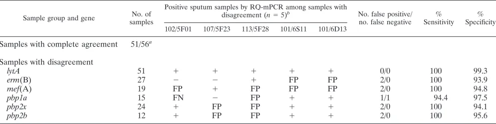
TABLE 3. Comparison of RQ-mPCR results between in pneumococcal positive sputum samples and inS. pneumoniaeisolates
Sample group and gene No. of
samples
Positive sputum samples by RQ-mPCR among samples with
disagreement (n⫽5)b No. false positive/
no. false negative % Sensitivity
% Specificity
102/5F01 107/5F23 113/5F28 101/6S11 101/6D13
Samples with complete agreement 51/56a
Samples with disagreement
lytA 51 ⫹ ⫹ ⫹ ⫹ ⫹ 0/0 100 99.3
erm(B) 27 ⫺ ⫺ ⫹ FP FP 2/0 100 93.9
mef(A) 19 FP ⫹ FP FP FP 2/0 100 94.8
pbp1a 15 FN ⫺ FP ⫹ ⫹ 1/1 94.4 97.5
pbp2x 24 ⫹ FP FP ⫹ ⫹ 2/0 100 94.1
pbp2b 12 ⫹ FP FP ⫹ ⫹ 2/0 100 95.6
aThat is, the number of samples in complete agreement with RQ-mPCR results inS. pneumoniaeisolates/number of all samples.
bFP, false-positive; FN, false-negative.
on May 16, 2020 by guest
http://jcm.asm.org/
ventilator-associated pneumonia: agreement of calibrated loop and serial
dilution. Am. J. Respir. Crit. Care Med.172:1229–1232.
2.Apfalter, P., B. Stoiser, W. Barousch, M. Nehr, L. Kramer, and H. Burg-mann.2005. Community-acquired bacteria frequency detected by means of quantitative polymerase chain reaction in nosocomial early-onset ventilator
associated pneumonia. Crit. Care Med.33:1492–1498.
3.Arbique, J. C., C. Poyart, P. Trieu-Cuot, G. Quesne, M. G. S. da Carvalho, A. G. Steigerwalt, R. E. Morey, D. Jackson, R. J. Davidson, and R. R. Facklam.2004. Accuracy of phenotypic and genotypic testing for
identifica-tion ofStreptococcus pneumoniaeand description ofStreptococcus
pseudo-pneumoniaesp. nov. J. Clin. Microbiol.42:4686–4696.
4.Baquero, F.1995. Pneumococcal resistance to-lactam antibiotics: a global
geographic overview. Microb. Drug Resist.1:115–120.
5.Bayram, A., E. Kocoglu, I. Balci, A. Filiz, and F. Eksi.2006. Real-time
polymerase chain reaction assay for detection ofStreptococcus pneumoniae
in sputum samples from patients with community-acquired pneumonia. J.
Microb. Immunol. Infect.39:452–457.
6.Clancy, J., J. Petitpas, F. Dib-Haji, W. Yuan, M. Cronan, A. V. Kamath, J. Bergeron, and J. A. Retsema.1996. Molecular cloning and functional
anal-ysis of a novel macrolide-resistance determinant,mefA, fromStreptococcus
pyogenes. Mol. Microbiol.22:867–879.
7.Clinical and Laboratory Standards Institute.2006. Methods for dilution antimicrobial susceptibility tests for bacteria that grow aerobically; approved standard, 7th ed. CLSI document M7–A7. Clinical and Laboratory Standards Institute, Wayne, PA.
8.Clinical and Laboratory Standards Institute.2006. Performance standards for antimicrobial susceptibility testing; 16th informational supplement. CLSI document M100–S16. Clinical and Laboratory Standards Institute, Wayne, PA.
9.Delclaux, C., F. E. Roupie, Blot, L. Brochard, F. Lemaire, and C. Brun-Buisson.1997. Lower respiratory tract colonization and infection during severe acute respiratory distress syndrome: incidence and diagnosis. Am. J.
Respir. Crit. Care Med.156:1092–1098.
10.Dowson, C. G., A. Hutchinson, J. A. Brannigan, R. C. George, D. Hansman, J. Lin´ares, A. Tomasz, J. M. Smith, and B. G. Spratt.1989. Horizontal transfer of penicillin-binding protein genes in penicillin-resistant clinical
isolates ofStreptococcus pneumoniae. Proc. Natl. Acad. Sci. USA86:8842–
8846.
11.Farrell, D. J., I. Morrissey, S. Bakker, L. Morris, S. Buckridge, and D. Felmingham.2004. Molecular epidemiology of multiresistantStreptococcus pneumoniaewith botherm(B)- andmef(A)-mediated macrolide resistance.
J. Clin. Microbiol.42:764–768.
12.Hakenbeck, R., T. Grebe, D. Zahner, and J. B. Stock. 1999.-Lactam
resistance inStreptococcus pneumoniae: penicillin-binding proteins and
non-penicillin binding proteins. Mol. Microbiol.33:673–678.
13.Ho, P. L., R. C. W. Wong, F. K. H. Chow, M. Y. M. Cheung, S. S. Y. Wong, W. C. Yam, and T. L. Que.2004. Application of a multiplexpbp2bandpbp2x
PCR for prediction of penicillin resistance inStreptococcus pneumoniae. J.
Antimicrob. Chemother.53:890–891.
14.Jalal, H., S. Organji, J. Reynolds, D. Bennett, E. O’Mason, Jr., and M. R. Millar. 1997. Determination of penicillin susceptibility of Streptococcus pneumoniaeusing the polymerase chain reaction. Mol. Pathol.50:45–50. 15.Kais, M., C. Spindler, M. Kain, A. Or¨tqvist, and C. G. Giske.2006.
Quan-titative detection ofStreptococcus pneumoniae,Haemophilus influenzae, and
Moraxella catarrhalisin lower respiratory tract samples by real-time PCR.
Diagn. Microbiol. Infect. Dis.55:169–178.
16.Laible, G., B. G. Spratt, and R. Hakenbeck.1991. Interspecies recombina-tional events during the evolution of altered PBP2X genes in
penicillin-resistant clinical isolates of Streptococcus pneumoniae. Mol. Microbiol.
5:1993–2000.
17.Lentino, J. R., and D. A. Lucks.1987. Nonvalue of sputum culture in the
management of lower respiratory tract infections. J. Clin. Microbiol.25:758–
762.
18.Mandell, L. A., J. G. Bartlett, S. F. Dowell, T. M. File, Jr., D. M. Musher, and C. Whitney.2003. Update of practice guidelines for the management of community-acquired pneumonia in immunocompetent adults. Clin. Infect.
Dis.27:1405–1433.
19.Miller, D. L., and R. Jones.1963. A study of techniques for the examination of sputum in a field survey of chronic bronchitis. Am. Rev. Respir. Dis.
88:473–483.
20.Morozumi, M., E. Nakayama, Y. S. Iwata, Aoki, K. Hasegawa, R. Kobayashi,
N. Chiba, T. Tajima, and K. Ubukata. 2006. Simultaneous detection of pathogens in clinical samples from patients with community-acquired pneu-monia by real-time PCR with pathogen-specific molecular beacon probes.
J. Clin. Microbiol.44:1440–1446.
21.Nagai, K., Y. Shibasaki, K. Hasegawa, T. A. Davis, M. R. Jacobs, K. Ubukata, and P. C. Appelbaum.2001. Evaluation of PCR primers to screen forStreptococcus pneumoniaeisolates and-lactam resistance, and to detect
common macrolide resistance determinants. J. Antimicrob. Chemother.48:
915–918.
22.Niederman, M. S., L. A. Mandell, A. Anzueto, J. B. Bass, W. A. Broughton, G. D. Campbell, N. Dean, T. File, M. J. Fine, P. A. Gross, F. Martinez, T. J. Marrie, J. F. Plouffe, J. Ramirez, G. A. Sarosi, A. Torres, R. Wilson, and V. L. Yu.2001. Guidelines for the management of adults with
community-acquired pneumonia. Am. J. Respir. Crit. Care Med.163:1730–1754.
23.Roberts, M. C., J. Sutcliffe, P. Courvalin, L. B. Jensen, J. Rood, and H. Seppala. 1999. Nomenclature for macrolide and macrolide-lincosamide-streptogramin B resistance determinants. Antimicrob. Agents Chemother.
43:2823–2830.
24.Sahm, D. F., M. E. Jones, M. L. Hickey, D. R. Diakun, S. Y. Mani, and C. Thornsberry. 2000. Resistance surveillance of Streptococcus pneumoniae,
Haemophilus influenzae, andMoraxella catarrhalisisolated in Asia and
Eu-rope, 1997–1998. J. Antimicrob. Chemother.45:457–466.
25.Sanbongi, Y., T. Ida, M. Ishikawa, Y. Osaki, H. Kataoka, T. Suzuki, K. Kondo, F. Ohsawa, and M. Yonezawa.2004. Complete sequences of six
penicillin-binding protein genes from 40Streptococcus pneumoniaeclinical
isolates collected in Japan. Antimicrob. Agents Chemother.48:2244–2250.
26.Sheppard, C. L., T. G. Harrison, R. Morris, A. Hogan, and R. C. George.
2004. Autolysin-targeted LightCycler assay including internal process control
for detection ofStreptococcus pneumoniaeDNA in clinical strains. J. Med.
Microbiol.53:189–195.
27.Skerrett, S. J.1999. Diagnostic testing for community-acquired pneumonia.
Clin. Chest Med.20:531–548.
28.Smith, A. M., and K. P. Klugman.1995. Alterations in penicillin-binding
protein 2B from penicillin-resistant wild-type strains ofStreptococcus
pneu-moniae. Antimicrob. Agents Chemother.39:859–867.
29.Smith, A. M., and K. P. Klugman.1998. Alterations in PBP 1A essential for
high-level penicillin resistance in Streptococcus pneumoniae. Antimicrob.
Agents Chemother.42:1329–1333.
30.Song, J. H., N. Y. Lee, S. Ichiyama, R. Yoshida, Y. Hirakata, W. Fu, A. Chongthaleong, N. Aswapokee, C. H. Chiu, M. K. Lalitha, K. Thomas, J. Perera, T. T. Yee, F. Jamal, U. C. Warsa, B. X. Vinh, M. R. Jacobs, P. C. Appelbaum, and C. H. Pai.1999. Spread of drug resistantStreptococcus pneumoniaein Asian countries: Asian Network for Surveillance of Resistant
Pathogens (ANSORP) study. Clin. Infect. Dis.28:1206–1211.
31.Sutcliffe, J., T. Grebe, A. T. Kamradt, and L. Wondrack.1996. Detection of erythromycin-resistant determinants by PCR. Antimicrob. Agents
Che-mother.40:2562–2566.
32.Tait-Kamradt, A., J. Clancy, M. Cronan, F. Dib-Hajj, L. Wondrack, W. Yuan, and J. Sutcliffe.1997.mefEis necessary for the erythromycin-resistant
M phenotype inStreptococcus pneumoniae. Antimicrob. Agents Chemother.
41:2251–2255.
33.Tang, Y. W., H. Li, J. P. Griffin, D. W. Haas, and E. M. D’Agata.2002.
Rapidly increasing prevalence of penicillin-resistant Streptococcus
pneu-moniae in middle Tennessee: a 10-year clinical and molecular analysis.
J. Clin. Microbiol.40:395–399.
34.Ubukata, K., Y. Asahi, A. Yamane, and M. Konno.1996. Combinational
detection of autolysin and penicillin-binding protein 2B genes of
Streptococ-cus pneumoniaeby PCR. J. Clin. Microbiol.34:592–596.
35.Ubukata, K., T. Muraki, A. Igarashi, Y. Asahi, and M. Konno.1997.
Iden-tification of penicillin and other beta-lactam resistance inStreptococcus
pneu-moniaeby polymerase chain reaction. J. Infect. Chemother.3:190–197. 36.Yang, S., S. Lin, A. Khalil, C. Gaydos, E. Nuemberger, G. Juan, J. Hardick,
J. G. Bartlett, P. G. Auwaerter, and R. E. Rothman.2005. Quantitative PCR assay using sputum samples for rapid diagnosis of pneumococcal pneumonia
in adults emergency department patients. J. Clin. Microbiol.43:3221–3226.
37.Whatmore, A. M., A. Efstratiou, A. P. Pickerill, K. Broughton, G. Woodard, D. Sturgeon, R. George, and C. G. Dowson.2000. Genetic relationships
between clinical isolates ofStreptococcus pneumoniae,Streptococcus oralis,
andStreptococcus mitis: characterization of “atypical” pneumococci and
or-ganisms allied toS. mitisharboringS. pneumoniaevirulence factor-encoding
genes. Infect. Immun.68:1374–1382.