0095-1137/11/$12.00
doi:10.1128/JCM.01914-10
Copyright © 2011, American Society for Microbiology. All Rights Reserved.
Evaluation of Twelve Real-Time Reverse Transcriptase PCR Primer-Probe
Sets for Detection of Pandemic Influenza A/H1N1 2009 Virus
䌤
Yaowu Yang,
1,2† Fang Huang,
3† Richard Gonzalez,
2,4Wei Wang,
2Guilan Lu,
3Yongjun Li,
2,4Guy Vernet,
4Qi Jin,
1* and Jianwei Wang
1,2*
State Key Laboratory for Molecular Virology and Genetic Engineering, Institute of Pathogen Biology (IPB), Chinese Academy of
Medical Sciences and Peking Union Medical College (CAMS and PUMC), Beijing 100730, China
1; Christophe Me
´rieux Laboratory,
IPB, CAMS-Fondation Me
´rieux, Beijing 100730, China
2; Beijing Centre for Disease Control and Prevention,
Beijing 100013, China
3; and Fondation Me
´rieux, 69365 Lyon, France
4Received 21 September 2010/Returned for modification 8 November 2010/Accepted 26 January 2011
Real-time reverse transcriptase PCR (rRT-PCR) assays have greatly contributed to the detection, control,
and prevention of the pandemic influenza A/H1N1 2009 virus. To improve the rRT-PCR assays for detection
of pandemic influenza A/H1N1 2009 virus, we evaluated the sensitivity, specificity, and performance of 12
rRT-PCR primer-probe sets [SW (a) to SW (l)] using a panel of virus strains and clinical specimens. These
primer-probe sets were derived from published work and designed for detecting the hemagglutinin (HA) or the
neuraminidase (NA) gene of the pandemic influenza A/H1N1 2009 virus. A primer-probe set, SW (CDC),
developed by the Centers for Disease Control and Prevention (U.S. CDC) to target the HA gene of pandemic
influenza A/H1N1 2009 virus, was used as a referee method. Our results demonstrated that although all
primer-probe sets in this study had as high as 98.4 to 100%
in silico
coverage, some of the primer-probe sets
had better specificity, sensitivity, and amplification efficiency than others. Two primer-probe sets, SW (h) and
SW (l), which target the NA gene of pandemic influenza A/H1N1 2009 virus, were highly sensitive (10
4copies/reaction), had high detection rates (56/60,
P
ⴝ
0.134, and 59/60,
P
ⴝ
1.000), and showed ideal specificity
compared with SW (CDC). In addition, a cocktail of primer-probe sets targeted to the HA and NA genes
displayed higher detection sensitivity than primer-probe sets targeting HA or NA alone, indicating that for
practical applications, a combination of primer-probes targeting HA and NA genes is the best option for the
detection of pandemic influenza A/H1N1 2009 virus.
The pandemic influenza A/H1N1 2009 virus, which was first
identified in Mexico and the United States in April 2009 (5, 9),
has rampaged globally for the past year. Although the World
Health Organization (WHO) has declared that pandemic
influenza A/H1N1 2009 virus is entering the postpandemic
period (http://www.who.int/mediacentre/news/statements/2010
/h1n1_vpc_20100810/en/index.html), its future pattern remains
to be determined. The pandemic influenza A/H1N1 2009 virus
has been predicted to be the predominant influenza A virus
causing seasonal influenza in winter/spring 2010-2011 (http:
//www.cdc.gov/h1n1flu/qa.htm;
http://www.ecdc.europa.eu/en
/healthtopics/H1N1/Documents/1003_RA_forward_look_influenza
.pdf). In addition, the recent identification of the re-sorting of
this virus in pigs poses further, unknown risks to humans (25).
Thus, detection, control, and prevention of an epidemic caused
by pandemic influenza A/H1N1 2009 virus is still a global
challenge, and an optimized detection method is needed in
preparation for another possible outbreak.
Numerous assays for pandemic influenza A/H1N1 2009 virus
have been reported, including virus isolation, enzyme-linked
immunosorbent assay (ELISA), immunofluorescence analysis,
and several kinds of molecular tests, such as conventional
re-verse transcriptase PCR (cRT-PCR), real-time rere-verse
tran-scriptase PCR (rRT-PCR), and sequence analysis (1, 3, 4, 11,
17, 18, 20, 22, 26). Among these, rRT-PCR has been widely
used in various clinical and public health laboratories because
of its unique advantages, including ease of performance, great
accuracy, high sensitivity and specificity, fast turnaround time,
high throughput capacity, and minimal carryover
contamina-tion (2, 23). In April 2009, the WHO released the Centers for
Disease Control and Prevention (U.S. CDC) rRT-PCR
proto-col for the detection of pandemic influenza A/H1N1 2009
virus
(http://www.who.int/csr/resources/publications/swineflu
/CDCRealtimeRTPCR_SwineH1Assay-2009_20090430.pdf). This
protocol has been widely used as the diagnostic standard in
many countries and as the referee method in multiple studies
(6, 14, 18, 24, 27). Based on this, many rRT-PCR assays have
been reported (2, 7, 11, 15, 18, 24, 27, 31–33). These assays
used different primer and TaqMan probe sets to detect the
hemagglutinin (HA) and/or the neuraminidase (NA) gene of
pandemic influenza A/H1N1 2009 virus. As the primer-probe
design plays a pivotal role in an rRT-PCR assay and the
pa-rameters of different primer-probe sets may differ (28, 30),
careful evaluation of the sensitivities, specificities, and
perfor-mances of different rRT-PCR primer-probe sets will help to
improve the rRT-PCR assays for detection of pandemic
influ-enza A/H1N1 2009 virus.
In this study, we used the SW (CDC) primer-probe set (SW
H1 Forward, SW H1 Reverse, and SW H1 Probe in the U.S.
CDC protocol) from the U.S. CDC’s rRT-PCR protocol (http:
//www.who.int/csr/resources/publications/swineflu/CDCRealtime
* Corresponding author. Mailing address: No. 9 Dong Dan San
Tiao, Dongcheng District, Beijing 100730, China. Phone and fax for J.
Wang: 86-10-67828516. E-mail: [email protected]. Phone for Q. Jin:
86-10-67877732. Fax: 86-10-67877736. E-mail: [email protected].
† Y.Y. and F.H. contributed equally to this work.
䌤Published ahead of print on 2 February 2011.
1434
on May 16, 2020 by guest
http://jcm.asm.org/
RTPCR_SwineH1Assay-2009_20090430.pdf) to target the HA
gene of pandemic influenza A/H1N1 2009 virus as a referee
method. We evaluated the sensitivities, specificities, and
performances of 12 primer-probe sets from rRT-PCR assays
developed by different groups using a panel of respiratory virus
strains and clinical samples. We demonstrate that these assays
show different sensitivities, specificities, and performances for
the detection of pandemic influenza A/H1N1 2009 virus.
MATERIALS AND METHODS
Virus stocks.A pandemic influenza A/H1N1 2009 virus strain (A/Beijing/01/
2009 [GenBank accession numbers GQ183617 to GQ183624]) was isolated from Beijing, China. Two subtypes of seasonal influenza A virus strains, A/Puerto Rico/8/1934 (H1N1) and A/Jiangxi/424/2004 (H3N2), together with an influenza virus B/Shanghai/361/2002 strain, were provided by the Chinese National Influ-enza Center (CNIC). Human coronavirus OC43 (OC43) was provided by the Beijing Union Medical College Hospital. Adenovirus serotype 35 (Ad35, Holden strain) was purchased from the American Type Culture Collection (ATCC) (Manassas, VA). All of the viruses used in this study were grown in continuous cell lines (influenza A virus strains in MDCK cells, the influenza B virus strain and OC43 in LLC-MK2 cells, and Ad35 in HeLa cells).
Clinical-sample panels.An archived panel of 110 nasopharyngeal-swab
spec-imens was used for the evaluation of rRT-PCR assays. Of those, 60 specspec-imens collected between November 2009 and February 2010 were positive for pan-demic influenza A/H1N1 2009 virus. These specimens were kindly provided by the Beijing Municipal Center for Disease Control and Prevention (Beijing CDC) and confirmed using U.S. CDC’s rRT-PCR protocol for pandemic influenza A/H1N1 2009 virus. The other 50 specimens were randomly selected from spec-imens that were collected from adult patients diagnosed with susceptible viral acute respiratory tract infections at the Beijing Union Medical College Hospital (PUMCH) between June 2007 and August 2009. Multiplex or single PCR assays were used to test these specimens for common respiratory viruses, including influenza A, B, and C viruses; enterovirus; rhinovirus; parainfluenza virus types 1 to 4; metapneumovirus; coronavirus (OC43, 229E, NL-63, and HKU1); and adenovirus (29). Influenza virus HA/NA, including those of pandemic influenza A/H1N1 2009 virus, were subtyped using multiplex rRT-PCR assays as described previously (33). The PCR results were confirmed by sequencing. The 50 speci-mens included 15 positive for an H3N2 subtype, 16 positive for a seasonal H1N1 subtype, 2 positive for enterovirus, 2 positive for rhinovirus, 2 positive for para-influenza virus, and 13 negative for the respiratory viruses we tested for as described above.
Viral RNA extraction.Viral RNA was extracted from clinical specimens or
cultured viruses using the NucliSens easyMag apparatus (bioMe´rieux, Marcy L’Etoile, France) according to the manufacturer’s protocol. The viral nucleic acids were stored at⫺80°C prior to use.
Pandemic influenza A/H1N1 2009 virus rRT-PCR assays.Twelve rRT-PCR
assays for pandemic influenza A/H1N1 2009 virus were selected from work pub-lished between July 2009 and July 2010. Details of these primer-probe sets are shown in Table 1. The thermocycling parameters for these primer-probe sets were based on original reports (7, 11, 18, 24, 27, 31–33). However, the number of PCR cycles was set at 45, which was the highest cycle number used in the 12 evaluated assays (Table 1). We used the SW (CDC) primer-probe set from the U.S. CDC’s rRT-PCR protocol to target the HA gene of pandemic influenza A/H1N1 2009 virus as a referee method. The thermocycling parameters for SW (CDC) were set according to the U.S. CDC’s rRT-PCR protocol (http://www.who.int/csr/resources/publications /swineflu/CDCRealtimeRTPCR_SwineH1Assay-2009_20090430.pdf). For the sake of data analysis, all assays were performed using the same apparatus, a LightCycler 480 instrument (Roche Diagnostics, Mannheim, Germany), and the same one-step RT-PCR kit, the SuperScript III One-Step RT-PCR System with PlatinumTaq
DNA polymerase (Invitrogen, Carlsbad, CA). For all tests, 2l of nucleic acid solution was added to 23l of master mixture containing the following components at the indicated final concentrations: 1⫻reaction buffer, 1l of SuperScript III RT/PlatinumTaqMix, 500 nM each primer, and 300 nM probe labeled at the 5⬘end with a reporter dye, 6-carboxyfluorescein (FAM), and at the 3⬘end with a nonfluo-rescent quencher, BHQ-1. Reverse transcription was performed for 30 min at 50°C and for 3 min at 95°C according to the manufacturer’s instructions. The baseline fluorescence threshold for each analysis was manually adjusted based on the back-ground fluorescence of the “no-template” control reaction using the baseline sub-tracted curve fit analysis mode in LightCycler 480 Software 1.5.0. For each analysis, a sample amplification curve exceeding baseline fluorescence with a corresponding
cycle threshold (CT) value (not exceeding 36 in a 45-cycle run) was considered positive. In addition, aCTvalue between 36 and 45 was considered weakly positive. An amplification curve lower than the baseline fluorescence was defined as negative.
In silicocoverage of the primer-probe sets.A total of 1,848 HA and 1,374 NA gene sequences of pandemic influenza A/H1N1 2009 virus were obtained from the Influenza Virus Resource (http://www.ncbi.nlm.nih.gov/genomes/FLU/) for
in silicocoverage analysis.In silicocoverage of primer-probes was calculated using an in-house program, as previously described (33). A sequence was defined as a “hit” if there were two or fewer substitutions within the probe or if there were no substitutions within 5 bases from the 3⬘end, one or fewer substitution within 10 bases from the 3⬘end, or 3 or fewer substitutions within the primers (16). For degenerate primers or probes, an acceptable “hit” was defined by the presence of an accurate match between the sequence data consensus (made by BioEDIT 7.01 software) and its primers or probes.
Sensitivity, reproducibility, and specificity of rRT-PCR assays.The titers
of the three known influenza A virus strains [A/Puerto Rico/8/1934 (H1N1), A/Jiangxi/424/2004 (H3N2), and A/Beijing/01/2009 (pandemic influenza A/H1N1 2009 virus)], B/Shanghai/361/2002 (influenza B virus), human coronavirus OC43, and Ad35 were measured by 50% tissue culture infective dose (TCID50) and
calculated by the Reed-Muench method (8). Copies of RNA extracts of the three known subtypes of influenza A virus strains and the other aforementioned vi-ruses were quantified by SYBR green rRT-PCR (10, 33). The quantifications were done using standard curves made by serial dilutions of plasmids containing partial genes of influenza A and B virus NP, coronavirus RNA polymerase, and adenovirus hexon (data not shown). The limit of detection (LOD) and repro-ducibility analysis were determined using 10-fold serial dilutions (107to 1 copies/
reaction or 4.0 to 4.0⫻10⫺7
TCID50/reaction) of the quantified viral RNA
extract of pandemic influenza A/H1N1 2009 virus. Stringent criteria for the LOD were adopted to evaluate the rRT-PCR assays: (i) to achieve the maximumCT
values, (ii) to positively detect all three parallel wells (amplification curves exceeding the baseline fluorescence in a 45-cycle run), and (iii) to keep the coefficient of variation (CV) of theCTvalues less than 10%. Viral RNA extracts were tested at high concentration and high TCID50(Table 2) to determine the
analytical specificity of the rRT-PCR assays. To further evaluate the specificities of different rRT-PCR assays in clinical testing, we used an additional 50 clinical nasopharyngeal-swab specimens, obtained from PUMCH as described above, for the cross-reaction assay (Table 3). All of the analytical assays were performed at least three times.
Statistical analysis.Statistical analysis for the rRT-PCR assays was performed
using McNemar’s chi-square test for matched pairs. APvalue ofⱕ0.05 was considered significant.
RESULTS
In silico
coverage of the primer-probe sets in this study.
To
evaluate the coverage of the 12 primer-probe sets derived from
the published work for pandemic influenza A/H1N1 2009 virus
strains, an
in silico
analysis was performed using an in-house
program. The results showed that the coverage of these
primer-probe sets ranged from 98.4% to 100% (Table 1), suggesting
that all 12 primer-probe sets target the conserved regions of
the HA or NA gene of pandemic influenza A/H1N1 2009 virus.
Specificities of rRT-PCR assays.
To assess the specificities
of the rRT-PCR assays, all of the sequences of the
primer-probe sets were analyzed by BLAST analysis. The actual
cross-reactivity of the primer-probe sets was examined using a panel
of virus strains and clinical specimens (Tables 2 and 3). Based
on BLAST analysis, the SW (j) and SW (c) primer-probe sets,
which target the HA gene of pandemic influenza A/H1N1 2009
virus, cross-react with the HA genes of swine H1N2 (such as
the A/swine/Hainan/1/2005 strain) (Fig. 1A) and seasonal
H1N1 (such as A/Puerto Rico/8/1934) (Fig. 1B), respectively.
The other 10 primer-probe sets tested do not share obvious
sequence homology, nor do they cross-react with other
respi-ratory viruses (data not shown). Subsequent experiments also
showed that the SW (c) set could detect the A/Puerto Rico/8/
1934 strain (mean
C
T, 21.8; standard deviation, 0.16; CV,
on May 16, 2020 by guest
http://jcm.asm.org/
TABLE
1.
Primer-probe
sets
and
in
silico
analysis
in
the
assays
Primer/probe Sequence (5 ⬘ 3 3 ⬘ ) Gene target/nucleotide positions Amplicon size (bp) Thermocycling Total no. of sequences a No. of hits b Coverage (%) c Reference or origin SW (a) H1 hemagglutinin (segment 4) 66 45 cycles of 95°C for 15 s, 55°C for 30 s, and 72°C for 1 min SwineHA-359_For AGCAATTGAGCTCAGTGTCATCA 359 3 381 1,848 1,842 99.7 27 SwineHA-405_Rev TGGGCCATGAACTTGTCTTG 424 3 405 1,848 1,847 99.9 27 SwineHA-386_Prove FAM-AAAGGTTTGAGATATTCC-BHQ-1 386 3 403 1,848 1,818 98.4 27 SW (b) H1 hemagglutinin (segment 4) 177 45 cycles of 95°C for 15 s and 60°C for 1 min Novel swine origin H1 Forward Primer TGTGAATCACTCTCCACAGCAAGC 250 3 273 1,848 1,834 99.2 31 Novel swine origin H1 Reverse Primer ATTGGGCCATGAACTTGTCTTGGG 426 3 403 1,848 1,847 99.9 31 Novel swine origin H1 Probe FAM-CAGACAATGGAACGTGTTACCCAGGA-BHQ-1 305 3 330 1,848 1,840 99.6 31 SW (c) H1 hemagglutinin (segment 4) 119 45 cycles of 95°C for 15 s and 60°C for 1 min H1 Universal Forward Primer ATTGCCGGTTTCATTGAAGG 1048 3 1067 1,848 1,848 100.0 31 H1 Universal Reverse Primer ATGGCATTYTGTGTGCTYTT d 1166 3 1147 1,848 1,847 99.9 31 Novel swine origin H1 probe FAM-ATGAGCAGGGGTCAGGATATGCAGCCGACC-BHQ-1 1115 3 1144 1,848 1,826 98.8 31 SW (d) H1 hemagglutinin (segment 4) 75 45 cycles of 95°C for 15 s and 60°C for 1 min H1-F GGTTTGAGATATTCCCCAAGACA 389 3 411 1,848 1,818 98.4 32 H1-R GAGGACATGCTGCCGTTACA 463 3 444 1,848 1,847 99.9 32 H1-TM FAM-TCATGGCCCAATCATGACTCGAACA-BHQ-1 415 3 439 1,848 1,830 99.0 32 SW (e) H1 hemagglutinin (segment 4) 122 45 cycles of 95°C for 5 s and 55°C for 20 s SwHA370F TCAGTGTCATCATTTGAAAGGTTTG 370 3 394 1,848 1,837 99.4 18 SwHA491R TTTTTGTAGAAGCTTTTTGCTCCAG 491 3 467 1,848 1,847 99.9 18 SwHA464Pb FAM-TGAGGACATGCTGCCGTTACACC-BHQ-1 464 3 442 1,848 1,846 99.9 18 SW (f) H1 hemagglutinin (segment 4) 107 45 cycles of 95°C for 5 s and 50°C for 40 s H1-Sw-1304F TTTGGACTTACAATGCCG 1304 3 1321 1,848 1,837 99.4 24 H1-Sw-1410R TAGCTGGCTTCTTACCT 1410 3 1394 1,848 1,845 99.8 24 H1-Sw-1357P FAM-GACTACCACGATTCAAATGTGAAGA-BHQ-1 1359 3 1383 1,848 1,842 99.7 24 SW (g) H1 hemagglutinin (segment 4) 90 45 cycles of 95°C for 5 s and 55°C for 40 s H1swl-sense GGCCATTGCCGGTTTCATTG 1044 3 1063 1,848 1,841 99.6 24 H1swl-antisense TATCCTGACCCCTGCTCATTTTG 1133 3 1111 1,848 1,848 100.0 24 H1swl-probe FAM-ATCCATCTACCATCCCTGTCCACCC-BHQ-1 1093 3 1069 1,848 1,848 100.0 24 SW (h) N1 neuraminidase (segment 6) 105 45 cycles of 95°C for 5 s and 50°C for 40 s MexFluN1-Fwd ACATGTGTGTGCAGGGATAACTG 865 3 887 1,374 1,367 99.5 24 MexFluN1-Rev TCCGAAAATCCCACTGCATAT 969 3 949 1,374 1,373 99.9 24 MexFluN1-Probe FAM-ATCGACCGTGGGTGTCTTTCAACCA-BHQ-1 899 3 923 1,374 1,362 99.1 24 SW (i) H1 hemagglutinin (segment 4) 97 45 cycles of 95°C for 15 s, 60°C for 30 s, and 68°C for 40 s NH1 forward TGAGATATTCCCCAAGACAAGTTC 393 3 416 1,848 1,831 99.1 11 NH1 reverse TTTGTAGAAGCTTTTTGCTCCAG 489 3 467 1,848 1,846 99.9 11 NH1 probe FAM-TCATGACTCGAACAAAGGTGTAACGG-BHQ-1 426 3 451 1,848 1,826 98.8 11 SW (j) H1 hemagglutinin (segment 4) 106 45 cycles of 95°C for 12 s, 55°C for 60 s, and 72°C for 15 s SWHA-440 AAGGTGTAACGGCAGCATGTC 440 3 460 1,848 1,823 98.6 7 SWHA-545 TAGGATTTGCTGAGCTTTGGGTAT 545 3 522 1,848 1,848 100.0 7 SWHA-465 FAM-AGAAGCTTTTTGCTCCAGCA-BHQ-1 484 3 465 1,848 1,847 99.9 7 SW (k) H1 hemagglutinin (segment 4) 179 45 cycles of 95°C for 10 s and 55°C for 40 s H1-2009-F TTATCATTTCAGATACACCAGT 845 3 866 1,848 1,824 98.7 33 H1-2009-R AATAGACGGGACATTCCT 1023 3 1006 1,848 1,847 99.9 33 H1-2009-P FAM-CCACGATTGCAATACAACT-BHQ-1 1045 3 1067 1,848 1,844 99.8 33 SW (l) N1 neuraminidase (segment 6) 93 45 cycles of 95°C for 10 s and 55°C for 40 s N1-2009-F CAGAGGGCGACCCAAAGAGA 1281 3 1300 1,374 1,370 99.7 32 N1-2009-R GGCCAAGACCAACCCACA 1373 3 1356 1,374 1,371 99.8 32 N1-2009-P FAM-CACAATCTGGACTAGCGGGAGCAGCAT-BHQ-1 1302 3 1328 1,374 1,367 99.5 32on May 16, 2020 by guest
http://jcm.asm.org/
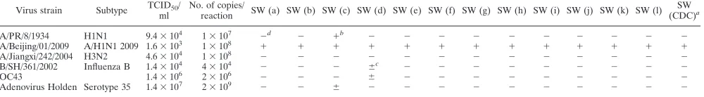
0.73%) (Table 2). The SW (j) set was not tested against the
swine H1N2 subtype because the strain was unavailable. In
addition, there were several other nonspecific, weakly positive
reactions, such as SW (c) to Ad35 and SW (d) to influenza B
virus and OC43 (Table 2). Other primer-probe sets, i.e., SW
(a), SW (b), SW (e), SW (f), SW (h), SW (k), SW (l), and SW
(CDC) (a primer-probe set in the U.S. CDC’s rRT-PCR
pro-tocol to target H1 of pandemic influenza A/H1N1 2009 virus),
did not obviously cross-react with other influenza virus
sub-types or other respiratory viruses.
Sensitivities of rRT-PCR assays.
The results of our LOD
analysis showed that the 12 rRT-PCR assays detected the
pan-demic influenza A/H1N1 2009 virus strain A/Beijing/01/2009
with different efficiencies (Table 4). Based on the stringent
conditions for the LOD, SW (d), SW (e), SW (g), and SW (k)
detected pandemic influenza A/H1N1 2009 virus most
sensi-tively, with an LOD of 10
3copies/reaction, followed by SW (c),
SW (h), SW (l), and SW (CDC), which had an LOD of 10
4copies/reaction.
Evaluation of rRT-PCR assays with clinical samples.
We
used a panel of 110 clinical samples to evaluate the
perfor-mances of different rRT-PCR assays in clinical-sample
detec-tion (Table 5). Among the assays tested, five primer-probe sets
[SW (c), (d), (e), (h), (i), and (l)] were not significantly
differ-ent from SW (CDC) (
2test;
P
⫽
0.480, 1.000, 0.480, 0.134,
1.000, and 1.000, respectively). Each set also had high
detec-tion rates for the 60 positive samples of pandemic influenza
A/H1N1 2009 virus (58, 59, 58, 56, 58, and 59, respectively).
Several sets nonspecifically detected other strains. SW (d)
nonspecifically detected H3N2 (2/15), SW (g) detected
sea-sonal H1N1 (11/16) and parainfluenza virus (1/2), and SW (i)
detected seasonal H1N1 (1/16) (Tables 3 and 5). In addition,
SW (g) reacted weakly with parainfluenza virus (1/2) and one
negative specimen (Table 3).
Based on the clinical-sample testing results (Tables 3 and 5)
and the specificity and sensitivity analyses (Tables 2 and 4 and
Fig. 1), SW (e), SW (h), and SW (l) were the ideal
primer-probe sets. In addition, the combination of SW (e) (HA
tar-geted) and SW (h) (NA tartar-geted) increased the detection rate
to 59/60 (
2test;
P
⫽
1.000), and the combination of SW (e)
(HA targeted) and SW (l) (NA targeted) increased the
detec-tion rate to 60/60 (
2test;
P
⫽
1.000) (Table 5), indicating that
a combination of primer-probe sets more effectively detects
target viruses than individual primer-probe sets alone.
DISCUSSION
Although rRT-PCR assays are more sensitive than
conven-tional reverse transcriptase PCR assays (21) and rapid
antigen-based tests (12, 13), each rRT-PCR assay may be different in
sensitivity, specificity, and performance for the detection of
pandemic influenza A/H1N1/2009 virus infection due to
differ-ent design criteria and target genes. In this study, we evaluated
12 primer-probe sets developed by different groups for the
detection of pandemic influenza A/H1N1 2009 virus. Our
re-sults demonstrated that although all primer-probe sets in this
study had as high as 98.4 to 100%
in silico
coverage (Table 1),
some of the primer-probe sets have better specificity,
sensitiv-ity, and amplification efficiency than others.
Based on the results of the specificity analysis in this study,
SW
(CDC)
e
H1
hemagglutinin
(segment
4)
116
45
cycles
of
95°C
for
15
s
and
55°C
for
30
s
SW
H1
Forward
GTGCTATAAACACCAGCCTYCCA
d
902
3
924
1,848
1,836
99.4
WHO
SW
H1
Reverse
CGGGATATTCCTTAATCCTGTRGC
d
1017
3
994
1,848
1,844
99.8
WHO
SW
H1
Probe
FAM-CAGAATATACA“T”(-BHQ-1)CCRGTCACAATTGGARAA
d
928
3
957
1,848
1,830
99.0
WHO
aTotal
sequences
for
HA
and
NA
were
obtained
from
Influenza
Virus
Resource.
bHits
were
calculated
using
an
in-house
program
described
in
Materials
and
Methods.
cCoverage
was
determined
by
the
following
formula:
100%
⫻
no.
of
hits/total
no.
of
sequences.
dR
⫽
50%
mixture
of
A
and
G;
Y
⫽
50%
mixture
of
C
and
T.
eThe
SW
(CDC)
primer-probe
set
was
from
U.S.
CDC’s
rRT-PCR
assay,
which
is
recommended
by
the
World
Health
Organization
and
has
been
used
for
clinical
sc
reening
as
the
diagnostic
gold
standard
for
pandemic
influenza
A/H1N1
2009
virus
in
many
countries
and
regions.
on May 16, 2020 by guest
http://jcm.asm.org/
the SW (j) (7) and SW (c) (31) sets are highly homologous to
the HA genes of swine H1N2 (Fig. 1A) and to those of the
human H1N1 strains (Fig. 1B and Table 2), respectively. The
high identity between SW (j) and swine H1N2 is consistent
with the notion that the pandemic influenza A/H1N1 2009
virus was a reassortant (swine H1N2
⫻
Eurasian swine H1N1)
that obtained its HA gene from swine H1N2 (19). We
exam-ined the cross-reactivities of primer-probe sets using a panel of
virus strains and clinical specimens and found several
primer-probe sets with high specificity [such as SW (a) (27), SW (h)
(24), and SW (l) (32)]. However, considering the numerous
HA and NA subtypes and the complicated evolutionary
rela-tionships between influenza A virus subtypes, the specificities
of the primer-probe sets need to be further evaluated.
As demonstrated in this report, it is often difficult to achieve
both high sensitivity and high specificity for viral nucleic acid
detection. The primer-probe sets SW (d) (32), SW (e) (18), SW
(g) (24), and SW (i) (11) are highly sensitive (10
3copies/
reaction) but react nonspecifically with other subtypes or
vi-ruses (Tables 2, 3, and 5). Given this, the two NA
gene-tar-geted sets, SW (h) and SW (l), and the HA gene-targene-tar-geted set
SW (e) appear to be the ideal primer-probe sets, because they
had relatively high sensitivity [10
4copies/reaction for SW (h)
and SW (l) and 10
3copies/reaction for SW (e)] and detection
rates [56/60,
P
⫽
0.134, for SW (h); 59/60,
P
⫽
1.000, for SW
(l); and 58/60,
P
⫽
0.480, for SW (e)], as well as ideal specificity
(Tables 2, 3, and 5). Surprisingly, our results showed that the
SW (f) (21) set had a relatively low sensitivity and detection
rate (Tables 4 and 5) for pandemic influenza A/H1N1 2009
virus, although the optimized annealing temperature, 50°C,
was selected from a series of temperatures. This may be
at-tributed to the lower melting temperature (
T
m) values of the
primers and probe. The
T
mvalues of the forward primer,
reverse primer, and probe of the SW (f) set were 47°C, 39°C,
and 54°C, respectively, which are lower than the
T
mvalues
recommended in the guidelines for designing primer-probe
sets for quantitative assays (primers, 58 to 60°C; probes, 68 to
70°C) (http://www.uic.edu/depts/rrc/cgf/).
To keep the PCR parameters consistent with the original
parameters and to derive accurate evaluation results, we did
not change any parameters except the number of PCR cycles.
Because the number of PCR cycles differed among the 12
assays compared in our study and the different numbers of
rRT-PCR cycles may affect the comparisons of sensitivity and
specificity, we set the number of PCR cycles to 45 in all assays.
This number corresponds to the largest cycle number used in
the 12 evaluated assays. The adjustment of cycle numbers did
not affect the sensitivities and specificities of the primer-probe
sets based on our comparisons (data not shown).
[image:5.585.43.543.85.154.2]Compared with the U.S. CDC’s rRT-PCR protocol for
pan-demic influenza A/H1N1 2009 virus, the SW (a), (b), (f), (j),
and (k) primer-probe sets are less sensitive and less specific.
Considering that there is no preferred standard method for
evaluating rRT-PCR assays used to detect pandemic influenza
A/H1N1 2009 virus, these assays should be further evaluated
TABLE 2. Analysis of the specificities of the different rRT-PCR assays using a panel of virus strains
Virus strain Subtype TCID50/ ml
No. of copies/
reaction SW (a) SW (b) SW (c) SW (d) SW (e) SW (f) SW (g) SW (h) SW (i) SW (j) SW (k) SW (l) SW (CDC)a
A/PR/8/1934 H1N1 9.4⫻104 1⫻107 ⫺d ⫺ ⫹b ⫺ ⫺ ⫺ ⫺ ⫺ ⫺ ⫺ ⫺ ⫺ ⫺
A/Beijing/01/2009 A/H1N1 2009 1.6⫻103 1⫻108 ⫹ ⫹ ⫹ ⫹ ⫹ ⫹ ⫹ ⫹ ⫹ ⫹ ⫹ ⫹ ⫹
A/Jiangxi/242/2004 H3N2 4.6⫻104 1⫻108 ⫺ ⫺ ⫺ ⫺ ⫺ ⫺ ⫺ ⫺ ⫺ ⫺ ⫺ ⫺ ⫺
B/SH/361/2002 Influenza B 1.4⫻104 4⫻104 ⫺ ⫺ ⫺ ⫾c ⫺ ⫺ ⫺ ⫺ ⫺ ⫺ ⫺ ⫺ ⫺
OC43 1.4⫻106 2⫻106 ⫺ ⫺ ⫺ ⫾ ⫺ ⫺ ⫺ ⫺ ⫺ ⫺ ⫺ ⫺ ⫺
Adenovirus Holden Serotype 35 1.4⫻107 2⫻109 ⫺ ⫺ ⫾ ⫺ ⫺ ⫺ ⫺ ⫺ ⫺ ⫺ ⫺ ⫺ ⫺
a
The SW (CDC) primer-probe set (SW H1 Forward, SW H1 Reverse, and SW H1 Probe in U.S. CDC protocol) was from U.S. CDC’s rRT-PCR assay, which is recommended by the World Health Organization and has been used for clinical screening as the diagnostic gold standard for pandemic influenza A/H1N1 2009 virus in many countries and regions.
b
The meanCTs, standard deviations, and CVs for “⫹” were all 8.8 to 21.8, 0.02 to 2.07, and 0.20 to 2.97%, respectively. c⫾,
CTs at 36.0 to 45.0.
d⫺, negative results in which the amplification curve values were lower than the baseline fluorescence values.
TABLE 3. Analysis of the specificities of the different rRT-PCR assays using a panel of clinical specimens positive for other viruses
Clinical specimen type/subtype
No. of samples
tested
No. positive
SW (a) SW (b) SW (c) SW (d) SW (e) SW (f) SW (g) SW (h) SW (i) SW (j) SW (k) SW (l) SW (CDC)a
Seasonal H3N2
15
0
0
0
2 (
⫹
)
b0
0
0
0
0
0
0
0
0
Seasonal H1N1
16
0
0
0
0
0
0
11 (
⫹
)
0
1 (
⫹
)
0
0
0
0
Enterovirus
2
0
0
0
0
0
0
0
0
0
0
0
0
0
Parainfluenza virus
2
0
0
0
0
0
0
1 (
⫾
)
c0
0
0
0
0
0
Rhinovirus
2
0
0
0
0
0
0
0
0
0
0
0
0
0
Negative samples
d13
0
0
0
0
0
0
1 (
⫾
)
0
0
0
0
0
0
a
The SW (CDC) primer-probe set (SW H1 Forward, SW H1 Reverse, and SW H1 Probe in U.S. CDC protocol) was from U.S. CDC’s rRT-PCR assay, which is recommended by the World Health Organization and has been used for clinical screening as the diagnostic gold standard for pandemic influenza A/H1N1 2009 virus in many countries and regions.
b⫹,
CTs were less than 36.0.
c⫾,
CTs were 36.0 to 45.0.
d
Negative samples were negative for the common respiratory viruses, such as influenza virus, enterovirus, rhinovirus, parainfluenza virus, metapneumovirus, coronavirus. and adenovirus.
on May 16, 2020 by guest
http://jcm.asm.org/
[image:5.585.42.542.569.668.2]by a third-party referee in the future. Our preliminary
conclu-sion may provide a basis for such future studies.
[image:6.585.42.536.73.273.2]Due to limitations of primer-probe sets targeting a single
gene and leading to false-negative diagnosis when sequence
variations exist in the primer- and/or probe-targeting region,
we evaluated two primer-probe sets to target the HA and NA
genes simultaneously in order to increase the sensitivity of the
rRT-PCR assay for detecting pandemic influenza A/H1N1
2009 virus. Our results show that from the standpoint of
sen-sitivity and specificity, the cocktail of SW(e) (HA) and SW(h)
(NA) or SW (e) (HA) and SW (l) (NA) is a recommended
improvement over the single set of primers for the detection of
FIG. 1. Sequence alignment of the primer-probe sets with nonspecific viruses. (A) Pairwise alignment of nucleotides between the SW (j) (7)
primer-probe set and the HA gene of a swine H1N2 strain (A/swine/Hainan/1/2005 [GenBank accession number EF556203]). (B) Pairwise
alignment of nucleotides between the SW (c) (31) primer-probe set and the HA gene of a human H1N1 strain (A/Puerto Rico/8/1934 [GenBank
accession number EF556203]).
TABLE 4. Analysis of the LODs of the different rRT-PCR assays
Assay
LODa
CT
No. of copies/
reaction TCID50/reaction Mean SD CV (%)
SW (a)
1.0
⫻
10
54.0
⫻
10
⫺228.27
0.78
2.76
SW (b)
1.0
⫻
10
64.0
⫻
10
⫺123.12
0.43
1.87
SW (c)
1.0
⫻
10
44.0
⫻
10
⫺331.98
0.08
0.26
SW (d)
1.0
⫻
10
34.0
⫻
10
⫺433.31
0.31
0.93
SW (e)
1.0
⫻
10
34.0
⫻
10
⫺435.44
1.23
3.48
SW (f)
1.0
⫻
10
74.0
33.28
1.86
5.59
SW (g)
1.0
⫻
10
34.0
⫻
10
⫺431.89
0.5
1.56
SW (h)
1.0
⫻
10
44.0
⫻
10
⫺329.51
0.08
0.26
SW (i)
1.0
⫻
10
44.0
⫻
10
⫺329.26
0.51
1.74
SW (j)
1.0
⫻
10
54.0
⫻
10
⫺226.9
0.08
0.3
SW (k)
1.0
⫻
10
34.0
⫻
10
⫺433.64
0.66
1.96
SW (l)
1.0
⫻
10
44.0
⫻
10
⫺333.45
0.61
1.82
SW (CDC)
b1.0
⫻
10
44.0
⫻
10
⫺332.37
0.42
1.31
aThe sufficient conditions of LODs were 3/3 amplification curves exceeding the baseline fluorescence in a 45-cycle run andCTCVs of less than 10%.
b
[image:6.585.300.540.357.649.2]The SW (CDC) primer-probe set (SW H1 Forward, SW H1 Reverse, and SW H1 Probe in U.S. CDC protocol) was from U.S. CDC’s rRT-PCR assay, which is recommended by the World Health Organization and has been used for clinical screening as the diagnostic gold standard for pandemic influenza A/H1N1 2009 virus in many countries and regions.
TABLE 5. Comparisons of the different rRT-PCR assays for the
detection of pandemic influenza A/H1N1 2009 virus
rRT-PCR assay Result
No. by SW (CDC)a
2
Pb
⫹ ⫺
SW (a)
⫹
48
0
10.08
0.002
⫺
12
50
SW (b)
⫹
44
0
14.06
0.000
⫺
16
50
SW (c)
⫹
58
0
0.50
0.480
⫺
2
50
SW (d)
⫹
59
2
0.00
1.000
⫺
1
48
SW (e)
⫹
58
0
0.50
0.480
⫺
2
50
SW (f)
⫹
36
0
22.04
0.000
⫺
24
50
SW (g)
⫹
58
12
5.79
0.016
⫺
2
38
SW (h)
⫹
56
0
2.25
0.134
⫺
4
50
SW (i)
⫹
58
1
0.00
1.000
⫺
2
49
SW (j)
⫹
50
0
8.10
0.004
⫺
10
50
SW (k)
⫹
51
0
7.11
0.008
⫺
9
50
SW (l)
⫹
59
0
0.00
1.000
⫺
1
50
SW (e)
⫹
SW (h)
⫹
59
0
0.00
1.000
⫺
1
50
SW (e)
⫹
SW (l)
⫹
60
0
0.00
1.000
⫺
0
50
a
The SW (CDC) primer-probe set (SW H1 Forward, SW H1 Reverse, and SW H1 Probe in U.S. CDC protocol) was from U.S. CDC’s rRT-PCR assay, which is recommended by the World Health Organization and has been used for clinical screening as the diagnostic gold standard for pandemic influenza A/H1N1 2009 virus in many countries and regions.
b
Statistical significance was determined via McNemar’s chi-square test for matched pairs. APvalue ofⱕ0.05 was considered significant. Boldface indicates values⬎0.05, representing no statistical significance for the results of compar-isons of the evaluated RT-PCR to the U.S. CDC’s rRT-PCR assay.
on May 16, 2020 by guest
http://jcm.asm.org/
[image:6.585.42.282.507.668.2]HA or NA alone. The detection rates of the combination of
SW (e) (HA targeted) and SW (h) (NA targeted) and the
combination of SW (e) (HA targeted) and SW (l) (NA
tar-geted) were 59/60 (
P
⫽
1.000) and 60/60 (
P
⫽
1.000),
respec-tively, while those of SW(e), SW (h), and SW (l) alone were
58/60, 56/60, and 59/60, respectively.
In summary, we evaluated 12 rRT-PCR primer-probe sets
for the detection of pandemic influenza A/H1N1 2009 virus.
The disparities in specificity, sensitivity, and amplification were
demonstrated by tests using virus strains and clinical samples.
Our study provides useful information on the optimization of
rRT-PCR for the detection of pandemic influenza A/H1N1
2009 virus showing that a combination of primers and probes
targeting the HA and NA genes may be the best option for the
detection of pandemic influenza A/H1N1 2009 virus.
ACKNOWLEDGMENTS
This work was supported in part by the National Basic Research
Program of China (2010CB534003), the International Science and
Technology Cooperation Program of China (2010 DFB33270), and an
intramural grant from the Institute of Pathogen Biology, Chinese
Academy of Medical Sciences (2006IPB05).
REFERENCES
1.Boggild, A. K., and A. J. McGeer.2010. Laboratory diagnosis of 2009 H1N1
influenza A virus. Crit. Care Med.38:e38–e42.
2.Butot, S., et al.2010. Evaluation of various real-time RT-PCR assays for the
detection and quantitation of human norovirus. J. Virol. Methods167:90–94.
3.Carr, M. J., et al.2009. Development of a real-time RT-PCR for the
detec-tion of swine-lineage influenza A (H1N1) virus infecdetec-tions. J. Clin. Virol. 45:196–199.
4.Centers For Disease Control And Prevention.10 August 2009. Interim
guid-ance for the detection of novel influenza A virus using rapid influenza diagnostic tests. http://www.cdc.gov/h1n1flu/guidance/rapid_testing.htm.
5.Centers For Disease Control and Prevention. 2009. Swine influenza A
(H1N1) infection in two children—Southern California, March-April 2009. MMWR Morb. Mortal. Wkly. Rep.58:400–402.
6.Cheung, T. K., et al.2010. Evaluation of novel H1N1-specific primer-probe
sets using commercial RT-PCR mixtures and a premixed reaction stored in a lyophilized format. J. Virol. Methods165:302–304.
7.Chidlow, G., et al.2010. Duplex real-time reverse transcriptase PCR assays
for rapid detection and identification of pandemic (H1N1) 2009 and seasonal influenza A/H1, A/H3, and B viruses. J. Clin. Microbiol.48:862–866.
8.Cunningham, C. H.1973. Quantal and enumerative titration of virus in cell
cultures, p. 527–532.InP. F. Kruse, Jr., and M. K. Patterson, Jr. (ed.), Tissue culture: methods and application. Academic Press, Inc., New York, NY.
9.Dawood, F. S., et al.2009. Emergence of a novel swine-origin influenza A
(H1N1) virus in humans. N. Engl. J. Med.360:2605–2615.
10.Dhanasekaran, S., T. M. Doherty, and J. Kenneth.2010. Comparison of
different standards for real-time PCR-based absolute quantification. J. Im-munol. Methods354:34–39.
11.Dong, H., et al.2010. Detection of human novel influenza A (H1N1) viruses
using multi-fluorescent real-time RT-PCR. Virus Res.147:85–90.
12.Drexler, J. F., et al.2009. Poor clinical sensitivity of rapid antigen test for
influenza A pandemic (H1N1) 2009 virus. Emerg. Infect. Dis.15:1662–1664.
13.Faix, D. J., S. S. Sherman, and S. H. Waterman.2009. Rapid-test sensitivity
for novel swine-origin influenza A (H1N1) virus in humans. N. Engl. J. Med. 361:728–729.
14.Ginocchio, C. C., et al.2009. Evaluation of multiple test methods for the
detection of the novel 2009 influenza A (H1N1) during the New York City outbreak. J. Clin. Virol.45:191–195.
15.Hall, R. J., M. Peacey, Q. S. Huang, and P. E. Carter.2009. Rapid method
to support diagnosis of swine origin influenza virus infection by sequencing of real-time PCR amplicons from diagnostic assays. J. Clin. Microbiol.47: 3053–3054.
16.He, J., et al.2009. Rapid multiplex reverse transcription-PCR typing of
influenza A and B virus, and subtyping of influenza A virus into H1, 2, 3, 5, 7, 9, N1 (human), N1 (animal), N2, and N7, including typing of novel swine origin influenza A (H1N1) virus, during the 2009 outbreak in Milwaukee, Wisconsin. J. Clin. Microbiol.47:2772–2778.
17.Hurt, A. C., et al.2009. Performance of influenza rapid point-of-care tests in
the detection of swine lineage A(H1N1) influenza viruses. Influenza Other Respi. Viruses3:171–176.
18.Jiang, T., et al.2010. Development of a real-time RT-PCR assay for a novel
influenza A (H1N1) virus. J. Virol. Methods163:470–473.
19.Kingsford, C., N. Nagarajan, and S. L. Salzberg.2009. 2009 Swine-origin
influenza A (H1N1) resembles previous influenza isolates. PLoS One 4:e6402.
20.Lalle, E., et al.2010. Design and clinical application of a molecular method
for detection and typing of the influenza A/H1N1pdm virus. J. Virol.
Meth-ods163:486–488.
21.Lam, W. Y., et al.2010. Development and comparison of molecular assays
for the rapid detection of the pandemic influenza A (H1N1) 2009 virus. J. Med. Virol.82:675–683.
22.Lu, Q., et al.2009. Detection in 2009 of the swine origin influenza A (H1N1)
virus by a subtyping microarray. J. Clin. Microbiol.47:3060–3061.
23.Mackay, I. M., K. E. Arden, and A. Nitsche.2002. Real-time PCR in virology.
Nucleic Acids Res.30:1292–1305.
24.Meijer, A., et al.2009. Preparing the outbreak assistance laboratory network
in the Netherlands for the detection of the influenza virus A(H1N1) variant. J. Clin. Virol.45:179–184.
25.Mitka, M.2010. H1N1 influenza virus reassorting in pigs, poses unknown
risk to human health. JAMA304:626–627.
26.Ninove, L., et al.2010. A simple method for molecular detection of
swine-origin and human-swine-origin influenza a virus. Vector Borne Zoonotic Dis. 10:237–240.
27.Pabbaraju, K., et al.2009. Design and validation of real-time reverse
tran-scription-PCR assays for detection of pandemic (H1N1) 2009 virus. J. Clin. Microbiol.47:3454–3460.
28.Proudnikov, D., et al.2003. Optimizing primer-probe design for fluorescent
PCR. J. Neurosci. Methods123:31–45.
29.Ren, L., et al.2009. Prevalence of human respiratory viruses in adults with
acute respiratory tract infections in Beijing, 2005–2007. Clin. Microbiol. Infect.15:1146–1153.
30.Rozen, S., and H. Skaletsky.2000. Primer3 on the WWW for general users
and for biologist programmers. Methods Mol. Biol.132:365–386.
31.Wang, R., Z. M. Sheng, and J. K. Taubenberger.2009. Detection of novel
(swine origin) H1N1 influenza A virus by quantitative real-time reverse transcription-PCR. J. Clin. Microbiol.47:2675–2677.
32.Whiley, D. M., et al.2009. Detection of novel influenza A(H1N1) virus by
real-time RT-PCR. J. Clin. Virol.45:203–204.
33.Yang, Y., et al.2010. Simultaneous typing and HA/NA subtyping of influenza
A and B viruses including the pandemic influenza A/H1N1 2009 by multiplex real-time RT-PCR. J. Virol. Methods167:37–44.
![FIG. 1. Sequence alignment of the primer-probe sets with nonspecific viruses. (A) Pairwise alignment of nucleotides between the SW (j) (7)primer-probe set and the HA gene of a swine H1N2 strain (A/swine/Hainan/1/2005 [GenBank accession number EF556203])](https://thumb-us.123doks.com/thumbv2/123dok_us/8277660.846059/6.585.300.540.357.649/sequence-alignment-nonspecic-pairwise-alignment-nucleotides-genbank-accession.webp)
