STUDY OF THE
COELIAC TRUNK AND
ITS BRANCHES IN
50
SPECIMENS
Dissertation submitted in partial fulfillment of the requirement
for the award of
M.S. DEGREE EXAMINATION (ANATOMY)
BRANCH V
MARCH – 2009
Institute of Anatomy Madurai Medical College
THE TAMILNADU Dr. M.G.R. MEDICAL UNIVERSITY
CHENNAI- 600 032
TAMILNADU
CERTIFICATE
This is to certify that the dissertation entitled “STUDY OF THE COELIAC TRUNK AND ITS BRANCHES IN 50 SPECIMENS” submitted by Dr.D.Devi Jansirani, postgraduate in Anatomy to the faculty of Anatomy, The Tamilnadu Dr. M.G.R Medical University, Chennai in partial fulfillment of the requirement for the award of M.S. Degree in Anatomy, is a bonafide work carried out by her under my direct supervision and guidance.
DECLARATION
I, Dr.D.Devi Jansirani solemnly declare that the dissertation
entitled “STUDY OF THE COELIAC TRUNK AND ITS
BRANCHES IN 50 SPECIMENS” has been prepared by me under the guidance and supervision of Dr.V.Rajaram, D.L.O., M.S., Director & Professor I/C, Institute of Anatomy, Madurai Medical college, Madurai in partial fulfillment of the requirement for the award of M.S. (Anatomy) Degree Examination of The Tamilnadu Dr. M.G.R Medical University, Chennai to be held in March 2009. This work has not formed the basis for the award of any other degree to me from any other university.
ACKNOWLEDGEMENT
I sincerely thank the Dean, Madurai Medical College, Madurai, for permitting me to use the college and department facilities to my study.
I profoundly thank Dr.V.Rajaram, D.L.O., M.S., Director & Professor I/C, Institute of Anatomy, Madurai Medical College, Madurai for his constant guidance, encouragement and help rendered throughout the period of the study.
I thank Dr.T.Hariharan, M.S, Director & Professor (Rtd), Institute of Anatomy for the selection of this topic.
I express my sincere thanks to Dr.K.Meiyazhagan, M.D., Professor & Head of the department, Department of Forensic Medicine, for prompt help in providing the autopsy specimens to my study.
CONTENTS
CHAPTER TITLE PAGE NO.
1 INTRODUCTION 1
2 AIM OF THE STUDY 4
3 REVIEW OF LITERATURE 5
4 MATERIAL AND METHODS 29
5 OBSERVATIONS 34
6 DISCUSSION 49
7 CONCLUSION 66
INTRODUCTION
A good knowledge of arterial supply of the upper abdominal organ is very much essential for the surgeons, interventional radiologists and anatomists. During surgery, arterial variations cannot be ignored for the risk of ligating the wrong vessel or severing an essential artery resulting ischemia or bleeding.
Identification of replaced arteries is more important because that may be the only blood supply for that region, ligation of which may lead to fatal necrosis. Knowledge of accessory arteries is also a must to anticipate the presence of an additional artery during surgery.
The liver donor shortage and higher demand on liver transplantation lead to advanced liver surgical techniques for which the sound knowledge of arterial anatomy and its variation is very much mandatory.
Variations had been noticed in the origin of the left gastric artery from aorta instead of from coeliac trunk. This alerts the surgeons during the gastric surgeries.
The varied origin of common hepatic artery as a direct branch from aorta and the origin of superior mesenteric artery from coeliac trunk must be known by the gastroenterologists during the surgical procedures. The rare origin of middle colic artery from coeliac trunk must also be known by the surgeons because careless ligation of which may lead to the ischemic necrosis of the right two-third of the transverse colon.
The presence of aberrant right hepatic artery from superior mesenteric artery and its unusual course as running posterior to the head of the pancreas and posterior to the portal vein should be borne in mind during portocaval shunt surgeries and care should be taken not to interfere with the blood supply of the right lobe of the liver.
A good knowledge of anatomy of splenic artery is very much essential during splenectomy, percutaneous interventional techniques such as partial splenic artery embolization, stent placement, etc.
AIM OF THE STUDY
REVIEW OF LITERATURE
Haller (1756) was the first to make an extensive work on coeliac trunk and its branches. He observed that the coeliac trunk divided into three branches the left gastric, the hepatic and the splenic artery and all these branches were found to take origin from a common point. Because of his work, such type of the coeliac trunk had long been known as the “Tripod of Haller”. He reported that the coeliac trunk may be derived from the superior mesenteric artery. The coeliac trunk gave only the splenic and left gastric arteries, the hepatic from the superior mesenteric artery. The left gastric artery took origin from the splenic artery or directly from abdominal aorta.
Eaton (1917) classified the 206 coeliac trunks into 4 types. They were:
Type I The left gastric artery and the splenic artery from a common trunk, the common hepatic artery arising from the aorta or from the superior mesenteric artery.
Type III Common trunk having the left gastric artery as its first branch showing accessory hepatic arteries and a pancreatic branch.
Type IV The left gastric artery arise separately from aorta. The common hepatic artery and the splenic artery forming a common trunk with a pancreatic branch.
He observed Tripod of Haller in 15.5% of specimens. He found that all three major branches of coeliac trunk arose from aorta by a common stem in 86% as complete trunk, coeliac trunk from aorta, but incomplete (without any of the three branches) in 12.5%, coeliaco-mesenteric trunk in 1 % and absent coeliac trunk in 0.5%. He reported 11.2% of specimens had dorsal pancreatic artery from coeliac trunk. Lipshutz (1917) dissected 83 cadavers and classified 4 types of coeliac
trunk. They were:
Type 1 Common trunk for left gastric, splenic and hepatic arteries (75%)
Type 2 Common trunk for hepatic and splenic arteries. The left gastric artery from aorta as separate branch (15%)
Type 4 Common trunk for left gastric artery and splenic artery, common hepatic artery from aorta separately (4%).
He reported 75% of specimens had complete coeliac trunk and 25% had incomplete coeliac trunk. He found that the origin of accessory hepatic artery arises from the left gastric artery and from superior mesenteric artery in 19% of cases. He reported that in 3 cases cystic artery took origin directly from the superior mesenteric artery.
Adachi (1928) dissected 252 Japanese cadavers. He distinguished 6 types of coeliac trunk. They were:
Type I The left gastric, the splenic and the hepatic artery from a common trunk (87.7%).
Type II The left gastric artery from abdominal aorta. The hepatic artery and the splenic artery from a common trunk (6.3 %).
Type III The left gastric as a direct branch of abdominal aorta. The hepatic, the splenic and the superior mesenteric artery from a common trunk (1.2%).
Type V The left gastric artery and the splenic artery from a common trunk. The hepatic and superior mesenteric artery arise from a common trunk (0.4 %).
Type VI The left gastric artery and the splenic artery from a common trunk, the hepatic artery is missing. The liver is supplied by an aberrant right or left hepatic artery or both (2%).
He reported that the origin of accessory left hepatic artery from left gastric artery in 17.9% of cases, accessory right hepatic artery from the superior mesenteric artery in 10.3% cases, accessory right hepatic artery from coeliac trunk in 2% of cases. He described the middle hepatic artery arising from the right hepatic artery in 50% of cases, from left hepatic artery in 40% of cases and from proper hepatic artery in 10% of cases. He considered the artery as the specific artery for quadrate lobe.
Pick and Anson (1940): observed inferior phrenic artery taking origin from coeliac trunk in 47.8% out of 200 cadavers.
and distribution of all the arteries in this region. Michels classified the coeliac trunk into 7 types. They were:
Type I Hepatolienogastric trunk – the typical text book pattern of coeliac trunk dividing into the hepatic, the splenic and the left gastric arteries in 89% of cases. Tripod of Haller is seen in 20% of cases. In 5% of cases, dorsal pancreatic artery is seen as a branch of coeliac trunk.
Type II Hepatolieneal trunk – coeliac trunk gives rise to hepatic and the splenic; the left gastric is displaced in 3.5% cases.
Type III Hepatolienomesentric trunk – in 0.5% cases. Here the left gastric artery arise separately at the level of coeliac trunk directly from the aorta. The hepatic, the splenic and the superior mesenteric artery arise from a common trunk.
Type IV Hepatogastric trunk – in 0.5% cases. The left gastric and the hepatic artery arise from a common trunk and the splenic artery arises from the superior mesenteric artery.
Type VI Coeliaco-mesenteric trunk (0.4%). The four arteries hepatic, splenic, left gastric and the superior mesenteric artery arise from abdominal aorta by a common trunk.
Type VII Coeliaco-colic Trunk (2 cases).In this type the middle colic (1 case) or the left colic artery (1 case) took origin from the coeliac trunk instead of from the superior mesenteric and inferior mesentric arteries.
In the textbook of Anatomy for Surgeons by Henry Hollinshead (1961), it was stated that the coeliac trunk is a large, short trunk of 1-3 cm long, which typically arises from the aorta just as this vessel enters the abdomen.
Dr. Kalavathy (1980) reported that the coeliac trunk gave origin to dorsal pancreatic artery in 10% out of 75 specimens.
Shoumura S et al (1991) studied the mode of branching of the coeliac trunk in 184 Japanese cadavers. The findings were as follows; 166 of the 184 cases belonged to Type I of Adachi's classification of variation in the arrangement of the branches of the coeliac trunk, 7 cases belonged to Type II, 2 cases belonged to Type III, 1 case belonged to Type IV, 3 cases belonged to Type V and 2 cases belonged to Type VI of which 1 case had a gastrolienal trunk and an accessory right hepatic artery arising from the superior mesenteric artery. The other one had a gastrolienal trunk and an accessory right hepatic artery arising from the gastrolienal trunk. Of the 184 cases, three could not be classified according to Adachi. 2 cases had a gastrolienal trunk and the hepatic artery arising from the abdominal aorta. 1 case had a lienomesenteric trunk and a gastrohepatic trunk.
Yamaki K et al (1995) reported a rare case of absence of coeliac trunk. In this case, the left gastric, the splenic and the common hepatic arteries arose independently in that order from the abdominal aorta and also the left aberrant hepatic artery from the left gastric artery.
Higashi N et al (1995) described that all the three branches of coeliac trunk arose directly from the abdominal aorta.
artery which arose from the aorta and supplied blood to the territory of both the coeliac and superior mesenteric arteries by giving rise to the splenic, the jejunal, the ileal, the pancreaticoduodenal, the proper hepatic and the left gastric arteries.
Cavdar S et al (1997) observed a rare variation of coeliacomesenteric trunk during the dissection of a 54-year-old male cadaver. The rare occurrence of this variation is stated to be 1% to 2.7%
Cavdar S et al (1998) described a case in which the left inferior phrenic artery and left gastric artery arose from the long coeliac trunk (4.3 cm.) via a common trunk.
Murakami T et al (1998) observed an anomaly of absent coeliac trunk in the postmortem of a Japanese adult male in which the left gastric, common hepatic, splenic arteries arose independently from the abdominal aorta.
Piao Dx et al (1998) dissected 68 Japanese cadavers and stated that inferior phrenic arteries arise from the coeliac trunk in 28.2% cases and from the left gastric artery in 2.9% cases.
aorta. In addition the common hepatic artery divided into the left hepatic, right hepatic and gastro duodenal arteries simultaneously.
Witte B et al (2001) observed during dissection of an 89-year-old female cadaver, in which the coeliac trunk gave off four arteries: the hepatic, splenic, left gastric arteries and an additional dorsal pancreatic artery.
Kahraman G et al (2001) reported a hepatomesenteric trunk, formed by the common hepatic and superior mesenteric arteries in a 50-year-old male cadaver. The left gastric and splenic arteries arose as a common trunk, the gastrosplenic trunk, from the abdominal aorta.
Nakamura Y et al (2003) presented three cases of the gastrosplenic and the hepatomesenteric trunks in Japanese cadavers. Especially, in Case 1, the left inferior phrenic artery arose from the gastrosplenic trunk and the left hepatic artery arose from the left gastric artery. In Cases 2 and 3, the common hepatic artery penetrated the pancreatic parenchyma before reaching liver. In Case 3, the right hepatic artery arose from the hepatomesenteric trunk.
Saeed M et al (2003) reported the coexistence of multiple anomalies: a short lienogastric trunk; a common hepatic artery arising directly from the abdominal aorta; a common inferior phrenic trunk arising from the coeliac trunk.
In the textbook of Gray’s Anatomy, The Anatomical basis of clinical practice (2005), it is stated that the coeliac trunk is the first anterior branch of aorta. It is 1.5 to 2 cm long. It divides into left gastric, common hepatic and splenic arteries.
the hepatogastric trunk, gave off the common hepatic and splenic arteries.
Ciçekcibaşi AE et al (2005) observed a rare variation, a coeliacomesenteric trunk. This trunk gave rise to the left gastric, the common hepatic, the splenic, the left gastroepiploic, the right and left inferior phrenic arteries.
Peschaud F et al (2006) found a variation of absent coeliac trunk. The left gastric artery and the splenic artery arose directly from the aorta, without coeliac trunk separation. The common hepatic artery was unusual in that it formed the first branch of the superior mesenteric artery, and passed in front of the portal vein to reach the hilum of the liver, where it divided into a right and a left branch.
Karakose M et al (2006) encountered the coeliac trunk divided into the left gastric, hepatic, splenic, and dorsal pancreatic arteries during a routine upper abdomen dissection of a 62-year-old male cadaver.
Yi SQ et al (2007) reported a rare variation, a common coeliacomesenteric trunk. The trunk gave rise to left gastric, common hepatic, splenic and superior mesenteric arteries.
Review of Literature for Hepatic Artery
Rossi and Cova (1904) dissected about 102 bodies and they had observed that the hepatic artery as a direct branch from the abdominal aorta in 4 cases.
Pedro Belour (1915) found that the aberrant right hepatic artery from superior mesenteric artery or from the coeliac trunk passed behind the portal vein after its origin.
Thompson (1933) made observations on 50 cadavers and found that aberrant hepatic arteries in 28% of cases.
Reginald H Jackson (1940) stated that in 20% of cases the blood supply of right lobe of liver comes from the superior mesenteric artery. John M Pierson (1943) described that an anomalous right hepatic artery arising from the superior mesenteric artery in 10% cases; Out of 10%, 8% is replaced right hepatic artery and 2% is accessory right hepatic artery. All these arteries passed dorsal to the head of the pancreas.
cases, as a branch of gastroduodenal artery in 1% cases, from coeliac trunk in 0.4% cases and from abdominal aorta in 0.2% cases. Regarding the left hepatic artery, they found its origin from proper hepatic artery in 87% cases, replaced left hepatic artery in 18% cases and accessory left hepatic artery in 35% cases. They also found the origin of right gastric artery from proper hepatic artery in 50% cases, from left hepatic artery in 32.4% cases, from right hepatic artery in 4% cases, from gastroduodenal artery in 13.2% cases and as a direct branch of coeliac trunk in 0.4% cases.
Dorvan A Moosman et al (1951) dissected 250 cadavers and found that normal right hepatic artery from coeliac axis in 85.6% cases (214 cases) and aberrant right hepatic arteries in 18.4% cases (46 cases). Out of these 46 cases, 36 were replaced right hepatic artery and 10 were accessory right hepatic artery. It was stated that cystic artery taking origin from right hepatic artery in 86% cases, from replaced right hepatic artery in 10% and origin from other sources 4%.
Gastroduodenal artery arise form the hepatic artery in all specimens studied. Right hepatic artery took origin from the proper hepatic artery in 32 specimens, as replaced right hepatic artery from superior mesenteric artery in 3 specimens (8.6%) and as accessory right hepatic artery in 4 specimens (11.4%). Left hepatic artery took origin as a branch of proper hepatic artery in 32 specimens. Right gastric artery out of 31 specimens recorded, it took origin from proper hepatic artery in 18 specimens, from left hepatic artery in 11 specimens, and from gastroduodenal artery in one specimen. In 7 specimens, aberrant right hepatic artery of superior mesenteric artery origin coursed posterior to portal vein.
Michels (1955) made a detailed study on the hepatic artery and its branches. He classified the hepatic artery into 10 types. They were:
Type-I The right, left and middle hepatic arteies-55%
Type-II The right and the middle hepatic artery; Left hepatic artery replaced form the left gastric artery -10%
Type-III The left and middle hepatic artery; Right hepatic artery replaced from the superior mesenteric artery-11%.
Type V The right, middle and the left hepatic artery. An accessory left hepatic artery from left gastric artery - 8%
Type VI The right, middle and the left hepatic artery. An accessory right hepatic artery form superior mesenteric artery - 7%.
Type VII The right, middle and the left hepatic arteries. An accessory left hepatic artery from left gastric artery and an accessory right hepatic artery form superior mesenteric artery - 1%.
Type VIII Combination pattern of the replaced right hepatic artery and an accessory left hepatic artery (or) an accessory right hepatic artery and replaced left hepatic artery - 2%.
Type IX Classical coeliacal hepatic artery was absent. The common hepatic artery form superior mesenteric artery - 4.5%
Type X The coeliacal hepatic artery is absent. The entire hepatic artery is derived from the left gastric artery - 0.5%.
hepatic artery in 10% of specimens. He observed the origin of cystic artery from right hepatic artery in 78%, from replaced right hepatic artery in 17%, from other sources in 5% of specimens .
Frederic et al (1980) analyzed the embryology of coeliac and superior mesenteric arteries. He stated that abnormal persistence or retrogression of primitive arterial segments account for the vascular anomalies. He discussed about the anomalous right hepatic artery of superior mesenteric origin in 18 to 20% of population and their importance in portocaval shunt surgery.
Eckmann I Krahn (1984) reported the origin of the right gastric artery from proper hepatic artery in 53% cases, from left hepatic artery in 15% cases, from the level of bifurcation of the proper hepatic artery in 20% cases, from gastroduodenal artery in 8% cases and from common hepatic artery in 4% cases.
Rang G.H. Stedelor WF (1987) studied in 120 patients by angiography and found that normal hepatic artery in 66% cases, aberrant right hepatic artery of superior mesenteric artery in 16% cases, aberrant left hepatic artery from left gastric artery by 11% cases.
Jonathan et al (1994) studied the anatomical variations of the hepatic arteries in the 1000 donor livers for transplantation and classified the variations into 5 types.
Type I Common hepatic artery from coeliac trunk and gave
gastroduodenal artery and proper hepatic arteries; proper hepatic artery divided into right and left hepatic arteries - 75% cases.
Type II A replaced or accessory left hepatic from left gastric artery - 9.7% cases.
Type III A replaced or accessory right hepatic artery from superior mesenteric artery - 10.6% cases.
Type IV The right hepatic artery from superior mesenteric artery and the left hepatic artery from left gastric artery - 4.5% cases.
Hiatt JR et al (1994) studied the anatomic variations in the hepatic arteries 1000 donor livers and observed that:
Type 1 (n = 757) Normal anatomy, with the common hepatic artery arising from the coeliac axis to form the gastroduodenal and proper hepatic arteries and the proper hepatic dividing distally into right and left branches.
Type 2 (n = 97), with a replaced or accessory left hepatic artery arising from the left gastric artery.
Type 3 (n = 106), with a replaced or accessory right hepatic artery originating from the superior mesenteric artery.
Type 4 (n = 23), with both right and left hepatic arteries arising from the superior mesenteric and left gastric arteries, respectively.
Type 5 (n = 15), with the entire common hepatic artery arising as a branch of the superior mesenteric artery.
Type 6 (n = 2), with the common hepatic artery originating directly from the aorta.
replaced right hepatic artery from superior mesenteric artery in 10.2% and from other sources in 4.8%.
Futura Ali et al (2001) investigated in 110 post mortem cadaver specimens in detail and found that right hepatic artery taking origin from proper hepatic artery in 66.3% specimens, common hepatic artery in 18.2% specimens, superior mesenteric artery in 8.2% specimens, coeliac trunk in 7.3% specimens. Accessory right hepatic artery taking origin from superior mesenteric artery in 7% specimens, gastroduodenal artery in 2% specimens, left hepatic artery in 1% specimen. Left hepatic artery taking origin from proper hepatic artery in 71.8% specimens, common hepatic artery in 16.4% specimens, coeliac trunk in 10.9% specimens, splenic artery in 0.9% specimen.
Bertevello PL et al (2002) disseceted in 60 cadavers and found that the right hepatic artery araised from coeliac trunk in 44 (73.3%) cases, and in 15 cases (25%) from superior mesenteric artery; it was accessory in 11 (18.3%) cases. The accessory left hepatic artery of left gastric artery in 2 (3.3%) cases. Hepatic artery trifurcation was found in 9 (15%) cases.
originating from the superior mesenteric artery in 11.5% cases and a replaced or accessory left hepatic artery originating from the left gastric artery in 5.5%. In 0.5% of the cases, there was a combination of variations of both right and left hepatic arteries. Variants of the common hepatic artery arising from the superior mesenteric artery were found in 0.5%.
Review of Literature for Splenic Artery
Arantius (1571) was the first to describe the tortuosity of the splenic artery.
Franz (1896) in his short series of 28 cases found that the splenic artery running along the upper border of the pancreas in 12 cases, behind it in 10 cases, somewhat above it in 4 cases and front of it in 2 cases.
Michels (1942) found the varying length of the splenic artery from 8 to 32 cm. Among 100 specimens, he found the origin of splenic artery from aorta in one specimen. He also reported an accessory splenic artery which was actually a superior polar artery running parallel to the splenic artery. He observed the presence of superior polar artery in 65% and inferior polar artery in 82% of specimens.
Trubel W et al (1988) studied the splenic arteries of 126 cadavers and found a cranio-sinistrally directed branch from splenic artery in 76.2%. It appeared as a posterior gastric artery (only stomach supply) in 27.7% and as a superior polar artery (only splenic supply) in 3.27%. In the most cases an intermediate type called as "gastrosplenic artery” having a posterior gastric and a superior polar branch simultaneously was detected. Such a "gastrosplenic artery" leaves the main trunk of the splenic artery in its middle segment.
Sylvester PA et al (1995) studied 29 cadaveric specimens and 44 coeliac angiograms. The straight distance from the origin of the splenic artery, from the coeliac trunk, to the point of commencement of the hilar branches was measured, as was the total length of the artery between these two points. The ratio of these two measurements is called the "index of tortuosity."
Liu DL et al (1996) observed 850 spleen specimens and found the existence of the superior and inferior polar arteries and of the coexistence of both polar arteries were 31.3%, 38.8%, and 13.3% respectively.
and found 76% of cases had superior polar artery and 24% had inferior polar artery.
Jauregui E (1999) found that the splenic artery originates, in all cases, from the coeliac trunk, and this artery is the most important with an average length of 10.6 centimeters.
The branches are noted to be superior polar artery appearing in 53% of the cases, inferior polar artery appearing in a 33%.
Daisy Sahni A.et al (2003) found that the posterior gastric artery arose from about the middle of the splenic artery.
Pandey SK et al (2004) studied the splenic artery in 320 cadavers. They found the origin of splenic artery from coeliac trunk (90.6%), aorta (8.1%) & other sites (1.3%). suprapancreatic course of the artery was commonly observed (74.1%) followed by enteropancreatic (18.5%), intrapancreatic (4.6%), and retropancreatic (2.8%) courses.
Review of Literature for Left Gastric Artery
Eaton (1917) in his study of vasculature of the stomach, found the origin of left gastric artery from aorta in 4.5% of specimens.
Reeves (1920) described the left gastric artery as commonly dividing, into an anterior and posterior branch to supply the stomach.
Michels (1952) in his study found that the incidence of the origin of left gastric artery from aorta was 2.5%.
Naidich JB et al (1978) studied 500 angiograms and reported that in 13 of 500 cases, the left gastric artery arose anomalously most often as a direct branch of the aorta; in 14 of 500 cases, the left gastric artery primarily supplied the liver with only minor contributions to the stomach. An aberrant origin of the left gastric artery necessarily influences the angiographic diagnosis and therapy of gastrointestinal hemorrhage.
Rao AK et al (1978) reported the origin of the left gastric artery from the aorta in the arteriographic evaluation in two cases.
Sawai K et al (1984) studied the coeliac angiography in 296 gastric cancer patients and found that left gastric artery emerged from the coeliac trunk in 94.9%, splenic artery in 2.7%, abdominal aorta in 2.1%, and common hepatic artery in 0.3%. The accessory hepatic arteries emerged from left gastric artery in 17.9% of the cases.
from the abdominal aorta. The rare occurrence of this variation is stated to be 0.5% -1.5%.
Ray CE Jr et al (1998) reported 2 cases, in which the left gastric artery had replaced origin from superior mesenteric artery.
MATERIAL AND METHODS
The study was conducted in the Institute of Anatomy, Madurai Medical College, Madurai- 625020.
Sample Study:
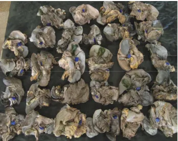
The study was done in 50 human cadaveric specimens. Of the 50 specimens, 22 were done in the dissection hall cadavers, 28 were collected from the post mortem among the Indian population irrespective of age and sex (Fig.1). Apart from these 50 specimens, a radiological study had been done in one patient. The radiological study was not included among the 50 specimens.
Collection of Specimens:
Post mortem specimens were collected from the Institute of
Forensic Medicine, Madurai Medical College, Madurai.-20. By an I-shaped incision extending from the suprasternal notch to pubic
vena cava and oesophagus were tied, cut and removed proximally. Distally the abdominal aorta and inferior vena cava were tied and cut below the level of origin of the renal arteries and removed along with abdominal diaphragm, liver, spleen, stomach and pancreas in toto. The specimens were washed in the running water. 300 – 400 ml of 10%
formalin was injected through one cut end of the abdominal aorta using a 20 ml syringe and then the specimens were completely immersed in the buckets containing 10% formalin solution and were preserved for 10 days.
Materials Used:
• Stainless steel student’s scalpel.
• Stainless steel forceps- toothed and non- toothed.
• Stainless steel long and short straight scissors.
• Knife and bone cutter.
• Black cream sheet, Rubber sheet, Graduated scale, HB pencil, 0.4mm thread and Cotton.
• Gloves and Apron
• Covered container for preserving specimens in formalin.
• 10% formalin.
• Sodium diatriazoate contrast medium.
METHOD OF STUDY
1. Gross Dissection:
a) In dissection hall specimens:
The stomach, right gastric and gastro-epiploic vessels were cut immediately to the left of the pylorus and turned to the left. The coeliac trunk was identified. The dense autonomic plexus around the trunk and its branches were removed and the branching pattern had been noticed. The splenic artery was traced along the superior border of pancreas and its branches were noticed. All the findings were recorded.
b) In postmortem specimens:
Manual dissection was done in the 28 post mortem specimens regarding the origin of the coeliac trunk and its branches. The arteries supplying the liver, stomach, pancreas and the spleen were dissected according to the above said procedure and the findings were recorded.
2. Radiological Study:
This study was done in one patient. It was not included in the present study. These images had been taken with the help of MDCT (multi-detector computerized tomography) with the administration of contrast medium.
rendering and volume rendering principle. This type of imaging provide better view of overlapping vessels.
The important structures that had been encountered in this current study are:
1. Coeliac Trunk:
o Pattern of the trunk
o Presence of Tripod of Haller o Length
o Supernumery branches like Inferior phrenic artery, Dorsal pancreatic artery, etc.
2. Branches of the Coeliac Trunk:
a) Hepatic artery:
o Origin of the hepatic artery
o Terminal branches of the hepatic artery viz. right hepatic artery and left hepatic artery.
o Aberrant branches - both accessory and replaced arteries.
o Other branches of hepatic artery like gastroduodenal artery, right gastric artery and cystic artery.
b) Splenic artery
o Length
o Tortousity index o Branches
c) Left gastric artery:
o Origin
OBSERVATIONS
By manual dissection of the coeliac trunk and its branches in 50 human specimens, the following observations were made.
COELIAC TRUNK 1. Origin:
In all the 50 specimens, coeliac trunk took origin from the ventral surface of the abdominal aorta just below the crura of the diaphragm. 2.Direction of Inclination:
In 49 specimens, coeliac trunk most inclined towards the right side and passed forwards and downwards. In 1 specimen, coeliac trunk inclined more towards left side. It was Lienogastric trunk with replaced common hepatic artery from aorta. This inclination to the left side was due to the absence of the pull exerted by coeliacal hepatic artery.
3. Pattern of Coeliac Trunk:
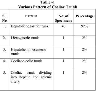
The various pattern of coeliac trunk observed in the current study were: (Table 1), (Chart 1)
(a) Hepatolienogastric trunk:
Table -1
Various Pattern of Coeliac Trunk
Sl. No
Pattern No. of
Specimens
Percentage
1. Hepatolienogastric trunk 46 92%
2. Lienogastric trunk 1 2%
3. Hepatolienomesenteric trunk
1 2%
4. Coeliaco-colic trunk 1 2%
5. Coeliac trunk dividing into hepatic and splenic artery
Chart - 1
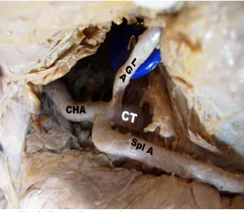
(b)Lienogastric trunk:
In one specimen, the coeliac trunk branched to the left gastric and splenic artery with left inferior phrenic artery as a supernumery branch. The common hepatic artery arose from the aorta a little below the origin of lienogastric trunk (Fig.3a). The aorta was opened and two separate ostia for the origin of coeliac trunk above and the common hepatic artery below were visualized (Fig. 3b).
(c) Hepatolienomesenteric trunk:
In one specimen, the common hepatic artery, splenic artery and superior mesenteric artery arose together as hepatolienomesenteric trunk with dorsal pancreatic artery as a supernumery branch from it. The left gastric artery had replaced origin directly from the aorta, a little above the origin of hepatolienomesenteric trunk and it gave both inferior phrenic arteries leading to the formation of the gastrophrenic trunk (Fig.4).
(d)Coeliaco-colic trunk:
down in the transverse mesocolon and supplied the right two-third of the transverse colon by its vasa recta (Fig.5).
Fig.3(a). Lienogastric trunk. Left inferior phrenic artery from
coeliac trunk.
Fig.3 (b). Luminal view of aorta: Same specimen showing two
separate ostia for the origin of coeliac trunk above (yellow
[image:50.612.137.435.138.345.2] [image:50.612.137.443.400.605.2]F
ig.4. Hepatolienomesenteric trunk with dorsal pancreatic
artery. Gastrophrenic trunk from aorta.
[image:51.612.139.430.439.650.2](e) Coeliac trunk dividing into hepatic and splenic arteries:
In one specimen, the coeliac trunk just divided into splenic artery and common hepatic artery. The left gastric artery took origin from the splenic artery (Fig.6).
4. Complete and Incomplete coeliac trunk:
Coeliac trunk giving origin to all the 3 branches i.e., left gastric, hepatic and splenic artery is known as complete trunk. Coeliac trunk without any of these 3 branches is known as incomplete coeliac trunk. In the present study, 96% of specimens had complete trunk and 4% of specimens had incomplete coeliac trunk (lienogastric & hepatolienomesenteric trunk) (Fig.3a,4), (Chart 2).
5. Tripod of Haller:
Coeliac trunk dividing into common hepatic, left gastric and splenic arteries simultaneously at a common point is known as Tripod of Haller. Such type of Tripod of Haller was observed in 19 specimens of the present study (Fig. 7).
In 24 specimens, supernumery branches from coeliac trunk were observed (Table 2), (Chart 3). The supernumery branches were:
Fig.6. Coeliac trunk dividing into hepatic artery and splenic
artery. Left gastric artery took origin from splenic artery.
Chart – 2
Table - 2
Supernumery Branches from Coeliac Trunk
Sl.No Name of the artery No. of specimens Percentage 1. Inferior phrenic artery
a. Left Inferior phrenic artery
b. Right Inferior phrenic artery c. Both right and left inferior phrenic artery - separate origin
d. Both right and left inferior phrenic artery – origin from a common trunk
16
10 2 3
1
32%
20% 4% 6%
2%
Chart - 3
(a) Inferior phrenic artery:
In 16 specimens, the inferior phrenic artery took origin from coeliac trunk. Out of these 16 specimens:
• In 10 specimens, the left inferior phrenic artery arose from coeliac trunk (Fig 8).
• In 2 specimens, the right inferior phrenic artery arose from coeliac trunk (Fig. 9).
• In 3 specimens, both right and left inferior phrenic arteries arose from coeliac trunk (Fig.10).
• In 1 specimen, coeliac trunk gave a common trunk which in turn divided into right and left inferior phrenic arteries (Fig.11).
(b) Dorsal pancreatic artery:
In 6 specimens, the origin of dorsal pancreatic artery from coeliac trunk was observed (Fig.12). In all the 6 specimens, the artery coursed posterior to the head of the pancreas and entered into the uncinate process.
(c) Superior mesenteric artery:
Fig.8. Left inferior phrenic artery from coeliac trunk.
Fig.10. Origin of both right and left inferior phrenic artery by a
common trunk from the coeliac trunk
.
(d) Middle colic artery:
In one specimen, the middle colic artery took origin from coeliac trunk (Fig.5). It coursed in the transverse mesocolon and supplied the right two third of the transverse colon by its vasa recta.
7. Length:
The length of the coeliac trunk ranged from 1.1 to 2.3 cm. The number of specimens having variable length was tabulated (Table 3). The maximum number of specimens had the length ranged from 1.1 to 2.0 cm.
Table 3
Length of the Coeliac Trunk
Length (cm) No. of specimens
1.0 to 1.5 24
1.6 to 2.0 22
2.1 to 2.5 4
HEPATIC ARTERY AND ITS BRANCHES 1. Common hepatic artery:
2.Proper Hepatic Artery:
Common hepatic artery after giving gastroduodenal artery ascended as proper hepatic artery which divided into right and left hepatic artery. This branching pattern was noticed in 36 specimens. In rest of the 14 specimens, the proper hepatic artery was not noted and the branching pattern differed as follows:
(i) Trifurcation of Common Hepatic Artery:
Out of 14 specimens, the common hepatic artery trifurcated in 4 specimens into gastroduodenal artery, right hepatic artery and left hepatic artery without the intervention of the proper hepatic artery (Fig.13).
(ii) Continuation of common hepatic artery as right hepatic artery
alone:
In 2 specimens, the common hepatic artery after giving gastroduodenal artery, it just continued as right hepatic artery alone, without the intervention of the proper hepatic artery. In those 2 specimens, the left hepatic artery had replaced origin.
(iii) Continuation of common hepatic artery as left hepatic artery
alone:
without the intervention of the proper hepatic artery. In those 8 specimens, the right hepatic artery had replaced origin.
3. Right Hepatic Artery:
The right hepatic artery took origin from proper hepatic artery in 37 specimens, from common hepatic artery in 4 specimens and in rest of the 9 specimens, it had replaced origin (Chart 4).
4. Aberrant Right Hepatic Artery:
Aberrant hepatic artery includes accessory and replaced hepatic arteries. In my study, aberrant right hepatic artery was noticed in 13 specimens, of which replaced right hepatic artery were seen in 9 specimens and accessory right hepatic artery were noticed in 4 specimens. The source of aberrant right hepatic arteries was noted as follows (Table 4):
(a)Replaced right hepatic artery:
Out of 9 specimens, the source of origin was as follows:
•In 5 specimens, it took origin from superior mesenteric artery (Fig 14).
•In 2 specimens, it took origin from gastroduodenal artery (Fig.15).
Chart -4
Table - 4
Aberrant Right Hepatic Artery
Replaced Right Hepatic Artery
Sl.
No Origin No. of specimens Percentage
1.
2.
3.
4.
Superior mesenteric artery
Gastroduodenal artery
Two replaced RHA from SMA and GDA
Replaced common hepatic artery
5 2 1 1 10% 4% 2% 2%
Total 8 16%
Accessory Right Hepatic artery
1. Gastroduodenal artery 3 6%
2. Proper hepatic artery 1 2%
Fig.14. Replaced RHA from superior mesentric artery, running
posterior to the portal vein.
• In 1 specimen, it took origin from proper hepatic artery of replaced common hepatic artery.
The replaced right hepatic artery of superior mesenteric artery origin, in all the above 6 specimens, ran unusually posterior to the head of the pancreas and ascended posterior to the portal vein in the right free margin of the lesser omentum (Fig.14).
(b) Accessory right hepatic artery:
Out of 4 specimens, the source of origin was as follows:
• In 3 specimens, it took origin from gastroduodenal artery
(Fig 17).
• In 1 specimen, it took origin from proper hepatic artery (Fig 18).
5. Left Hepatic Artery:
The left hepatic artery took origin from proper hepatic artery in 43 specimens, from common hepatic artery in 4 specimens and in the rest of the 3 specimens, it had replaced origin (Chart 5).
6. Aberrant Left Hepatic Artery:
In 3 specimens, aberrant left hepatic artery was observed (Table 5). All were replaced left hepatic artery, out of which,
Fig.17. Accessory RHA from gastroduodenal artery
.
•
•
•
•
•
•
•
•
Chart - 5
Table – 5
Aberrant Left Hepatic Artery
Replaced Left Hepatic Artery
Sl.
No
Origin No. of
specimens
Percentage
1. Left gastric artery 2 4%
2. Replaced common
hepatic artery
1 2%
• In 1 specimen, it took origin from the proper hepatic artery of replaced common hepatic artery.
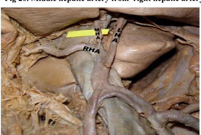
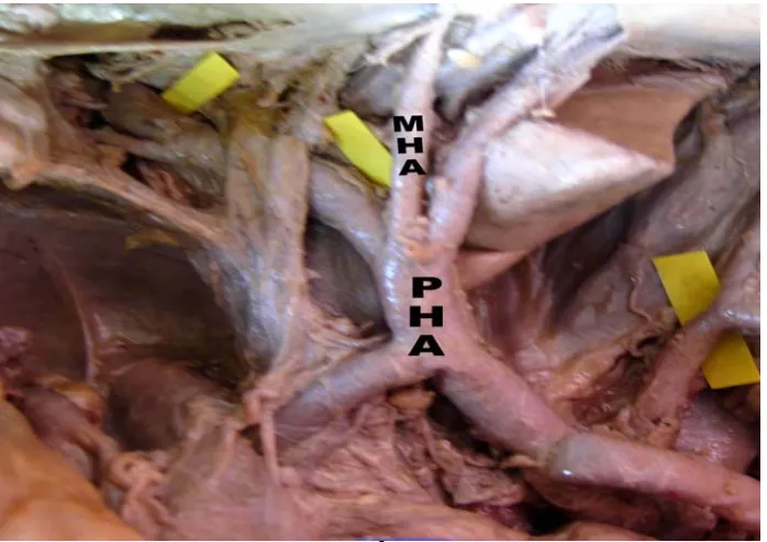
7.Middle Hepatic Artery:
In 31 specimens, middle hepatic artery was seen. Out of which,
• In 16 specimens, it took origin from right hepatic artery (Fig 20).
• In 11 specimens, it took origin from left hepatic artery (Fig 21).
• In 4 specimens, it took origin from proper hepatic artery (Fig 22).
8. Other Branches of Hepatic Artery:
(a)Gastroduodenal Artery:
In all the 50 specimens, it took origin from common hepatic artery. Among these 50 specimens, in 1 specimen, it took origin from the common hepatic artery of aortic origin. In all the specimens, it gave right gastroepiploic artery that run along the greater curvature of the stomach.
• In 3 specimens, it gave origin to replaced right hepatic artery (Fig 15).
Fig 20. Middle hepatic artery from right hepatic artery.
[image:76.612.136.474.349.576.2].
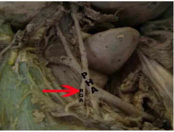
(b)Right Gastric Artery:
The origin of right gastric artery was observed as follow as: • From proper hepatic artery in 25 specimens (Fig. 23).
• From left hepatic artery in 15 specimens (Fig. 24).
• From gastroduodenal artery in 6 specimens (Fig. 25).
• From right hepatic artery in 3 specimens (Fig. 26).
• From common hepatic artery in 1 specimen.
(c) Cystic artery:
The origin of cystic artery was observed as:
• In 39 specimens, it took origin from right hepatic artery.
• In 10 specimens, it took origin from aberrant right hepatic artery. Out of which in one specimen, two cystic arteries arose separately from the two replaced right hepatic artery of gastroduodenal artery and superior mesenteric artery (Fig. 25).
• In 1 specimen, it took origin from gastroduodenal artery (Fig. 27).
SPLENIC ARTERY: 1. Origin:
Fig. 23. Right gastric artery from Proper hepatic artery.
Fig.25. Two cystic arteries from replaced RHA of GDA &
replaced RHA of SMA origin. RGA from GDA.
[image:80.612.140.491.358.618.2]2. Length:
The length of the splenic artery ranged from 8 cm to 13.5 cm. Its mean length was 9.11 cm. The number of specimens having variable length was tabulated (Table 6).
Table 6
Length of the Splenic Artery
Length (cm) No. of
specimens 8.0 to 10.0 26
10.1 to 12.0 18
12.1 to 13.5 6
3. Tortuosity Index:
Splenic artery is famous for its tortousity. In some specimens, it was highly tortuous and in few it was almost straight. The ratio of the curved length of the splenic artery to its straight distance from its origin to the point of commencement of hilar branches is called as tortuosity index. It ranged from 1.02 to 1.29.
2.Relation with the Pancreas:
specimens (Fig. 28). In 10 specimens, it passed behind the pancreas (Fig. 29). In 2 specimens, it passed inside the substance of pancreas (Fig. 30).
2. Branches:
In all the specimens, the usual branches of splenic artery such as pancreatic branches, short gastric arteries, and the left gastroepiploic artery were observed. The artery terminated by dividing into two or more splenic branches that entered into the hilum of the spleen. Apart from these usual branches, some of the peculiar branches were noted. They were:
(a) Polar Arteries:
A separate branch from the splenic artery was observed to enter into the superior or inferior poles according to which they are named as superior polar artery and inferior polar artery respectively. In the present study,
• Superior polar artery was observed in 15 specimens (Fig.31).
• Inferior polar artery was observed in 18 specimens (Fig.32).
Fig.28. Splenic artery running along the upper border of
pancreas.
Fig 30. Intrapancreatic course of splenic artery.
[image:85.612.136.489.385.632.2]Fig.32. Inferior polar artery from splenic artery.
(b) Dorsal pancreatic artery:
In 9 specimens, dorsal pancreatic artery took origin from the proximal few centimeter of origin of the splenic artery (Fig.34).
(c) Posterior gastric artery:
In 14 specimens, the posterior gastric artery took origin from the middle of the splenic artery and it coursed upwards to enter into the posterior surface of the stomach (Fig.35).
(d) Gastrosplenic artery:
In 12 specimens, the gastrosplenic artery took origin from the splenic artery. This artery divided into two branches, one entered into the posterior surface of the stomach and the other entered into the inferior pole of the spleen (Fig.36). This artery is considered to be the intermediate type between posterior gastric artery and inferior polar artery.
(e) Left Gastric Artery:
In one specimen, the left gastric artery took origin from the splenic artery (Fig.6).
(f) Accessory Splenic Artery:
Fig 34. Dorsal pancreatic artery from splenic artery.
[image:88.612.138.487.366.620.2]Fig 36. Gastrosplenic artery dividing into 2 branches to supply
the posterior surface of stomach & inferior pole of spleen.
[image:89.612.136.487.392.630.2](g) A branch to the Accessory Spleen:
In 1 specimen, a branch from the splenic artery supplied an accessory spleen which was present in the lienorenal ligament (Fig.38). LEFT GASTRIC ARTERY
1. Origin:
In 48 specimens, it took origin from the coeliac trunk. In one specimen, it took origin directly from the aorta, a little above the origin of coeliac trunk as Gastrophrenic Trunk, which gave both right and left inferior phrenic arteries (Fig.4). In another one specimen, it took origin from the splenic artery (Fig.6).
2. Division and Distribution:
In one specimen, the artery divided into two branches at proximal few centimeters of its origin as an anterior and posterior branch to supply the respective surfaces of the stomach (Fig 39).
3. Supernumery Branches:
Apart from the gastric and oesophageal branches, the left gastric artery gave following supernumery branches.
(a) Inferior Phrenic Artery:
Fig.38. Splenic artery giving a branch to accessory spleen.
[image:91.612.137.488.292.542.2]• In 1 specimen, the left gastric artery gave origin to left inferior phrenic artery (Fig. 40).
• In 1 another specimen, the left gastric artery taking origin from aorta gave both right and left inferior phrenic arteries as Gastrophrenic trunk (Fig. 4).
(b) Replaced Left Hepatic Artery:
In 2 specimens, the left gastric artery gave replaced left hepatic artery (Fig. 19).
Radiological Study
Fig 40. Left inferior phrenic artery from left gastric artery.
[image:93.612.136.487.463.681.2]Fig.42
Reconstructed
volumetric 3D
rendered CT
angiogram in AP
projection showing
origin of coeliac trunk
and its branches.
Fig.43
Reconstructed
volumetric 3D
rendered CT
DISCUSSION
The anatomy of coeliac trunk and its branches studied in 50 specimens were compared with the previous studies.
COELIAC TRUNK 1. Origin:
In the textbook of Gray’s Anatomy, it was stated that the coeliac trunk is the ventral branch of the aorta dividing into the left gastric, common hepatic and splenic arteries. In my study also the coeliac trunk took origin from the ventral surface of the aorta in all the 50 specimens.
Rossi (1904), Piquand (1910), Yamaki et al (1995), Higashi N et al (1995) Basar et al (1995), Marakami T et al (1998) Peschaud F et
(2006) had noticed absence of coeliac trunk and all the three branches took separate origin from aorta. But in the present study, coeliac trunk was not absent in any of the 50 specimens.
2. Direction of Inclination:
3. Pattern:
The various pattern of coeliac trunk were: (Table 7), (Chart 6)
a) Hepatolienogastric Trunk:
Lipshutz (1917) had observed this type of trunk in 75% out of 83 cadavers, Adachi (1928) in 87.7% out of 252 specimens, Michels (1955) in 89% out of 200 specimens, Shoumura S et al (1991) in 90.2% out of
184 specimens. In my study, its incidence is 92% out of 50 specimens, which is closely similar to Shoumura S et al study.
b) Lienogastric Trunk:
Lipshutz (1917) found this type in 4% specimens, Michels (1955) in 2%, Shoumura S et al (1991) in 1.09% of specimens. In the current
study, it was noticed in 1 specimen – 2% incidence, which is similar to Michels study.
c) Hepatolienomesenteric Trunk:
This type of trunk had been reported by Adachi (1928) in 1.2%, Michels (1955) in 0.5%, Shourmura S et al (1991) in 1.09%. This trunk
Table – 7
Various Pattern of Coeliac Trunk
Sl. No
Name of the authors Year of Study No. of specimens Hepatolieno gastric trunk Lienogastric trunk Hepatolieno mesenteric trunk Coeliaco colic trunk 1. Lipshutz 1917 83 75% 4% - - 2. Adachi 1928 252 87.7% - 1.2% - 3. Michels 1955 200 89% 2% 0.5% 0.5% 4. Shoumura S
et al 2001 184 90.2% 1.09% 1.09% -
5. Present
Chart - 6
Various Pattern of Coeliac Trunk
Percenta
d) Coeliaco-colic Trunk:
This rare variation had been reported by Michels (1955) in which the origin of middle colic artery from coeliac trunk was found in 1 out of 200 specimens - 0.5%. In my study, it was noticed in 1 out of 50 specimens - 2%, which is closely similar to the above study.
e) Others:
Hepatogastric trunk was observed by Lipshutz (1917) in 6%, Michels (1955) in 0.5%. Coeliaco mesenteric trunk was reported by Adachi (1928) in 2.4%, Michels (1955) in 0.4%. In my study, hepatogastric and coeliaco mesenteric trunk was not observed.
4. Complete and Incomplete Coeliac Trunk:
Eaton (1917) observed complete coeliac trunk in 86% and incomplete trunk in 12.5% of specimens. Lipshutz (1917) reported complete coeliac trunk in 75% and incomplete in 25%. In the current study, complete coeliac trunk was seen in 96% and incomplete in 4% of specimens.
5. Tripod of Haller:
6. Supernumery Branches:
a) Inferior Phrenic Artery:
Pickand Anson (1940) in 47.8% out of 200 cadavers observed the origin of inferior phrenic artery from coeliac trunk. Piao Dx et al (1998)
stated that the incidence of inferior phrenic artery taking origin from coeliac trunk was 28.2%. In my study, in 32% of the specimens, the inferior phrenic artery took origin from the coeliac trunk as supernumery branch, which is closer to Piao Dx study.
Saeed M et al (2003) reported a case in which a common inferior
phrenic artery taking origin from coeliac trunk and then dividing into right and left inferior phrenic artery. Such type of common inferior phrenic artery was observed in 1 in 50 specimens (2%) in the my study.
Cicekcibasi AE et al (2005) reported a case in which both inferior
phrenic arteries took origin from coeliac trunk separately. In my study, the same variation had been recorded in 3 specimens - 6%.
b) Dorsal Pancreatic Artery:
c) Superior mesenteric artery:
Adachi (1928) in 1.2%, Michels (1955) in 0.5% of specimens observed the superior mesenteric artery arising from coeliac trunk. In my study, the same was observed in one specimen - 2%, which is closely similar to Adachi’s study.
d) Middle colic artery:
Michels (1955) observed the origin of middle colic artery from coeliac trunk in 0.5%. In my study too, it was observed in one specimen (2%).
7. Length of the Coeliac Trunk:
In the textbook of Gray’s Anatomy, it was stated that the length of the coeliac trunk is 1.5 to 2 cm. In the textbook of Anatomy for Surgeons by Hollinshead, it was stated that its length is 1 to 3 cm. In my study also, the length of the coeliac trunk ranged from 1.1 to 2.3 cm. 8. Michel’s Classification of Coeliac Trunk:
HEPATIC ARTERY
1. Origin of Common Hepatic Artery:
Daseler et al (1947) reported that the incidence of origin of the
common hepatic artery from coeliac trunk was 83.2%. In the present study the incidence was 98%.
Origin of common hepatic artery from aorta was reported by Rossi and Cova (1904) in 3.9%, Daseler et al (1947) in 0.2%, Jonathen
et al (1984) in 0.2%, by Shoumura S et al (1991) in 1.08%, Hirari JR et
al (1994) in 0.2%. In my study, in 2% of specimens the common hepatic
artery took origin from aorta which is similar to that of Shoumura S et al
study (Chart 7).
2. Trifurcation of Common Hepatic Artery:
Trifurcation of common hepatic artery into right hepatic, left hepatic and gastroduodenal artery was observed by Margaret Kemeny et al (1986) in 9% cases, by Bartevello P.L et al (2002) in 15%. In this
study, the incidence was 8%, which is similar to that of Margaret Kemeny et al study.
3. Right Hepatic Artery:
E.R.Flint (1922-23) in 79%, Daseler et al (1947) in 83.2% and
Dorvan A Moosman et al (1951) in 85.6%, Arjhansiri K et al (2006) in
Chart – 7
Replaced Origin of Common Hepatic Artery from Aorta
Percent
a
g
normal coeliacal hepatic artery. In my study, the incidence was noted in 82% of the specimens which is similar to Daseler et al study.
4. Left Hepatic Artery:
Daseler et al (1947) in 87%, Edward V Johnson et al (1952) in
91.4% of the specimens observed the origin of left hepatic artery from normal coeliacal hepatic artery. In my study, the incidence was noted to be 94% of the specimens, which is nearer to that of the Edward V Johnson study.
3. Aberrant Hepatic Artery:
Thompson (1933) found aberrant hepatic arteries in 28% of cases. In my study, the incidence was 32%, which is closely similar to the above study.
4. Aberrant Right Hepatic Artery:
Daseler et al (1947) in 24% out of 500 specimens, Dorvan A
Moosman et al (1951) in 18.4% out of 250 specimens, Edward V
Johnson et al (1952) in 20% out of 35 specimens, observed the presence
of aberrant right hepatic artery. In the current study, it was noticed in 13 out of 50 specimens - 26% which is closely similar to Daseler et al
(a) Replaced Right Hepatic Artery:
Dorvan A Moosman et al (1951) observed replaced right hepatic
artery in 36 out of 250 specimens - 14.4%. Margaret M Kemeny et al
(1986) observed it in 20 out of 100 specimens - 20%. In my study, it was found in 9 out of 50 specimens - 18%, which is similar to Margaret M Kemeny et al study.
(b)Replaced Right Hepatic Artery of Superior Mesenteric Artery:
Daseler et al (1947) in 11.2%, Edward V Johnson et al (1952) in
8.6%, Michels (1955) in 11%, Nakayasu et al (2000) in 10.2%,
Arjhansiri K et al (2006) in 11.5% observed the replaced right hepatic
artery from superior mesenteric artery. In my study, the incidence was 12%, which is similar to Daseler et al and Arjhansiri et al study and
closer to Michels study (Table 8), (Chart 8).
COURSE: John M Pierson (1943) and Michles (1955) observed that all the aberrant right hepatic arteries of superior mesenteric artery origin coursed behind the pancreas. Higashi N Hirari (1955), Kahraman G et al (1984) reported that aberrant right hepatic artery of superior
Table – 8
Replaced Right Hepatic Artery of Superior Mesenteric Artery Origin
Sl. No .
Name of the authors
Year of study
No. of specimens
Percentage
1. Daseler et al 1947 500 11.2%
2. Edward V Johnson
1952 35 8.6%
3. Michels 1955 200 11%
4. Nakayasu 2000 166 10.2%
5. Arjhansiri et al
2006 200 11.5%
Chart - 8
Replaced Right Hepatic Artery of Superior Mesenteric Artery
Percenta
In my study also, all the aberrant right hepatic artery of superior mesenteric origin (12%) coursed posterior to the head of the pancreas and posterior to the portal vein.
(c) Replaced Right Hepatic Artery from Replaced Common Hepatic
Artery of Aorta:
Daseler et al (1947) reported this type of variation in 1 out of 500
specimens - 0.2%. In my study, it was noticed in 1 out of 50 specimens (2%).
(d)AccessoryRight Hepatic Artery:
Daseler et al (1947) in 7.2%,Dorvan A Moosman et al (1951) in
4% of specimens observed the presence of accessory right hepatic artery. In my study, it was observed in 8% which is similar to Daseler et al study.
(e) Accessory Right Hepatic Artery from Gastroduodenal Artery:
Daseler et al (1947) noticed this artery in 5 out of 500 specimens -
1%. Futura Ali et al (2001) noticed it in 2% of the specimens. In my
5. Replaced Left Hepatic Artery from Left Gastric Artery:
Margaret M Kemeny et al (1986) observed the presence of
replaced left hepatic artery from left gastric artery in 4 out of 100 specimens - 4%. Arjhansiri K et al (2006) observed it in 5.5% of cases.
In the present study, it was observed in 2 out of 50 specimens - 4%, which coincides with the result of the Margaret M Kemeny et al study. 6. Middle Hepatic Artery:
Adachi (1928) reported the origin of middle hepatic artery from right hepatic, left hepatic and proper hepatic artery in 50%, 40% and 10% respectively.
Michles (1955) observed its origin from right hepatic, left hepatic and proper hepatic artery in 45%, 45% and 10% respectively.
In my study, the origin of middle hepatic artery was noticed in 31 specimens (62%). Out of which, its origin from right hepatic, left hepatic and proper hepatic artery was seen in 32%, 22%, and 8% of the specimens respectively and in rest of the 38% of the specimens, middle hepatic artery was not noticed.
7.Classification of Hepatic Artery:
out of 50 specimens, 70% belonged to Type I, 4% to Type II, 12% to Type III, 2% to Type V and Type IV specimens were not noticed in any of the specimens. Rest of the 12% of specimens could not be classified under Jonathen study, out of which right and left hepatic artery arose from common hepatic artery in 8% and right hepatic artery took replaced origin from gastroduodenal artery in 4% of specimens.
8. Other Branches of Hepatic Artery:
(a) Gastroduodenal Artery:
Daseler et al (1947) observed that this artery took origin from
common hepatic artery in 75.4%, from replaced common hepatic artery in 3.6%, from right hepatic artery in 7%, from coeliac trunk in 2.5%, from aorta in 0.2% and absent in 2.8%. Edward V Johnson et al (1952)
reported its origin from common hepatic artery in all the cases.
In my study, it took origin from the common hepatic artery in all the specimens; among which in one of the specimens, it took origin from the replaced common hepatic artery of aorta.
(b) Right Gastric Artery:
The origin of right gastric artery from proper hepatic artery, left hepatic artery, gastroduodenal artery, right hepatic artery was observed by Daseler et al (1947) in 50%, 32.4%, 13.2%, 4%, by Michels (1955)
proper hepatic artery, left hepatic artery, gastroduodenal artery and right hepatic artery was observed to be 50%, 30%, 12%, and 6% respectively. The findings of the present study are similar to that of Daseler et al
study (Table 9).
Eckmann I Krahn (1984) observed its origin from common hepatic artery in 4% of specimens. In my study, in 1 specimen - 2%, it took origin from the common hepatic artery, which is closely similar to the above study.
(c) Cystic Artery:
Daseler et al (1947) observed the origin of cystic artery from right
hepatic artery in 84.4%, aberrant right hepatic artery in 12.7% and from other sources in 3%. Dorvann A Moosman et al (1951) observed the
origin of cystic artery from right hepatic artery in 86%, aberrant right hepatic artery in 10% and from other sources in 4%. Michels (1955) observed the origin of cystic artery from right hepatic artery in 78%, aberrant right hepatic artery in 18% and from other sources in 5%.
Table – 9
Origin of Right Gastric Artery
Origin of Right gastric artery
Sl. No
Name of the authors
Year of
study hepatic Proper artery Left hepatic artery Gastro duodenal artery Right hepatic artery Coeliac trunk Common hepatic artery 1. Daseler 1947 50% 32.4% 13.2% 4% 0.4% - 2. Michels 1955 40% 40.5% 8%