0022-538X/04/$08.00⫹0 DOI: 10.1128/JVI.78.23.13153–13162.2004 Copyright © 2004, American Society for Microbiology. All Rights Reserved.
SYNCRIP, a Member of the Heterogeneous Nuclear Ribonucleoprotein
Family, Is Involved in Mouse Hepatitis Virus RNA Synthesis
Keum S. Choi,
1Akihiro Mizutani,
2and Michael M. C. Lai
1*
Department of Molecular Microbiology and Immunology, Keck School of Medicine, University of Southern California, Los Angeles, California,1and Department of Molecular Neurobiology, Institute of Medical Science,
University of Tokyo, Tokyo, Japan2
Received 2 June 2004/Accepted 20 July 2004
Several cellular proteins, including several heterogeneous nuclear ribonucleoproteins (hnRNPs), have been shown to function as regulatory factors for mouse hepatitis virus (MHV) RNA synthesis as a result of their binding to the 5ⴕand 3ⴕ untranslated regions (UTRs) of the viral RNA. Here, we identified another cellular protein, p70, which has been shown by UV cross-linking to bind both the positive- and negative-strand UTRs of MHV RNA specifically. We purified p70 with a a one-step RNA affinity purification procedure with the biotin-labeled 5ⴕ-UTR. Matrix-assisted laser desorption ionization (MALDI)-mass spectrometry identified it as synaptotagmin-binding cytoplasmic RNA-interacting protein (SYNCRIP). SYNCRIP is a member of the hnRNP family and localizes largely in the cytoplasm. The p70 was cross-linked to the MHV positive- or negative-strand UTR in vitro and in vivo. The bacterially expressed SYNCRIP was also able to bind to the 5ⴕ-UTR of both strands. The SYNCRIP-binding site was mapped to the leader sequence of the 5ⴕ-UTR, requiring the UCUAA repeat sequence. To investigate the functional significance of SYNCRIP in MHV replication, we expressed a full-length or a C-terminally truncated form of SYNCRIP in mammalian cells expressing the MHV receptor. The overexpression of either form of SYNCRIP inhibited syncytium formation induced by MHV infection. Furthermore, downregulation of the endogenous SYNCRIP with a specific short interfering RNA delayed MHV RNA synthesis; in contrast, overexpression or downregulation of SYNCRIP did not affect MHV translation. These results suggest that SYNCRIP may be directly involved in MHV RNA replication as a positive regulator. This study identified an additional cellular hnRNP as an MHV RNA-binding protein potentially involved in viral RNA synthesis.
Mouse hepatitis virus (MHV) belongs to theCoronaviridae
family and contains a single-stranded, 31-kb, positive-sense RNA (15). The viral genome is composed of a series of open reading frames (ORFs 1 to 7), flanked by untranslated regions (UTRs) at the 5⬘ and 3⬘ ends. MHV RNA replication and transcription take place in the cytoplasm and are mediated by its own RNA-dependent RNA polymerase and other viral and cellular proteins. Six to seven subgenomic mRNAs share 5⬘ and 3⬘ends with the genomic RNA and are translated through a cap-dependent mechanism. Regulation of transcription, rep-lication, and translation of viral RNA involves severalcis- and
trans-acting RNA elements, including intergenic sequence, leader sequence, and the 3⬘-UTR of viral RNA (19, 20, 36, 37) and viral and cellular proteins. The leader RNA can function in viral RNA synthesis both incisand intransin virus-infected cells (36). Severaltrans-acting factors, including viral and cel-lular proteins, have been shown to bind to this region (6).
Besides the cis- and trans-acting RNA elements, cellular proteins have been increasingly recognized to play important roles in virus replication, transcription, and translation, as well as virus entry, assembly, and release (16). For example, polio-virus translation and replication are coordinated by the inter-action of host factors with viral factors at the 5⬘and/or 3⬘end of viral RNA. These factors include host poly(C)-binding
pro-tein, poly(A)-binding protein and the viral polymerase precur-sor 3CD. Poly(C)-binding protein and poly(A)-binding protein bind to the 5⬘and 3⬘ends, respectively, thus promoting trans-lation early in the infection. As 3CD accumulates later in infection and binds to the cloverleaf structure in the 5⬘end of viral RNA, poly(C)-binding protein, poly(A)-binding protein, and 3CD interact with each other to induce the circularization of poliovirus RNA. This circular RNP complex has been shown to be required for positive-strand RNA synthesis, thus affecting viral replication (1, 10). In addition, it has been reported that La autoantigen (22), poly(C)-binding protein, polypyrimidine tract-binding protein (PTB), and Unr (2) are involved in po-liovirus internal ribosome entry site (IRES) sequence-depen-dent translation.
For MHV, several cellular proteins have been identified to bind to the untranslated regions of viral RNA by UV cross-linking methods (6). These include polypyrimidine tract-bind-ing protein (PTB) (17), heterogeneous nuclear ribonucleo-proteins A1 (hnRNP A1) (18), mitochondrial aconitase (25), poly(A)-binding protein (32), and several other unidentified proteins. PTB binds to the UCUAA repeat sequence within the 5⬘-UTR and the sequence complementary to the 3⬘-UTR (14, 17). PTB binding induced a conformational change in RNA structure (14). Site-directed mutagenesis of the PTB-binding site in the sequence either 5⬘or complementary to the 3⬘-UTR inhibited the replication and transcription of MHV genomic and defective interfering RNA, suggesting that PTB may play a role in regulating viral RNA synthesis. Moreover, the study of a dominant-negative PTB mutant showed that
* Corresponding author. Mailing address: Department of Molecular Microbiology and Immunology, University of Southern California, Keck School of Medicine, 2011 Zonal Ave., HMR 401, Los Angeles, CA 90033-1054. Phone: (323) 442-1748. Fax: (323) 442-1721. E-mail: [email protected].
13153
on November 8, 2019 by guest
http://jvi.asm.org/
PTB affected MHV RNA transcription and replication, but not translation (3). In contrast, hnRNP A1 binds to the leader and intergenic sequence of the negative strand and 3⬘-UTR region of the positive strand (18).
Mutations of the intergenic sequence that caused reduced binding of hnRNP A1 also inhibited the transcription of MHV defective interfering RNA to the corresponding extents (18, 36). The effect of hnRNP A1 on MHV RNA transcription was further confirmed in cell lines expressing a dominant-negative mutant of hnRNP A1 (29). Although MHV can replicate in cell lines deficient in hnRNP A1 (28), a recent study showed that multiple type A/B hnRNPs substituted for the functions of hnRNP A1 in these cell lines (30).
In this study, we have attempted to identify and character-ize additional MHV RNA-binding proteins that interact with either the 5⬘-UTR or c5⬘-UTR of MHV. By RNA affinity pu-rification, we were able to identify a 70-kDa, novel MHV binding protein, synaptotagmin-binding cytoplasmic RNA-interacting protein (SYNCRIP), which belongs to the hnRNP family. Our results showed that SYNCRIP binds to either MHV 5⬘-UTR or c5⬘-UTR in vitro, and the overexpressed SYNCRIP was able to bind to viral RNA in the MHV-infected cells. In an in vivo study, a truncated form of SYNCRIP with the deletion of its C terminus functioned as a dominant-neg-ative mutant of viral replication, and delayed syncytium for-mation caused by virus infection. Furthermore, down-regula-tion of SYNCRIP by a specific short interfering RNA (siRNA) retarded syncytium formation, viral protein synthesis, and viral RNA replication. Since SYNCRIP does not have any effect on MHV RNA translation, we suggest that SYNCRIP is directly involved in MHV RNA synthesis.
MATERIALS AND METHODS
Cells, virus, and antibodies.DBT cells, a mouse astrocytoma cell line (11), were cultured in Eagle’s minimal essential medium supplemented with 7% new-born calf serum, 10% tryptose phosphate broth, and streptomycin-penicillin. The 293A cell line was cultured in Dulbecco’s modified Eagle’s medium supple-mented with 10% fetal bovine serum. MHV-JHM was amplified in DBT cells and maintained in minimal essential medium minimal essential medium contain-ing 1% newborn calf serum.
The polyclonal anti-SYNCRIP antibody was made in rabbits by injecting a peptide (amino acids 140 to 152) of SYNCRIP (23). Anti-PTB antibody (BB7) and anti-hnRNP A1 antibody were purchased from the American Type Culture Collection (Manassas, Va.) and Aves Labs, Inc. (Tigard, Oreg.), respectively. The monoclonal anti-hemagglutinin (HA) antibody was purchased from the microcore facility at the University of Southern California. Anti-actin antibody was obtained from Sigma (St. Louis, Mo.). The mouse monoclonal antibody against the N protein of MHV has been described previously (5).
UV cross-linking assay.The UV cross-linking assay was performed as
previ-ously described (6). Briefly, 20g of DBT cytoplasmic extract was incubated for
10 min at 30°C with 20g of tRNA and 40 U of RNasin. Next, in
vitro-transcribed and32
P-labeled (106
cpm) MHV RNA was added and incubated for 10 more minutes. Samples were placed on ice and exposed to UV in a
Strata-linker (Stratagene, La Jolla, Calif.) for 10 min, followed by digestion with 400g
of RNase A per ml for 30 min at 37°C. The protein-RNA complexes were separated by sodium dodecyl sulfate (SDS)-polyacrylamide gel electrophoresis (PAGE) and visualized by autoradiography.
RNA affinity purification.For biotinylation of RNA, either the 5⬘-UTR or
c5⬘-UTR of MHV was in vitro transcribed from pNX1-182 (6) by T7 or T3 RNA
polymerase with biotin-UTP (Roche, Indianapolis, Ind.) with Maxiscript kit from Ambion (Austin, Tex.). DBT cytoplasmic extract (10 mg) was incubated with 30
g of biotinylated RNA, 400 U of RNasin, 400g of yeast tRNA in the binding
buffer (5 mM KCl, 1 mM HEPES pH 7.6, 0.4 mM MgCl2, 0.1 mM EDTA, 0.04%
glycerol, 0.4 mM dithiothreitol), overnight at 4°C. RNA-protein complexes were pulled down with streptavidin-agarose beads (Sigma) by incubating for 1 h at
4°C. The beads were washed with binding buffer containing 200 mM KCl five times and then eluted with 2 M KCl. The eluates were separated by SDS-PAGE and stained with Coomassie brilliant blue. Individual bands were excised from the gel and analyzed by matrix-assisted laser desorption ionization (MALDI)-mass spectrometry in the W. M. Keck Facility at Yale University, New Haven, Conn.
Plasmid construction.The cDNA of SYNCRIP open reading frame (23) was amplified and cloned into pET28a (Novagen, Madison, Wis.) with the His tag or pcDNA3.1 (Invitrogen, Carlsbad, Calif.) with the HA tag. The truncated form of SYNCRIP was similarly constructed with a PCR-amplified fragment that repre-sents SYNCRIP amino acids 1 to 407.
Immunoprecipitation and RNase protection assay.The immunoprecipitation and RNase protection assays were performed as previously described (3). Briefly, 293A cells were transfected with vector, pcDNA3.1/F-SYN or pcDNA3.1/N-SYN with Fugene 6 transfection reagent (Roche, Indianapolis, Ind.). At 24 h post-transfection, cells were infected with MHV-JHM at a multiplicity of infection of 10. At 8 h postinfection, cells were lysed with buffer K (20 mM Tris-HCl pH 7.5, 100 mM NaCl, 0.2 mM EDTA pH 8.0). The supernatant was incubated with various antibodies for 2 h at 4°C and then with protein A-Sepharose beads (Zymed Laboratories Inc., San Francisco, Calif.) for an additional 2 h. After washing with buffer K five times, RNA was extracted with elution buffer (0.3 M
Na acetate pH 5.2, 0.2% SDS, 1 mM EDTA, pH 8.0, 10g of proteinase K per
ml) for 10 min at 65°C, followed by phenol-chloroform extraction and ethanol precipitation.
To prepare the RNase protection assay probe, the 5⬘-UTR region (nucleotides
1 to 251) was amplified into DNA from the purified MHV-JHM viral RNA by reverse transcription-PCR with appropriate primers containing 19 nucleotides of T7 promoter and 19 nucleotides of noncomplementary sequence. The DNA product was directly used for in vitro transcription (Ambion). RNase protection assay was performed accordining to the manufacturer’s guide (Ambion).
RNA interference analysis.Target sequences of RNA interference duplex were chosen with the siRNA target finder software from Ambion (www.ambion .com/techlib/misc/siRNA_finder.html) and chemically synthesized from Inte-grated DNA Technologies, Inc. (Coralville, Iowa). Nonspecific siRNA was pur-chased from Ambion.
DBT cells were grown in the appropriate media without antibiotics. For transfection, cells were plated to 30% confluency in a 24-well plate. On the following day, 3 ul of the 20 uM stock of siRNA duplex was mixed with 47 ul of Opti-MEM (Invitrogen, Carlsbad, Calif.). In a separate tube, 3 ul of Lipo-fectamine 2000 (Invitrogen, Carlsbad, Calif.) was resuspended with 47 ul of Opti-MEM, followed by incubation at room temperature for 7 min. The two mixtures were combined and allowed to sit for 20 min at room temperature. After the incubation, 100 ul of mixture was directly added to the well containing 500 ul of growth medium. Cells were grown and harvested at 2, 4, and 6 days posttransfection for further analysis.
Kinetic analysis of MHV RNA synthesis.DBT cells were transfected with RNA interference as described above and, at 2 days posttransfection, infected with MHV-JHM at a multiplicity of infection of 1. To label newly synthesized
viral RNA, cells were treated with 5g of actinomycin D per ml for 1 h prior to
the addition of [3
H]uridine (100 uCi/ml) (New England Nuclear, Boston, Mass.).
After incubation with [3H]uridine for 1 h, cytoplasmic extracts were prepared,
spotted onto 3MM paper, and washed with 10% trichloroacetic acid. The radio-activity on the paper was counted with a Beckman scintillation counter. Exper-iments were repeated three times with duplication of samples.
Translation study.The translation study was performed as previously de-scribed (3). In brief, rabbit reticulocyte lysate (Promega, Madison, Wis.) was incubated with radiolabeled and in vitro-transcribed MHV defective interfering RNA (either DE25 or MHV-UTR/LUC) with increasing amounts of bacterially
purified SYNCRIP protein for 90 min at 30°C. The35S-labeled translation
products were separated by SDS-PAGE and visualized by autoradiography.
For in vivo translation studies, 2.5g of in vitro-transcribed MHV-UTR/LUC
RNA was transfected into either 293A cells that transiently overexpressed SYNCRIP or DBT cells transfected by the SYNCRIP siRNA, with DMRIE-C transfection reagent (Invitrogen, Carlsbad, Calif.). At 8 h posttransfection, cells were harvested and used for luciferase assay (Promega). Experiments were repeated with triplicate samples.
RESULTS
p70 is purified by the one-step RNA affinity method and identified as SYNCRIP. With either 32P-labeled 5⬘-UTR or c5⬘-UTR (complementary to the 5⬘-UTR) as a probe, the UV
on November 8, 2019 by guest
http://jvi.asm.org/
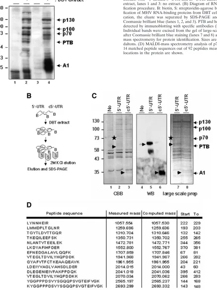
FIG. 1. Specific association of cellular proteins with MHV 5⬘-UTR and c5⬘-UTR and RNA affinity purification of MHV RNA-binding proteins from DBT cells. (A) UV cross-linking of DBT cytoplasmic extracts with32P-labeled 5⬘-UTR and c5⬘-UTR. Lanes 2 and 4: DBT extract, lanes 1 and 3: no extract. (B) Diagram of RNA affinity puri-fication procedure. B: biotin, S: streptavidin-agarose beads. (C) Puri-fication of MHV RNA-binding proteins from DBT cells. After purifi-cation, the eluate was separated by SDS-PAGE and stained with Coomassie brilliant blue (lanes 1, 2, and 3). PTB and hnRNP A1 were detected by immunoblotting with specific antibodies (lanes 5 and 6). Individual bands were excised from the gel of large-scale preparation after Coomassie brilliant blue staining (lanes 7 and 8) and analyzed by mass spectrometry for protein identification. Sizes are shown in kilo-daltons. (D) MALDI-mass spectrometry analysis of p70 protein. The 14 matched peptide sequences out of 92 peptides measured and their locations in the protein are shown.
on November 8, 2019 by guest
http://jvi.asm.org/
cross-linking experiment with DBT cell extract showed that several proteins bound to MHV-UTR RNA specifically (Fig. 1A). These proteins include not only PTB, which binds to 5⬘-UTR, and hnRNP A1, which binds to c5⬘-UTR, but also additional proteins of 70, 100, and 130 kDa. Specific binding of the 70-kDa protein to MHV RNA has been suggested in our previous studies (6, 14), but it was not clear whether the bind-ing of the 100- and 130-kDa proteins was specific to MHV RNA.
To purify unidentified cellular proteins that interact with 5⬘ -UTR, we developed a one-step purification procedure based on the RNA affinity method (Fig. 1B). Biotinylated 5⬘-UTR or c5⬘-UTR RNA was incubated with DBT extract and pulled down with streptavidin-agarose beads, and the bound RNP complexes were eluted by 2 M KCl. By this procedure, we were able to detect PTB and hnRNP A1 with 5⬘-UTR and c5⬘-UTR as a probe, respectively, confirming that this purification pro-cedure worked properly (Fig. 1C). In addition to PTB and hnRNP A1, 70-, 100-, and 130-kDa and several other minor proteins were pulled down with both 5⬘-UTR and c5⬘-UTR specifically (Fig. 1C). The 70-kDa protein was identified as synaptotagmin-binding cytoplasmic RNA-interacting protein (SYNCRIP) by MALDI-mass spectrometry analysis, while the 100-kDa protein was HSP90, and the 130-kDa protein was an unknown protein.
Fourteen matched peptides out of 92 measured peptides of
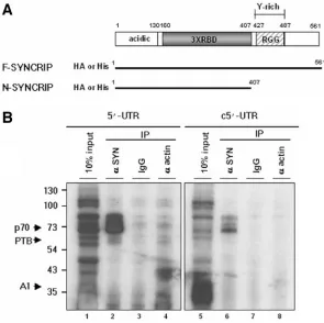
the 70-kDa protein are shown in Fig. 1D. The coverage of the known sequence for the 70-kDa protein was 32%, which is more than the 25% coverage typically considered reliable. Since our previous study based on RNA competition experi-ments showed that the binding of the 70-kDa protein to MHV UTR was specific (14), we focused on the characterization of SYNCRIP in this study. SYNCRIP is a member of the hnRNP family, and its human homologue is hnRNP Q, which was previously named NS1-associated protein (NSAP1) or glycine and tyrosine-rich RNA-binding protein (Gry-rbp) (9, 12). SYNCRIP has been suggested as a cytoplasmic counterpart of hnRNP R (23). As presented in Fig. 2A, SYNCRIP has an acidic domain at N terminus, followed by three sets of RNA-binding domains and another type of RNA-RNA-binding motif (RGG box). Furthermore, a tyrosine-rich motif is present in the C terminus, which putatively mediates proteprotein in-teractions.
[image:4.585.144.439.69.363.2]To validate that SYNCRIP was indeed the 70-kDa protein cross-linked to the MHV-UTR, we performed immunoprecipi-tation of DBT extract cross-linked to32P-labeled 5⬘-UTR or c5⬘-UTR, with anti-SYNCRIP antibody. As shown in Fig. 2B, the 70-kDa protein cross-linked to either 5⬘-UTR or c5⬘-UTR was pulled down with anti-SYNCRIP antibody, but not with the control immunoglobulin G or antiactin antibody. The up-per two protein bands of approximately 85 and 74 kDa may be the contaminating hnRNP R and its degradation product, both
FIG. 2. Schematic diagram of SYNCRIP and confirmation of p70 as a SYNCRIP protein. (A) Structural organization of SYNCRIP. Acidic: protein domain rich in acidic amino acids. RBD: RNA-binding domain. RGG box: RGG RNA-binding domain. Y-rich: protein-protein-interacting domain. The diagrams of F-SYN and N-SYN constructs are shown below the diagram. His: 6X His, HA: eight amino acids of the HA tag. (B) Immunoprecipitation of UV cross-linked p70 with anti-SYNCRIP antibody. After UV cross-linking of DBT extracts with32P-labeled 5⬘-UTR or c5⬘-UTR RNA and digestion with RNase A, RNA-protein complexes were immunoprecipitated with anti-SYNCRIP antibody (lanes 2 and 6) or nonspecific antibody (lanes 3, 4, 7, and 8). Lanes 1 and 5 represent 10% of the UV cross-linked lysates used for each analysis.
on November 8, 2019 by guest
http://jvi.asm.org/
of which weakly cross-reacted with anti-SYNCRIP antibody (23). From this experiment, we established that SYNCRIP is indeed the UV cross-linked 70-kDa protein.
SYNCRIP binds to both MHV 5ⴕ-UTR and c5ⴕ-UTR in vitro.
To test whether SYNCRIP is able to bind to MHV UTR, we performed in vitro binding analysis. His-tagged, recombinant SYNCRIP was expressed inEscherichia coli, and purified by Ni-nitrilotriacetic acid-agarose columns. We generated two different forms of SYNCRIP, full-length (F-SYN) and C-ter-minally-truncated (N-SYN) forms (Fig. 2A). N-SYN includes the N-terminal RNA-binding motif but lacks the C terminus, which is responsible for protein-protein interaction. Both F-SYN (70 kDa) and N-F-SYN (54 kDa) were expressed inE. coli
and purified to similar purity. The UV cross-linking experi-ment showed that both forms of SYNCRIP bound to MHV 5⬘-UTR but not nonspecific RNA, although the binding of N-SYN was slightly stronger than that of F-SYN (Fig. 3A). The slow migrating molecular band detected with F-SYN may have resulted from nonspecific binding.
Since N-SYN showed a stronger binding activity, we used N-SYN for further in vitro binding studies. To map the SYNCRIP-binding site in 5⬘-UTR, several deletion mutants were used for the UV cross-linking experiment (Fig. 3B). N-SYN bound strongly to the 5⬘ end 184 nucleotides or 112 nucleotides of viral RNA. The truncated mutant,⫹56, which does not include the UCUAA repeats, retained a weak binding ability. ⌬4R, which is identical to ⫹184 RNA except for a deletion of four copies of UCUAA repeats, bound to N-SYN only weakly. Therefore, the UCUAA repeat sequence is cru-cial for efficient SYNCRIP binding. However, the four copies of UCUAA repeats (4R) alone were not sufficient for binding,
indicating that the neighboring sequence is also important for SYNCRIP binding, probably because it induces conforma-tional changes in 5⬘-UTR and/or stabilizes the structure of 5⬘ -UTR.
In addition, the binding of N-SYN to radiolabeled 5⬘-UTR was competed away with excess amounts of both unlabeled 5⬘-UTR and c5⬘-UTR, but not with 3⬘-UTR, sequence com-plementary to the 3⬘-UTR, or nonspecific RNA transcribed from pcDNA3.1 (Fig. 3C), indicating that SYNCRIP also binds to c5⬘-UTR. This result is consistent with the previous finding that SYNCRIP was pulled down with either 5⬘-UTR or c5⬘-UTR by RNA affinity purification (Fig. 2B).
Overexpressed SYNCRIP binds to MHV 5ⴕ-UTR in virus-infected cells. Next, we asked whether SYNCRIP bound to MHV 5⬘-UTR in virus-infected cells. To address this question, we performed immunoprecipitation of SYNCRIP from the lysates of MHV-infected cells with anti-SYNCRIP antibody; viral RNAs were isolated from the immunoprecipitated com-plex and detected by RNase protection assays with the 5⬘-UTR as the probe (Fig. 4A). For this purpose, HA-tagged, full-length SYNCRIP or a truncated form of SYNCRIP (Fig. 2A) was expressed together with an MHV receptor in 293A cells and then infected with MHV. As shown in lanes 2 and 3 in Fig. 4B, MHV RNA was detected with this probe only in virus-infected cells, but not in unvirus-infected cells.
[image:5.585.85.501.70.302.2]Immunoprecipitation of SYNCRIP with anti-HA antibody pulled down the viral RNA (Fig. 4B, lane 5). Anti-glyceral-dehyde-3-phosphate dehydrogenase (GAPDH) antibodies yielded only background signals similar to that obtained with the beads only (lanes 4 and 7). Anti-N (MHV nucleo-capsid protein) antibody also precipitated the viral RNA
FIG. 3. In vitro binding of the recombinant SYNCRIP and analysis of its binding sites on 5⬘-UTR. (A) Specific binding of recombinant F-SYN and N-SYN to 5⬘-UTR. Purified recombinant F-SYN and N-SYN (10 ng) were cross-linked to32P-labeled 5⬘-UTR (lanes 1 and 2) or vector pcDNA3.1 RNA transcript (200 nucleotides) (lanes 3 and 4). (B) Binding region of N-SYN in 5⬘-UTR. Purified N-SYN (10 ng) was cross-linked with 30 pmol of32P-labeled probes. R: UCUAA repeats. (C) Competition experiments.32P-labeled 5⬘-UTR (30 pmol) was UV cross-linked with 10 ng of purified N-SYN in the presence of unlabeled probes, 5⬘-UTR, c5⬘-UTR, 3⬘-UTR, c3⬘-UTR, and nonspecific RNA in serial dilutions (1-, 5-, 20-, and 50-fold). (-), UV cross-linked N-SYN without added unlabeled RNA.
on November 8, 2019 by guest
http://jvi.asm.org/
(lane 6), consistent with the finding that N protein binds to the 5⬘-UTR of MHV RNA (33). N-SYN had a lower binding ability than F-SYN (compare lanes 5 and 8). Given that N-SYN binds better than F-SYN in vitro (Fig. 3A), this finding suggests that in vivo binding of SYNCRIP to MHV UTR may require other factors, since N-SYN lacks the protein-interacting domain. Alternatively, N-SYN may not be properly folded so that it lost some of the RNA-binding ability. This result was also confirmed by detecting the bind-ing of endogenous SYNCRIP to MHV 5⬘-UTR with the same immunoprecipitation approach, although the background sig-nal was slightly higher than that with the exogenously ex-pressed SYNCRIP (data not shown).
These results together indicate that SYNCRIP binds to MHV RNA in virus-infected cells.
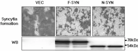
Syncytium formation is delayed when F-SYN or N-SYN is overexpressed. To address the biological significance of SYNCRIP in MHV replication, we overexpressed either F-SYN or N-F-SYN together with the MHV receptor in 293A cells; at 24 h posttransfection, cells were infected with MHV-JHM at a multiplicity of infection of 1. Cells were immunostained with anti-N antibody at 12 h postinfection. Compared with the vec-tor-transfected cells, the appearance of syncytium formation was significantly delayed in N-SYN-transfected cells (Fig. 5). However, by 24 h postinfection, N-SYN-transfected cells were fully infected and formed syncytia to the same extent as the vector-transfected cells (data not shown).
We examined the amount of MHV receptor and the viral spike protein on the cell surface to rule out the possibility that the delay of syncytium formation was due to down-regulation of MHV receptor or spike protein. There was no significant difference among those cells. Surprisingly, overexpression of F-SYN also caused delayed syncytium formation. Thus, both N- and F-SYN had a dominant-negative effect on MHV rep-lication. These dominant-negative effects may be caused by the “squelching effect” commonly observed with the overexpres-sion of transcription factors (34); it was also observed previ-ously with the full-length PTB in MHV RNA transcription (3). Since we have previously shown that the overexpression of hnRNP A1 accelerated MHV transcription (29), the domi-nant-negative effect observed with F-SYN was most likely not due to the indirect global effects caused by the overexpres-sion of proteins. These results show that overexpresoverexpres-sion of SYNCRIP may impair virus replication by titrating out the factors involved in virus replication. Therefore, these results suggest that SYNCRIP may be involved in MHV replication.
In vivo knockdown of SYNCRIP delayed MHV replication.
To further examine the role of the endogenous SYNCRIP in MHV replication, we attempted to knock down the endoge-nous SYNCRIP with the RNA interference method (4, 8). Three different short interfering RNAs (siRNAs) were de-signed against SYNCRIP (Fig. 6A). Transfection of DBT cells with any one of the SYNCRIP-specific siRNAs (Fig. 6B, 1, 2, and 3) but not with nonspecific siRNA (Fig. 6B) resulted in transient reduction of endogenous SYNCRIP. The three SYNCRIP siRNAs showed reduction of the endogenous SYNCRIP to a similar level, i.e., around 25 to 40% at 2 days posttransfection and around 40 to 65% at 4 days posttransfec-tion. The level of SYNCRIP returned to almost the normal level at 6 days posttransfection.
[image:6.585.44.284.70.261.2]To examine the effect of reduction of endogenous SYNCRIP on MHV replication, DBT cells transfected with either SYNCRIP siRNA #1 or nonspecific siRNA were infected with MHV at a multiplicity of infection of 1 at 2 days posttransfec-tion. First, we investigated the morphological changes induced by virus infection. Compared with the nonspecific siRNA-trans-fected cells, the SYNCRIP siRNA-transsiRNA-trans-fected cells showed significantly delayed syncytium formation, with at least a 4-h
FIG. 4. In vivo binding of overexpressed SYNCRIP in MHV-in-fected 293A cells. (A) Structure of the probe used. The probe consists of 184 nucleotides of 5⬘-UTR, 67 nucleotides of ORF1a 5⬘end, and 19 nucleotides of noncomplementary sequence. After RNase digestion, the protected band migrated as 251 nucleotides. (B) 293A cells were cotransfected with HA-tagged F-SYN (lanes 4, 5, 6, and 7) or N-SYN (lane 8) and MHV receptor and then infected with MHV-JHM at a multiplicity of infection of 10. At 8 h postinfection, the cell lysates were immunoprecipitated with various antibodies, anti-HA antibody (lanes 5 and 8), anti-N antibody (lane 6), and anti-GAPDH antibody (lane 7), followed by extraction of RNA from the beads. Viral RNAs were detected by RNase protection assay. Lane 1 indicates the 10% unhy-bridized probe. As a control, total RNA from uninfected and MHV-JHM-infected 293A cells without prior immunoprecipitation was used directly for the RNase protection assay (lanes 2 and 3).
FIG. 5. Syncytium formation of 293A cells overexpressing F-SYN or N-SYN. 293A cells were transfected as in Fig. 4 and, at 24 h post-transfection, were infected with MHV-JHM at a multiplicity of infec-tion of 1. At 12 h postinfecinfec-tion, cells were incubated with an antibody against the viral N protein, followed by incubation with -galactosi-dase-conjugated secondary antibody. Syncytium formation was visual-ized by 5-bromo-4-chloro-3-indolyl--D-galactopyranoside (X-Gal) staining. The bottom panel shows the expression of F-SYN or N-SYN by Western blotting (WB) with anti-HA antibody. The vector-transfected cell was used as the control.
on November 8, 2019 by guest
http://jvi.asm.org/
[image:6.585.301.539.543.632.2]delay (Fig. 7A). Next, we examined the viral protein and RNA synthesis by immunoblotting and [3H]uridine labeling, respec-tively. The kinetics of both viral protein and RNA synthesis were delayed in SYNCRIP siRNA-transfected DBT cells (Fig. 7B and C). The synthesis of viral protein and RNA peaked at 13 h postinfection in cells transfected with the nonspecific siRNA, but peaked at 20 h postinfection in SYNCRIP siRNA-transfected cells. From these results, we conclude that SYNCRIP is a positive regulator of MHV protein and RNA synthesis.
SYNCRIP directly regulates viral RNA synthesis but not viral translation.The finding that the reduction of endogenous SYNCRIP delayed both viral RNA and protein synthesis could be due to the possibilities that SYNCRIP is directly involved in both viral transcription and translation or that SYNCRIP af-fects transcription, which, in turn, afaf-fects translation, or vice versa. To distinguish these possibilities, we designed experi-ments to separate viral translation from viral RNA transcrip-tion with defective-interfering RNA. DE25 is a natural de-fective interfering RNA, in which part of ORF1a is fused to ORF7 encoding the N protein (Fig. 8A, top diagram) (21). This protein can be directly translated from DE25 RNA. We also used a reporter defective interfering RNA, MHV-UTR/ LUC, which contains the authentic MHV 5⬘and 3⬘-UTR and expresses an ORF1a-luciferase (LUC) fusion protein (Fig. 8A, bottom) (3). Therefore, the mechanism of translation of lucif-erase faithfully reflects that of the natural MHV RNA.
The use of both defective interfering RNAs made it possible to examine the direct effect of SYNCRIP on viral translation by excluding the possible transcriptional effect. First, we per-formed in vitro translation with rabbit reticulocyte lysate,
which was incubated with increasing amounts of recombinant SYNCRIP, and then in vitro-transcribed defective interfering RNAs were added for translation. The translation of DE25 and MHV-UTR/LUC was not affected until the amount of recombinant SYN reached 15 nM, when there was nonspecific inhibition. Similar nonspecific inhibition by SYN was observed with the control RNA, EF1a.
We further tested the possible translational effect of SYNCRIP in vivo. For this study, we overexpressed F-SYN or N-SYN transiently in 293A cells, followed by transfection of MHV-UTR/LUC RNA. Both F-SYN and N-SYN were ex-pressed to similar levels in cells (Fig. 5B). At 8 h posttrans-fection of RNA, luciferase activity was assayed (Fig. 8C, left). No difference in luciferase activity was observed between the vector-transfected cells and F-SYN- or N-SYN-overexpressing cells. We performed a similar experiment in cells in which SYNCRIP was knocked down by the specific siRNA. No dif-ference in luciferase activity was observed between cells trans-fected with the SYN-specific siRNA or the nonspecific siRNA (Fig. 8C, right).
Therefore, based on the in vitro and in vivo translation studies, we conclude that SYNCRIP does not affect viral trans-lation directly, implying that SYNCRIP most likely plays a direct role in MHV RNA synthesis.
DISCUSSION
[image:7.585.112.473.70.338.2]Since most viruses carry relatively small numbers of genes in their genome, most steps in virus replication, including virus entry, gene expression, RNA synthesis, assembly, budding, and
FIG. 6. RNA interference-mediated reduction of SYNCRIP in DBT cells. (A) Targeted regions of SYNCRIP siRNA and their sequences (1, 2, and 3). Nonspecific siRNA from Ambion was used as the negative control. (B) Immunoblotting from RNA interference-transfected DBT cells. DBT cells were transfected with SYNCRIP-specific siRNA (1, 2, and 3) or a nonspecific siRNA (⫺) and harvested at 2, 4, and 6 days posttransfection; 20g of total cell lysates was immunoblotted with anti-SYNCRIP antibody and antiactin antibody separately. The percentage of SYNCRIP reduction was quantified with a densitometer.
on November 8, 2019 by guest
http://jvi.asm.org/
release, require the participation of host factors, which interact with viral RNA and/or viral proteins (1, 16). We have focused on the host factors involved in the regulation of MHV RNA replication, transcription, and translation. By a classical UV cross-linking method, we have been able to define a set of spe-cific MHV RNA-binding proteins (Fig. 1A). So far, we have characterized two MHV RNA-binding proteins, PTB and hnRNP A1, and several other hnRNP A1-related proteins, which bind to the 5⬘and 3⬘ends of opposite RNA strands (17, 18, 30). PTB and hnRNP A1 may mediate 5⬘-3⬘crosstalks of viral RNA by interacting with each other (13), causing circularization of viral genome. This genomic circularization and its impor-tance in viral RNA replication have been reported for polio-virus (10), although they have not been unequivocally proven in the case of MHV. Moreover, the studies of dominant-neg-ative mutants of either PTB or hnRNP A1 have shown that these two hnRNP proteins are important for the modulation of MHV RNA synthesis (3, 29). Besides PTB and hnRNP A1-related proteins, several other proteins, including poly(A) binding protein (32) and mitochondrial aconitase (25), have been reported by other laboratories astrans-regulatory factors for MHV replication.
SYNCRIP represents yet another factor in MHV RNA syn-thesis. The one-step RNA affinity purification used in this study was highly specific, since we were able to detect PTB and hnRNP A1 with positive- and negative-strand RNA probes, respectively, but not vice versa. SYNCRIP was originally found as a binding partner of the ubiquitous synaptotagmin isoforms (23), and its human homologue, NASP1 or gry-rbp, has been identified from a two-hybrid screen as an interacting partner of NS1, the major nonstructural protein of parvovirus (9). It was subsequently renamed hnRNP Q (27). Recent database searches revealed that there are three isoforms of hnRNP Q, 1, 2, and 3, which migrate at 55, 60, and 70 kDa, respectively; SYNCRIP corresponds to hnRNP Q3 (24). It has been sug-gested that hnRNP Q is involved in mRNA processing and transport (9), translation-coupled mRNA turnover (7), and mRNA splicing (26). SYNCRIP is 81.2% similar to hnRNP R but lacks ⬇70 carboxyl-terminal amino acids that contain a nuclear localization motif and has been suggested as a cyto-plasmic counterpart of hnRNP R (23). Cytocyto-plasmic localiza-tion of SYNCRIP is further confirmed by immunofluorescence staining of SYNCRIP (23). Moreover, it has been reported that SYNCRIP is part of a cytoplasmic multiprotein complex that binds to the major determinant of instability of the c-fos
[image:8.585.47.284.72.677.2]proto-oncogene mRNA and regulates its stability and translat-ability (7). The cytoplasmic localization of SYNCRIP and its putative role in the cytoplasm are particularly noteworthy, since MHV replication occurs in the cytoplasm. It is also note-worthy that most of the MHV RNA-binding proteins identified so far are hnRNPs; SYNCRIP is yet another hnRNP. This fact
FIG. 7. Retardation of MHV replication by reduction of endoge-nous SYNCRIP. (A) Syncytium formation in SYNCRIP-specific siRNA-transfected DBT cells. At 2 days posttransfection of siRNA, cells were infected with MHV-JHM at a multiplicity of infection of 1
and maintained in virus growth medium. Pictures were taken at 0, 10, 13, and 16 h postinfection. (B) Kinetics of expression of viral N pro-tein. Cell lysates were taken at different time points and subject to immunoblotting to detect viral N protein. (C) Kinetics of MHV RNA synthesis. [3H]uridine labeling was done at different time points after virus infection. Incorporation of [3H]uridine is shown.
on November 8, 2019 by guest
http://jvi.asm.org/
underscores the importance of RNA processing machinery in MHV RNA synthesis.
The importance of SYNCRIP in MHV replication was es-tablished in our study with the RNA interference approach. Reduction of SYNCRIP in DBT cells resulted in the retarda-tion of syncytium formaretarda-tion and viral protein and RNA syn-thesis. This finding was further supported by the dominant-negative effect of the truncated form of SYNCRIP on MHV replication. These results suggest that SYNCRIP is a positive regulatory factor in MHV replication. On the other hand, it is puzzling why the overexpression of full-length SYNCRIP also inhibited MHV replication in a dominant-negative manner. Similar inhibitory effects on MHV replication were observed when PTB was overexpressed in cells (3). It is possible that the recombinant SYNCRIP, which has an HA tag, may be less ef-ficient in its biological activity than the endogenous SYNCRIP. Furthermore, overexpression of SYNCRIP may titrate out one or more replication components, resulting in the inhibition of replication and transcription, in a mechanism akin to the squelching effects frequently observed for overexpressed tran-scription factors (34).
So far, SYNCRIP, PTB, and hnRNP A1, all of which are hnRNPs, have been demonstrated to bind to 5⬘-UTR of both strands. It is likely that they form a large protein complex to regulate viral RNA replication. Interestingly, it has been re-ported that these hnRNPs are detected in the complex of spliceosome (26), and SYNCRIP/NSAP1 was detected in the same complex including poly(A) binding protein (7), which binds to the 3⬘end of MHV RNA. Therefore, it is not surpris-ing that they may form an RNP complex with viral proteins in
MHV-infected cells to regulate MHV RNA synthesis. The exact molecular mechanism of this complex in MHV RNA replication remains to be clarified.
Our result also showed that SYNCRIP was able to bind to c5⬘-UTR. Previous reports have shown that 5⬘-UTR, inter-genic sequences,and their binding proteins on the negative strand are important in regulating MHV RNA transcription (18, 20, 29). During transcription, production of several sub-genomic RNAs is regulated by the proper interaction be-tween leader sequence and intergenic sequences. Therefore, SYNCRIP may mediate the interaction between c5⬘-UTR and intergenic sequence, thus regulating the transcription of sub-genomic RNAs.
As a positive regulator of MHV replication, SYNCRIP may play a role through several mechanisms, which are not mutu-ally exclusive. First, SYNCRIP may recruit transcription/ replication factors to the replication site, through its pro-teproteinteracting domain. Second, SYNCRIP may in-duce conformational changes in the highly structured viral RNA into a structure that is more favorable for replication and transcription, probably functioning as an RNA chap-eron. Third, SYNCRIP may mediate 5⬘-3⬘ crosstalk by in-teracting with other proteins, causing the circularization of viral genome, which may help viral replication.
Recently, comparative genomic analysis of coronaviruses revealed the presence of several domains for putative RNA-processing enzymes in ORF1 of the viral genome (31). If the virus indeed encodes RNA-processing enzymes, SYNCRIP may act in concert with these RNA-processing enzymes to affect the cellular RNA-processing machinery or the viral
rep-FIG. 8. Effect of SYNCRIP on the translation of defective interfering RNA. (A) Structures of defective interfering RNAs used for the translation study. DE25 produces an 80-kDa ORF1a-N fusion protein, while MHV-UTR-LUC produces a 70-kDa ORF1a-luciferase fusion protein. (B) In vitro translation. Rabbit reticulocyte lysates were incubated with35S-Translabel mixtures, in vitro-transcribed defective interfering RNAs, and increasing amounts of purified recombinant F-SYN (up to 30 nM). EF1a was used for the negative control. (C) In vivo translation of the reporter RNA in SYNCRIP-overexpressing 293A cells or SYNCRIP-specific siRNA-transfected DBT cells. The cells were prepared as in Fig. 4 and 6. In vitro-transcribed MHV-UTR/LUC defective interfering RNAs were transfected into the various cells, and luciferase activity was assayed at 8 h posttransfection.
on November 8, 2019 by guest
http://jvi.asm.org/
[image:9.585.47.534.68.307.2]lication process. Alternatively, SYNCRIP may have a protec-tive role against either virus-encoded or cell-encoded RNA-processing enzymes.
Finally, our study showed that SYNCRIP is not directly implicated in MHV translation. The 5⬘-UTR has been shown to regulate the translation of MHV RNA, specifically, the binding of N protein to leader sequence of 5⬘-UTR enhanced the translation of MHV RNA (35). However, since genomic and subgenomic mRNAs of MHV are capped, polyadenyl-ated, and translated by a cap-dependent translation mech-anism, it may not need noncanonical factors for efficient trans-lation. Nevertheless, we could not rule out the possibility that SYNCRIP may regulate MHV translation under certain con-ditions.
ACKNOWLEDGMENT
This work was supported by National Institute of Health research grant AI19244.
REFERENCES
1.Andino, R., N. Boddeker, D. Silvera, and A. V. Gamarnik.1999. Intracellular
determinants of picornavirus replication. Trends Microbiol.7:76–82.
2.Boussadia, O., M. Niepmann, L. Creancier, A. C. Prats, F. Dautry, and H. Jacquemin-Sablon.2003. Unr is required in vivo for efficient initiation of translation from the internal ribosome entry sites of both rhinovirus and
poliovirus. J. Virol.77:3353–3359.
3.Choi, K. S., P. Huang, and M. M. C. Lai.2002. Polypyrimidine-tract-binding protein affects transcription but not translation of mouse hepatitis virus
RNA. Virology303:58–68.
4.Elbashir, S. M., J. Harborth, W. Lendeckel, A. Yalcin, K. Weber, and T. Tuschl.2001. Duplexes of 21-nucleotide RNAs mediate RNA interference in
cultured mammalian cells. Nature411:494–498.
5.Fleming, J. O., S. A. Stohlman, R. C. Harmon, M. M. Lai, J. A. Frelinger, and L. P. Weiner.1983. Antigenic relationships of murine coronaviruses:
analysis using monoclonal antibodies to JHM (MHV-4) virus. Virology131:
296–307.
6.Furuya, T., and M. M. C. Lai.1993. Three different cellular proteins bind to
complementary sites on the 5⬘-end-positive and 3⬘-end-negative strands of
mouse hepatitis virus RNA. J. Virol.67:7215–7222.
7.Grosset, C., C. Y. Chen, N. Xu, N. Sonenberg, H. Jacquemin-Sablon, and A. B. Shyu.2000. A mechanism for translationally coupled mRNA turnover: interaction between the poly(A) tail and a c-fos RNA coding determinant via
a protein complex. Cell103:29–40.
8.Hannon, G. J.2002. RNA interference. Nature418:244–251.
9.Harris, C. E., R. A. Boden, and C. R. Astell.1999. A novel heterogeneous nuclear ribonucleoprotein-like protein interacts with NS1 of the minute virus
of mice. J. Virol.73:72–80.
10.Herold, J., and R. Andino.2001. Poliovirus RNA replication requires
ge-nome circularization through a protein-protein bridge. Mol. Cell7:581–591.
11.Hirano, N., K. Fujiwara, S. Hino, and M. Matumoto.1974. Replication and plaque formation of mouse hepatitis virus (MHV-2) in mouse cell line DBT
culture. Arch. Gesamte Virusforsch.44:298–302.
12.Hresko, R. C., and M. Mueckler.2002. Identification of pp68 as the Ty-rosine-phosphorylated Form of SYNCRIP/NSAP1. A cytoplasmic
RNA-binding protein. J. Biol. Chem.277:25233–25238.
13.Huang, P., and M. M. C. Lai.2001. Heterogeneous nuclear
ribonucleopro-tein a1 binds to the 3⬘-untranslated region and mediates potential 5⬘-3⬘-end
cross talks of mouse hepatitis virus RNA. J. Virol.75:5009–5017.
14.Huang, P., and M. M. C. Lai.1999. Polypyrimidine tract-binding protein
binds to the complementary strand of the mouse hepatitis virus 3⬘
untrans-lated region, thereby altering RNA conformation. J. Virol.73:9110–9116.
15.Lai, M. M., and D. Cavanagh.1997. The molecular biology of coronaviruses.
Adv. Virus Res.48:1–100.
16.Lai, M. M. C.1998. Cellular factors in the transcription and replication of viral RNA genomes: a parallel to DNA-dependent RNA transcription.
Vi-rology244:1–12.
17.Li, H. P., P. Huang, S. Park, and M. M. C. Lai. 1999. Polypyrimidine tract-binding protein binds to the leader RNA of mouse hepatitis virus and
serves as a regulator of viral transcription. J. Virol.73:772–777.
18.Li, H. P., X. Zhang, R. Duncan, L. Comai, and M. M. C. Lai.1997. Heter-ogeneous nuclear ribonucleoprotein A1 binds to the transcription-regulatory
region of mouse hepatitis virus RNA. Proc. Natl. Acad. Sci. USA94:9544–
9549.
19.Lin, Y. J., X. Zhang, R. C. Wu, and M. M. C. Lai.1996. The 3⬘untranslated region of coronavirus RNA is required for subgenomic mRNA transcription
from a defective interfering RNA. J. Virol.70:7236–7240.
20.Makino, S., M. Joo, and J. K. Makino.1991. A system for study of corona-virus mRNA synthesis: a regulated, expressed subgenomic defective
inter-fering RNA results from intergenic site insertion. J. Virol.65:6031–6041.
21.Makino, S., C. K. Shieh, L. H. Soe, S. C. Baker, and M. M. C. Lai.1988. Primary structure and translation of a defective interfering RNA of murine
coronavirus. Virology166:550–560.
22.Meerovitch, K., Y. V. Svitkin, H. S. Lee, F. Lejbkowicz, D. J. Kenan, E. K. Chan, V. I. Agol, J. D. Keene, and N. Sonenberg.1993. La autoantigen enhances and corrects aberrant translation of poliovirus RNA in reticulocyte
lysate. J. Virol.67:3798–3807.
23.Mizutani, A., M. Fukuda, K. Ibata, Y. Shiraishi, and K. Mikoshiba.2000. SYNCRIP, a cytoplasmic counterpart of heterogeneous nuclear ribonucle-oprotein R, interacts with ubiquitous synaptotagmin isoforms. J. Biol. Chem.
275:9823–9831.
24.Mourelatos, Z., L. Abel, J. Yong, N. Kataoka, and G. Dreyfuss.2001. SMN interacts with a novel family of hnRNP and spliceosomal proteins. EMBO J.
20:5443–5452.
25.Nanda, S. K., and J. L. Leibowitz.2001. Mitochondrial aconitase binds to the
3⬘untranslated region of the mouse hepatitis virus genome. J. Virol.75:
3352–3362.
26.Neubauer, G., A. King, J. Rappsilber, C. Calvio, M. Watson, P. Ajuh, J. Sleeman, A. Lamond, and M. Mann.1998. Mass spectrometry and EST-database searching allows characterization of the multi-protein spliceosome
complex. Nat. Genet.20:46–50.
27.Rossoll, W., A. K. Kroning, U. M. Ohndorf, C. Steegborn, S. Jablonka, and M. Sendtner.2002. Specific interaction of Smn, the spinal muscular atrophy determining gene product, with hnRNP-R and gry-rbp/hnRNP-Q: a role for
Smn in RNA processing in motor axons? Hum. Mol. Genet.11:93–105.
28.Shen, X., and P. S. Masters.2001. Evaluation of the role of heterogeneous nuclear ribonucleoprotein A1 as a host factor in murine coronavirus discon-tinuous transcription and genome replication. Proc. Natl. Acad. Sci. USA
98:2717–2722.
29.Shi, S. T., P. Huang, H. P. Li, and M. M. C. Lai.2000. Heterogeneous nuclear ribonucleoprotein A1 regulates RNA synthesis of a cytoplasmic
virus. EMBO J.19:4701–4711.
30.Shi, S. T., G. Y. Yu, and M. M. C. Lai.2003. Multiple type A/B heteroge-neous nuclear ribonucleoproteins (hnRNPs) can replace hnRNP A1 in
mouse hepatitis virus RNA synthesis. J. Virol.77:10584–10593.
31.Snijer, E. J., P. J. Bredenbeek, J. C. Dobbe, V. Thiel, J. Ziebuhr, L. L. M. Poon, Y. Guan, M. Rozanov, W. J. M. Spaan, and E. Gorbalenya.2003. Unique and conserved features of genome and proteome of SARS-corona-virus, an early split-off from the coronavirus group 2 lineage. J. Mol. Biol.
331:991–1004.
32.Spagnolo, J. F., and B. G. Hogue.2000. Host protein interactions with the 3⬘
end of bovine coronavirus RNA and the requirement of the poly(A) tail for
coronavirus defective genome replication. J. Virol.74:5053–5065.
33.Stohlman, S. A., R. S. Baric, G. N. Nelson, L. H. Soe, L. M. Welter, and R. J. Deans.1988. Specific interaction between coronavirus leader RNA and
nu-cleocapsid protein. J. Virol.62:4288–4295.
34.Strasser, K., and E. Hurt.2001. Splicing factor Sub2p is required for nuclear
mRNA export through its interaction with Yra1p. Nature413:648–652.
35.Tahara, S. M., T. A. Dietlin, G. W. Nelson, S. A. Stohlman, and D. J. Manno.
1998. Mouse hepatitis virus nucleocapsid protein as a translational effector
of viral mRNAs. Adv. Exp. Med. Biol.440:313–318.
36.Zhang, X., and M. M. C. Lai.1995. Interactions between the cytoplasmic proteins and the intergenic (promoter) sequence of mouse hepatitis virus RNA: correlation with the amounts of subgenomic mRNA transcribed. J.
Vi-rol.69:1637–1644.
37.Zuniga, S., I. Sola, S. Alonso, and L. Enjuanes. 2004. Sequence motifs involved in the regulation of discontinuous coronavirus subgenomic RNA
synthesis. J. Virol.78:980–994.