JOURNALOF VIROLOGY,
JUlY
1987, P.2143-2149 0022-538X/87/0702143-07$02.00/0Copyright C) 1987, American Society for Microbiology
The
T-Antigen-Binding
Domain
of the
Simian Virus
40
Core
Origin
of
Replication
SUMITRA DEB, SHANLI TSUI, ANDREW KOFF, ANGELO L. DELUCIA, RAMON PARSONS,
AND PETERTEGTMEYER*
Department ofMicrobiology, State UniversityofNew York, StonyBrook, New York 11794 Received5January 1987/Accepted26March 1987
The simian virus 40 origin of replication contains a 27-base-pair palindrome with the sequence 5'-CA-GAGGC-C-GAGGC-G-GCCTC-G-GCCTC-TG-3'. The four 5'-GAGGC-3'/5'-GCCTC-3' pentanucleotides
are known contact sites for simian virus 40 T-antigen binding in vitro. We used oligonucleotide-directed cassette mutagenesis to identify features of this palindrome that are important for the initiation of DNA replication invivo. Each base pair ofapentanucleotide is crucial for DNA replication.Incontrast, sequences
adjacent to pentanucleotides have little or no effect on replication. Thus, the pentanucleotide is the basic
functional unit, not only for T-antigen bindingbut also for DNA replication. All four pentanucleotidesare indispensable inthe initiation process. The spacing of pentanucleotides is crucial because duplication ofthe single base pairbetweenbindingsites hasafargreatereffectonreplication than does substitution of thesame
base pair. Inversion of any pentanucleotide blocks DNA synthesis. Thus, the pentanucleotide is not a functionally symmetricalunit. Weproposethat each pentanucleotide positionsa monomerof T antigenatthe
properdistance,rotation, andorientationrelativetoother T-antigenmonomersand tootherorigin domains and thatsuchpositioningleadstosubsequenteventsin replication.
The initiation of DNA replication at a unique origin
requires recognition signals for the binding ofone or more
replication proteins. Site-specific binding, inturn, organizes
theproteins for site-specific unwinding of duplex DNA for subsequentsyntheticevents(2, 9, 22). Insome cases,these
steps in the initiation of replication are accomplished by a
specialized complex ofdifferentproteins (9). Inthe case of tumor virus simian virus 40 (SV40), however, a single
initiatorprotein recognizes and unwinds origin DNA (4a, 8, 28). Hence, the interaction ofSV40 T antigen with origin
DNAprovides asimplesystemforthecharacterization ofa
general mechanism that is of fundamental importance in
growth regulation.
The replication origin ofSV40 consists of multiple ele-ments(1,7, 17, 25, 26). Ancillary regionsatboth ends of the
originincreasetheefficiencyof DNAreplication butarenot
absolutelyessential for basalfunctioning. Theseregionsare
part of the SV40 early promoter and operator DNAs (29); possible mechanisms for their action in replication include theregulationoftranscriptionorthe maintenance ofanopen chromatin structure (3, 10, 14, 16). Unlike these ancillary elements, a64-base-pair (bp) core originofreplication
can-notbe deletedwithout loss of basalfunctioning (5).We have
investigated the domain structure within thecore origin by
using single-base-pair mutagenesis. For operational
pur-poses,wedefine afunctional domainas acontiguous setof
sequences in which base substitutions cause a significant
decrease in replication. In contrast to functional domains,
spacer sequencestolerate base substitutionswithouta
dras-tic effect on function but do not tolerate insertions or
deletions. We have previously identified and mapped two domains that extend from theouterlimitsof the coreorigin
toward a central T-antigen-binding palindrome from which theyare separated by apparent spacer sequences(5, 6).
* Correspondingauthor.
In thepresent study, we undertook an extensive genetic
analysis ofthe T-antigen-binding central domain to define
features critical forthe initiation of DNAreplication. These features determine the precise arrangement of bound T
antigen (8, 28)and its subsequentfunction in the T-antigen-induced unwinding of origin DNA (4a) to allow primer synthesis (13). The T-antigen-binding central palindrome
consists of a cluster of four repeated
5'-GAGGC-3'/5'-GCCTC-3' pentanucleotides separatedfromeach otherbya
single base pair. The pentanucleotides are arranged as two pairs of directrepetitions that areinverted relative to each other. We have proposed that each repeat serves as a
recognition and contact site for a monomerof Tantigen in
vitro (8, 20). Oligonucleotide-directed mutagenesis of the
coreoriginallowedustomanipulatethepentanucleotidesat will. We foundthat thecorrectsequence, orientation,
spac-ing,androtation of all fourpentanucleotidesareessential for DNA replication. We discuss possible interactions of the
T-antigen-binding domain with other functional domains of the coreorigin.
MATERIALS AND METHODS
Construction ofthewild-type plasmid. Thewild-type (WT) plasmidpOR1 has beendescribed previously (7).It consists ofpML2 (19) sequence 653 to 4361 with a HindIll linker attached to the 653 position and a polylinker (NcoI, Sall,
BamHI, XmaI, and EcoRI) attached to the 4361 position.
SV40 sequences from the HindIll site at 5171 through the
NcoI siteat39areinserted betweenthecorrespondingsites of theplasmid.ThisSV40segmenthasaninternaldeletion of
T-antigen-binding region I from nucleotides 5178 through
5208to producea small core origin capableof autonomous replication.
Oligonucleotide-directed mutagenesis. WTand mutant ori-gins were synthesized as previously described (6, 12). The
2143
Vol. 61,No. 7
on November 10, 2019 by guest
http://jvi.asm.org/
2144 DEB ET AL.
I
. . it1 I'.:
I
:,
p:: "' C
-
;
!4T
i
j
I27
WT
WI
_ 90U3 *-- ...: .#_ _ _ _
tw
0
Pr- cCcs jVFIG. 1. Mutationalmapping of theT-antigen-binding domain. The sequence ofthe27-bpcentralpalindromeoftheSV40coreorigin of
replication is shownatthe topofthefigure. The numbers indicate nucleotidepositionsin theSV40 genome(29).Single exchangesofA T andC G base pairsweresynthesizedateveryotherposition in thepalindrome(shownby boxes).Theautoradiogramshows thereplication
efficiency ofWTandmutantplasmids in COS-1 cells inarepresentative experiment. Input and progenyDNAsweredistinguishedby their MboI sensitivities. The data are quantitatedandsummarized inFig.6.
region ofthe coreorigin betweentheHindlIl andNcolsites
wasreconstructedwith12overlapping oligonucleotides that hadbeenphosphorylated in 70 mMTrishydrochloride (pH 7.5)-10mM
MgCl2-5
mMdithiothreitol-1 mMATPwithT4polynucleotidyl kinase. The oligonucleotides (0.5 pmol)
wereannealed in9 ,ul ofthe samebufferfor1h at37°C and
then for 1 h at23°C to produce adouble-stranded segment
with
HindIll
andNcoIprotrudingends. The annealedoligo-nucleotideswereligatedto eachotherandto 0.1 pmolofthe
large, dephosphorylated,HindIII-NcoI fragment of plasmid pOR1 by the addition of 400 U of T4 DNA ligase (New England BioLabs, Inc.) in the same buffer and incubation overnightat12°C.Thetransformation ofHB101cells and the
preparation ofcloned plasmid DNA have been described
previously (7). The construction of directed mutants
re-quiredthesynthesis ofonly two additionaloligonucleotides
withcomplementary base changes for ligation with 10 of the 12 WT oligonucleotides. All mutations were verified by
dideoxy sequencing.
DNAreplication. The assayforDNAreplication inCOS-1
cells has been described previously (7). To ensure equiva-lenceintransfection efficiencieswith each plasmid
prepara-tion, we quantitated the input DNAs by densitometry of form I DNA separated in agarose gels and stained with ethidium bromide.
RESULTS
Identification and mapping offunctionalsequences within
the T-antigen-binding domain. Gluzman et al. (11) and
Shortle and Nathans (23) showed that deletions or base
substitutions within the 27-bppalindrome inhibit DNA
rep-lication to variable extents. However, the number of mu-tantsexaminedwas insufficient to establish the substructure of this T-antigen-binding domain. We first screened the
entire region by exchanging A -T and C G base pairs at
everyother position in the palindrome (Fig. 1). This pattern of mutations allowed aninspection oftheequivalent second and fourthpositions within each of the fourpentanucleotide repeats and of every position adjacent to apentanucleotide. The A T-to-C G exchanges were chosen because these substitutionswould be expected to have the greatesteffects on potential interactions with proteins (21). We tested the replication efficiency of the mutant origins in COS-1 cells, which constitutively express T antigen and support the
replication of WT core origins. After 72 h of incubation at 36°C, DNA was extracted and digested with MboI to
distin-guish methylated input DNA from unmethylated progeny DNA. DNAs wereanalyzed by gelelectrophoresis,blotting, and hybridization with radiolabeled pBR322 DNA. The residualinputDNA, shown in theautoradiograms, served as an internal standard for the amount ofplasmidDNAadded tothe cells. We usedMboIprogenyfragmentsto quantitate theefficiency ofreplication.
Our results revealed a distinct four-subdomain arrange-ment. All mutations withinpentanucleotides drastically re-duced replication, whereas all mutations outside penta-nucleotides had lesser effects. Mutations ateitherend of the
pentanucleotide cluster allowed replication at WT levels.
Mutations between pentanucleotides reducedreplication to
approximately one-half of WT levels. Thus, the same
pentanucleotide sequence that is the simplest sequence common to known T-antigen-binding sites (8, 30) also is a functional subdomain in the central origin palindrome. Se-quences between the subdomains may contribute to
T-antigen binding and DNA replication also, but to a much lesser extent. The lesser role of theseintervening sequences is probably related to T-antigen binding rather than to cruciformformationbecause the centralbase pair at position 5243would have no effecton the inverted symmetry of the
palindrome. Ifcruciform structures areinvolved in replica-J. VIROL.
ALw&
w
on November 10, 2019 by guest
http://jvi.asm.org/
[image:2.612.164.459.72.291.2]T-ANTIGEN-BINDlNG DOMAIN OF SV40 ORIGIN 2145
tion, the singlebase substitutions of nucleotides5231, 5237, 6, and 12 musthave little effect ontheir structures in vivo.
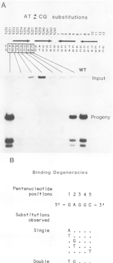
Todetermine whethereverybasepair inapentanucleotide
is important in replication, we made single transversions of each base pair in the first pentanucleotide (Fig. 2A). All mutations reduced replication more than 10-fold. Mutations in the first two positions of the 5'-GAGGC-3' sequence allowed low but detectable levels of replication. This func-tional hierarchy of bases within the pentanucleotide for
replication is similar to the hierarchy that we previously
A
AT > CG slubstitutirms.
WT
I-am b P., i
B
Binding Degeneracies
Pentanuc eotide
positions 1234 5 51 - GA G G C - 3'
Substitutions
observed
Single
DoubIe
A . .. .
T . ...
. G . . . . T. .
. . .
.T
T G. . .
FIG. 2. Finemappingofasingle pentanucleotidesite. To
deter-mine the precise structure and limits of subdomains within the
T-antigen-binding palindrome, wemade A T and C Gexchanges
at the seven consecutive positions indicated by the boxes in the sequence. (A) Autoradiogram showingthe replication efficiencyof
mutants in arepresentative experiment. The dataare quantitated
and summarizedinFig.6.(B)Alternativerecognitionsequencesfor
T-antigen bindingtakenfromprevious studies(8, 30).
identified for the
binding
ofTantigen
topentanucleotides
that occur innonorigin
DNA(Fig.
2B).
We found that Tantigen
is bound topentanucleotides
with alternative basesat
positions
1, 2,
and 5 butnot atpositions
3 and 4(8, 30).
To
investigate
further the functionalimportance
ofalter-nativebasesat thefirstandfourth
positions
ina5'-GAGGC-3'
pentanucleotide,
we made additional substitutions(Fig.
3A).
The G C-to-T- A mutation at the firstposition
re-duced
replication
10- to 20-fold. In contrast, the GC-to-A T substitution at the same location failed to reduce
replication.
Thiscomparison
suggeststhat,
in the firstposi-tion of the 5'-GAGGC-3' sequence, T
antigen
makescon-tacts with molecular sites common to G C and A - Tbase
pairs
and does notdepend
on contacts with sitesunique
to the G C basepair.
Forexample, Fig.
3B showspotential
binding
sites for amino acidson G C and A T basepairs.
T
antigen
could bind theequivalent
N7 atoms of either theguanine
oradenine bases andisunlikely
todepend
onbondsto sites
unique
to the G C basepair (shown by
thelarge
arrowheads).
The results werequite
different at the fourthposition,
whereall threepossible
base substitutionsreducedreplication
more than 100-fold.Thus,
at least one essentialcontactsite withT
antigen
mustbeunique
totheG C basepair.
This kind ofgenetic
analysis
will be ofimportance
indetermining
thefidelity
ofcocrystals
ofTantigen
andorigin
DNA when
they
become available.Importance
ofspacing
within theT-antigen-binding
do-main. The above studies of
single-base-pair
mutations have identified fourpentanucleotide
subdomains in the27-bp
central
palindrome
ofthe coreorigin.
We wishedtoinvesti-gate
possible
functional interactions between theseprotein-binding
sitesby
changing
theirspatial
relationships.
Dupli-cation of the
single
basepair
between these subdomains allowed the smallestpossible change
in the distance androtationamongthe
pentanucleotides (Fig.
4A).
The duplica-tion maintained the proper sequenceadjacent
to eachpentanucleotide,
whereas other insertions or deletionswouldnot. Each ofthe three insertions
drastically
reducedreplication. Thus,
theposition
of eachprotein
recognition
site relativeto the other three sites iscrucial to the
replica-tion funcreplica-tion. The same result has been shown for adupli-cation of the central base
pair
in thepalindrome
in thecomplete
viralorigin
rather than in thecoreorigin
(4).
Figure
4B shows a
proposed
arrangement of four monomers ofTantigen
bound to each of the fourpentanucleotides
of thecore
origin (27).
Thecloseproximity
oftherecognition
siteswouldlead to
precise
positional
relationships
among boundT-antigen
monomers;single-base-pair
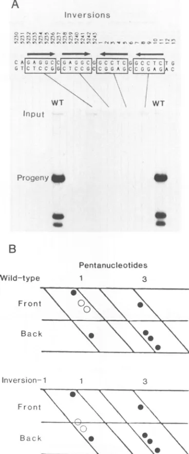
insertions would in-terferewith these interactions in three dimensions.Importance
of the orientation ofpentanucleotides
in theT-antigen-binding
domain.Thepentanucleotide
isapartially
symmetrical
sequence. Inversion of the sequence wouldcreate three base substitutions at the center of each
pentanucleotide.
Our results withsingle-base-pair
substitu-tions
suggested
that inversion of apentanucleotide
in thecore
origin
wouldseverely
inhibitreplication.
However, onthe basis ofa
comparison
ofT-antigen binding
to differentarrangements of
pentanucleotides
inorigin
regions
Iand I1,Jones and
Tjian
havesuggested
that asingle
species
ofTantigen
may interact with theguanine
clustersofarecogni-tion sequence in either orientation
(15).
Thus,
itwasimpor-tant to test
rigorously
theimportance
ofpentanucleotide
polarity by
using
asensitive in vivo functional assay.Figure
SA showsthatinversion ofanyoneof the four
pentanucleo-tides abolished
replication completely.
We concluded thatthiseffectwas not caused
by
lossoftheinvertedsymmetry
VOL.61, 1987
on November 10, 2019 by guest
http://jvi.asm.org/
[image:3.612.80.271.201.646.2]A
Atterna-|t:it s bs tu ..1
B
V
AFIF ,A A 9A .
A~ ~ ~ ~
X
s[.ra
~~~~~~~~~~~~~~~~~~..
T A1WTWT TA CC A
-I
,. . E_e a.
__m
Pta;'ra\yV
A
A
FIG. 3. Sequence specificity for the first and fourth positionsinapentanucleotide. Alternative base substitutionsweresynthesizedat two
positions (shown by boxes) in the first pentanucleotidetoexamine thenatureof theprotein-DNAcontacts. (A)Autoradiogramshowingthe replication efficiency ofmutants in arepresentative experiment. The data arequantitated and summarizedin Fig. 6. (B) Unique(large
arrowheads) andcommon(smallarrowheads) binding sites for amino acids shown for G Cand A Tbase pairs(21).
A
*.V*>.g*.. ' , ! st rt. §. t f;,r
B
WT WT
9faft"
T antigen
Recognition Pentanucleotides
T antigen
-w PrPf ,f, r
2146
V
Q
I
=
j :
Q
,
/ 6'CbC4~~~~~~~~~~~~~~
A
0t
I
on November 10, 2019 by guest
http://jvi.asm.org/
[image:4.612.79.545.52.423.2]T-ANTIGEN-BINDING DOMAIN OF SV40 ORIGIN 2147 of the entire 27-bp palindrome, because the simultaneous
inversion of the first and last pentanucleotides also
de-stroyed origin function completely (data not shown).Thus, a
pentanucleotide is not functionally symmetrical in DNA replication. Figure 5B shows a cylindrical projectionof the
firstand third pentanucleotides in the core origin. Only the positions of the guanines, known to contact T antigen by dimethyl sulfate protection studies (8), are shown. Inversion of the first pentanucleotide would change the positions of two of the four guanines (shown by open circles). This
inverted arrangement could have a number of different effects, including the following: failure to bind T antigen, inversion of one monomer of bound T antigen, or distortion of bound aminoacids projecting into the major groove. Any
ofthese alterations could block a subsequent step in DNA replication.
DISCUSSION
We have shown that the central 27-bp palindrome of the SV40 core origin of replication can be divided into four
distinct5'-GAGGC-3' subdomains that are essential for the initiation of DNA replication. Furthermore, each of the 5 bp in the first pentanucleotide repeat is important for the
replication function. This organizational pattern is entirely consistent with the results of previous studies of T-antigen
bindingtooriginandnonoriginDNAs. Footprinting analysis with dimethyl sulfate indicatedthat all binding sites contain one or more pentanucleotides (8, 30). Furthermore, both replication and binding studies revealed the samehierarchy of sequence importance at various positions within the
5'-GAGGC-3' repeat. This striking correlation between in
vivo and in vitro assays strongly argues that the pentanu-cleotide is aprimarycomponent common to both replication and T-antigen binding. Our finding that all five positions in thepentanucleotide are important for DNA replication indi-cates that a number of amino acids interact with each
pentanucleotide.
Our present results do not support the need for a
cruciformstructureformed by thecentralpalindromein the process of replication. Our mutations would introduce
py-rimidine-pyrimidineorpurine-purinebasepairs intothe stem
ofthe potential cruciform DNA. These mismatches cause
maximaldisruptionwithinknown cruciform structures (18). Two ofthe mutations at nucleotide positions 5231 and 12 have no effect on DNA replication, even though these
substitutions shorten the stem of aputative cruciform by 2
bp or more. Two other mutations, those at positions 5237 and 6, interrupt the stem ofa potential cruciform near its center. These mutations decrease DNA replication by
ap-proximately 50%. The mutation at position 5243 also de-creases DNAreplication to one-half ofWTefficiency. This
mutation does not affect the stem structure. Thus, these latter three mutations probably have a weak effect on
T-antigen binding or subsequent events rather than on the
formation ofaDNAcruciform.
A
nrvers ons
- - -
-i,
jcy
jjC7GA
WT WT
In put
Progeny
to
a,
B
Pentanucleotides Wild-type
Fronn
Back
Inversion-1
Front
Back
3 1
0
S
S 'S
S
3
a \
0
S
0
FIG. 5. Importance ofpentanucleotide orientation. We synthe-sizedcoreorigins with inversions ofsinglepentanucleotides (shown by boxes). (A)Autoradiogram showingthereplicationefficiency of WTandmutantplasmidsinarepresentativeexperiment.Prolonged exposure of the autoradiogram failed to reveal any replication
signal.(B)Cylindricalprojection showingtheguaninecontactsites (openandclosedcircles) forT-antigen bindingtothefirst andthird pentanucleotides. The first projection has a WT sequence; the second hasaninversionofthe firstpentanucleotide.
FIG. 4. Positional constraints within the T-antigen-binding palindrome. Each of the single base pairs between pentanucleotides was duplicatedtocreate asingle-base-pair insertionwhilemaintainingthesequenceintegrityimmediatelyadjacenttoeach
pentanucleotide.
(A)Autoradiogramshowingthereplicationefficiencyofmutantsinarepresentativeexperiment. (B)ModelforthebindingofTantigentothecore
origintakenfromTegtmeyeretal.(27).Thefourpentanucleotides,numbered inanearly-to-latedirection,areshownassmall boxesonthe DNA,andtheirorientationsareshownbyarrowheads. Thelargerectanglesrepresentmonomersof T
antigen
boundtoeachpentanucleotide.Theirlengthsapproximatetheextent towhichtheyinterfere with DNase I ina
protection
assay(27,30)and theirphysical lengthsdetermined by scanning transmissionelectronmicroscopy (20).Theorientationof eachpentanucleotide
determinestheorientation of the bound subunit ofTantigen.VOL.61, 1987
1
on November 10, 2019 by guest
http://jvi.asm.org/
[image:5.612.339.523.71.513.2]2148 DEB ET AL.
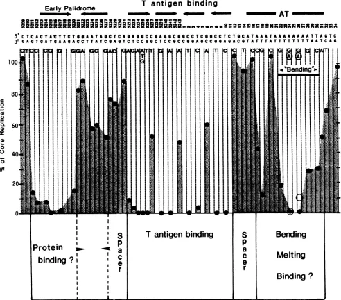
Early Palidrome
T antigen binding
I
AT
£12=2223itttj
!R§3!;;;;a,,;Xf-uzzvo* g*e "5'CTCACTAtTTCTooAATAoCTCoAoAoCCoAooCooCCTCOOCCTCTocATAAATAAAAAAAATTAcTC 3'^AGToATcAAoACCTTATCOAoTCTCCooCTCCoCCooAoCCQAnAnCOTATTTATTTTTTTTAATCA¢
3 BA A CA ATCAAA CSSSAC T T T T T T T A ATT C A
.4Bending~
c 0 o C._
C.) 0 0
aR
Protein
binding
I
I
I I
I
a
I I !
I I
I I
P
I I
I I
I I
4
T
antigen
binding
FIG. 6. Domain structure ofthe SV40 core origin ofreplication. The core origin consists of nucleotides 5211 through 31. Structural featuresofthe DNA are shown at the topofthefigure. Singlebasesubstitutionscorrespondingtothelower strandof SV40DNAareindicated below the WT sequence. Thehistogramshows themeanreplicationefficiencyof thesemutantsdeterminedin threeindependentexperiments. The opencirclesatnucleotides24and 26indicatethereplicationlevelsofadenine-to-thyminesubstitutions. Datacorrespondingtotheearly andlateends ofthe coreoriginweretakenfromourprevious studies (5, 6).
We propose that theprimary roleof the sequences within
theinverted repetitions that contain the pentanucleotides is
tobindTantigen and to position it in the proper location and orientation fora subsequent functional activity. The drastic effect of minimal changes in spacing between pentanucleo-tides or ofinversion of pentanucleotides strongly supports this idea. Mastrangelo et al. (20) showed that the core origin binds four monomer equivalents of T antigen. Previous studies showed that T antigen binds to the core DNA in a
stepwise fashion(27).Thus, the four pentanucleotides within thepalindrome of the core origin may assemble monomers
or dimers in the tightly packed, symmetrical arrangement shown in Fig. 4B. The spacing between the centers of the
bindingsites would placealternating monomers of T antigen
on opposite faces of the DNA. Such an arrangement may
allow T antigen to separate double-stranded DNA in and
around itscontactsites.Dean etal. (4a)showedrecently that purifiedTantigen unwinds plasmid DNA in the presence of
single-stranded binding protein and topoisomerase only if the plasmid contains afunctional origin. Presumably, melt-ing begins within origin sequences, but the initial meltmelt-ing site remains to be determined. This origin-specific unwinding activity may be relatedtoalessspecific helicase function of
Tantigen that is important in the subsequent propagation of
DNA replication (24).
The correct binding arrangement and orientation of T
antigen would promote interactions with other proteins bound to adjacent functional domains of the core origin.
Figure6summarizes the domain structure of the origin that hasbeen determined in our present and previous studies (5,
S
p
a
c
e
r
Bending
Melting
Binding
?
I
J. VIROL.
on November 10, 2019 by guest
http://jvi.asm.org/
[image:6.612.72.564.74.506.2]T-ANTIGEN-BINDING DOMAIN OF SV40 ORIGIN 2149 6). Three segments of contiguous or nearly contiguous DNA
encode sequence-specific core functions. These are sepa-ratedby spacer DNAs with relaxed sequence requirements but with precise positional constraints (5). The early func-tional domain extends 10 bp from positions 5211 through 5220. It corresponds to one arm of an imperfect inverted repetition. We do not know the function of thissegment, but it may be a recognition site or part of a recognition site for a cellular protein. This sequence-specific domain is followed
by an 11-bp segment frompositions 5221through5231.This
segment serves a spacer function (5) but also has a minor sequence-specific domain that corresponds to the second arm of the early imperfect palindrome. Perhaps it is the second and weaker binding site for a protein dimer that
interacts with the early palindrome. The central domain consists of four T-antigen-binding pentanucleotides which
play a key role in T-antigen binding and DNA replication. The central domain is followed by a 5-bp apparent spacer segment without a demonstrable sequence requirement. A late sequence-specific. A. T-rich, bipartite domain extends from nucleotides 17 through 31. The subdomain from
nucle-otides 20 through 31 corresponds exactly to a locus that determines DNA bending and may be astructural signal for protein binding or DNAmelting (6). Thus,theoriginconsists of three or more sequence-specific functional domains that are interaction sites forproteins, structural signals, or both. The orientation of the domains and the spacers between
them would position these functional regions at the proper distances, rotations, and directions to allow coordinated interactions in theinitiation ofDNA replication.
ACKNOWLEDGMENTS
We thank Mary Andersonfortechnical assistance.
This work was supported by Public Health service grants CA-18808, CA-38146, and CA-09176 from the National Cancer Institute.
LITERATURECITED
1. Bergsma, D. J., D. M. Olive, S. W. Hartzell, and K. N. Subramanian. 1982. Territoriallimits andfunctionalanatomy of the simian virus 40 replication origin. Proc. Natl. Acad. Sci. USA79:381-385.
2. Campbell, J. L. 1986.EukaryoticDNAreplication. Annu. Rev. Biochem. 55:733-771.
3. Cereghini, S., and M. Yaniv. 1984. Assembly of transfected DNA intochromatin: structural changes in the origin-promoter-enhancer region upon replication. EMBO J.3:1243-1253. 4. Cohen, G. L., P. J.Wright, A. L. DeLucia, B. A. Lewton, M. E.
Anderson, and P. Tegtmeyer. 1984. Critical spatialrequirement within the origin of simian virus 40 DNA replication. J. Virol. 51:91-96.
4a.Dean, F. B., P. Bullock, Y. Murakami, C. R. Woobe, L. Weissbach,and J.Hurwitz. 1987. SV40DNAreplication: SV40 large T antigen unwinds DNA containing the SV40 origin of replication. Proc. Natl. Acad. Sci. USA 84:16-20.
5. Deb, S.,A. L.DeLucia,C.-P.Baur,A. Koff, and P. Tegtmeyer. 1986. Domain structure of the simian virus 40 core origin of replication. Mol. Cell. Biol. 6:1663-1670.
6. Deb, S.,A. L.DeLucia,A. Koff, S.Tsui,and P. Tegtmeyer. 1986. Theadenine-thymine domain of the simian virus 40 coreorigin directs DNA bending and coordinately regulates DNA replica-tion. Mol. Cell. Biol. 6:4578-4584.
7. DeLucia, A. L., S. Deb, K. Partin, and P. Tegtmeyer. 1986. Functional interactions of the simian virus 40 core origin of replication with flanking regulatory sequences. J. Virol. 57: 138-144.
8. DeLucia,A. L., B. A. Lewton, R. Tjian, and P. Tegtmeyer. 1983. Topography of simian virus 40 A protein-DNA complexes: arrangement ofpentanucleotideinteraction sites at theorigin of
replication.J. Virol. 46:143-150.
9. Echols, H. 1986. Multiple DNA-protein interactions governing
high precisionDNAtransactions. Science 233:1050-1056. 10. Gerard, R. D., B. A. Montelone, C. F. Walter, J. W. Innis, and
W. A.Scott. 1985.Roleof specific simian virus 40 sequencesin the nuclease-sensitive structure in viral chromatin. Mol. Cell. Biol.5:52-58.
11. Gluzman,Y., R. J. Frisque,andJ.F. Sambrook. 1980.
Origin-defectivemutants ofSV40. Cold Spring Harbor Symp. Quant. Biol.44:293-299.
12. Grundstrom, T., W. M. Zenke, M. Wintzrith, H. W. D. Matthes, A. Staub, and P. Chambon. 1985. Oligonucleotide-directed mutagenesis by microscale "shotgun" gene synthesis. Nucleic Acids Res. 13:3305-3316.
13. Hay, R. T., and M. L. DePamphilis. 1982. Initiation ofSV40 DNA replication in vivo: locationand structure of5' ends of DNAsynthesized in theori region. Cell 28:767-779.
14. Innis, J. W., and W. A. Scott. 1984. DNA replication and chromatin structureof simian virus 40 insertion mutants. Mol. Cell. Biol. 3:1499-1507.
15. Jones, K. A., and R. Tjian. 1984. Essential contact residues within SV40 large T antigen binding sites I and IIidentified by alkylation interference. Cell 36:155-162.
16. Jongstra, J., T. L. Reudelhuber, P. Ouden, C. Benoist, C.-B. Chae, J.-M. Jeltsch, B. J. Mathis, and P. Chambon. 1984. Induction of altered chromatin structures by simian virus 40 enhancer and promoter elements. Nature (London) 307:708-714.
17. Li, J.-J., K. W. C. Peden, R. A. F. Dixon, and T. Kelly. 1986. Functional organization of the simian virus 40 origin of DNA replication. Mol. Cell. Biol. 6:1117-1128.
18. Lilley, D. M. 1982. Hairpin loops in supercoiled DNA, p. 173-198. In S. Neidle (ed.), Topics in nucleic acid structures. Part II. John Wiley & Sons, Inc., New York.
19. Lusky, M., and M.Botchan. 1981.Inhibition ofSV40replication in simiancells by specific pBR322 sequences. Nature (London) 293:79-81.
20. Mastrangelo, I. A., P. V. C. Hough, V. G. Wilson, J. S. Wall, J. F. Hainfeld, and P. Tegtmeyer. 1985. Monomers through trimers of large tumor antigenandmonomersthroughtetramers bind in region II of simian virus 40 origin ofreplication DNA as stable structures in solution. Proc. Natl. Acad. Sci. USA 82:3626-3630.
21. McClarin, J. A., C. A. Frederick, B.-C.Wang, P.Greene, H. W. Boyer, J. Grable, and J. M. Rosenberg. 1986. Structure of the DNA-EcoRI endonuclease recognition complex at 3 A resolu-tion. Science 234:1526-1541.
22. Mukherjee, S., I. Patel, and D. Bastia. 1985. Conformational changes in a replication origin induced by an initiator protein. Cell 43:189-197.
23. Shortle, D., and D. Nathans.1979.Regulatory mutants of simian virus 40: constructed mutants with base substitutions at the originofDNAreplication. J. Mol. Biol. 131:801-817.
24. Stahl, H., P. Droge, and R. Knippers. 1986. DNA helicase activityof SV40 large tumorantigen. EMBO J. 5:1939-1944. 25. Stillman, B., R. D. Gerard, R. A. Guggenheimer, and Y.
Gluzman. 1985. Tantigen and templaterequirementsforSV40 DNA replication invitro. EMBO J. 4:2933-2939.
26. Subramanian, K. N., and T. Shenk. 1978. Definition of the boundaries of the origin ofDNAreplication in simian virus 40. NucleicAcids Res. 5:2051-2060.
27. Tegtmeyer, P., B. A. Lewton, A. L.DeLucia,V.G.Wilson, and K.Ryder. 1983.Topography ofsimian virus 40 Aprotein-DNA complexes: arrangement of protein bound to the origin of replication. J. Virol. 46:151-161.
28. Tjian, R. 1978. Thebinding site ofSV40 DNA for a T-antigen-related protein. Cell 13:165-179.
29. Tooze, J. (ed.). 1980. DNA tumor viruses. Part 2. Molecular biologyoftumor viruses. ColdSpringHarborLaboratory, Cold SpringHarbor, N.Y.
30. Wright, P. J., A. L. DeLucia, and P. Tegtmeyer. 1984. Se-quence-specific binding of simian virus 40 Aproteintononorigin andcellularDNA. Mol. Cell. Biol. 4:2631-2638.
VOL.61, 1987