0022-538X/96/$04.0010
Copyrightq1996, American Society for Microbiology
Enzymatic Characterization of Hepatitis C Virus NS3/4A
Complexes Expressed in Mammalian Cells by Using the Herpes
Simplex Virus Amplicon System
ZHI HONG,
1* ERIC FERRARI,
1JACQUELYN WRIGHT-MINOGUE,
1ROBERT CHASE,
1CHRISTINE RISANO,
1GAIL SEELIG,
2CHEE-GUN LEE,
3ANDANN D. KWONG
1Antiviral Chemotherapy
1and Structural Chemistry
2Departments, Schering-Plough Research Institute, Kenilworth, New
Jersey 07033-0539, and Graduate Program in Molecular Biology, Memorial Sloan-Kettering
Cancer Center, Sloan-Kettering Institute, New York, New York 10021
3Received 11 January 1996/Accepted 27 March 1996
The hepatitis C virus (HCV) NS3 protein possesses three enzymatic activities: an N-terminal serine protease
activity, a C-terminal RNA-stimulated NTPase activity, and an RNA helicase activity. To characterize them, the
full-length NS3
631/4A and three C-terminal truncated proteases (NS3
201/4A, NS3
181/4A, and NS3
155/4A) were
expressed in mammalian cells with HSV amplicon-defective viruses. Our results revealed that all of the NS3/4A
proteins produced in mammalian cells (except NS3
155/4A) are active in processing both
cis
and
trans
cleavage
sites. Temperature optimization studies revealed that the protease is more active at temperatures ranging from
4 to 25
&
C and is completely inactive at 42
&
C. The RNA-stimulated ATPase activity was characterized with a
partially purified NS3
631/4A fraction and has a higher optimal temperature at 37 to 42
&
C. The effects of
detergents on both NS3 protease and RNA-stimulated ATPase were similar. Nonionic detergents such as
Triton X-100, Nonidet P-40 and Tween 20 did not affect the activities, while anionic detergents such as sodium
dodecyl sulfate and deoxycholic acid were inhibitory. Zwitterionic detergent such as
3-[(3-cholamidopropyl)-dimethyl-ammonio]-1-propanesulfonate (CHAPS) inhibited protease activity at a concentration of 0.5% (8
mM), which had no effect on ATPase activity. Finally, RNA-unwinding activity was demonstrated in the
NS3
631/4A fraction but not in the similarly purified NS3
181/4A and NS3
201/4A fractions. NS3
631/4A unwinds
RNA duplexes with 3
*
but not 5
*
single-stranded overhangs, suggesting that the NS3 RNA helicase functions
in a 3
*
-to-5
*
direction.
Hepatitis C virus (HCV) is the major etiological agent of
posttransfusion as well as community-acquired non-A non-B
hepatitis (1, 7, 26). Chronic and persistent infection by HCV
often leads to liver cirrhosis and hepatocellular carcinoma (4,
37).
HCV is believed to be a small enveloped virus containing a
positive-stranded linear RNA genome of approximately 9.5 kb
in size (23). It has been classified as a separate genus in the
family of Flaviviridae, on the basis of its genome organization
and hydropathy profile, which are similar to those of
flavivi-ruses and pestiviflavivi-ruses (8, 35, 42).
The HCV genome encodes a single large polyprotein of
approximately 3,010 amino acids (8, 42). Proteolytic processing
by both host signal peptidases and viral proteases results in at
least 10 viral proteins in the following order: NH
2-C-E1-E2-p7-NS2-NS3-NS4A-NS4B-NS5A-NS5B-COOH (16, 17, 32). C,
E1, and E2 are believed to be viral structural proteins which
are encoded by the 5
9
one-fourth of the long open reading
frame (ORF), and the role of p7 has not been established. The
remaining viral proteins (NS2, NS3, NS4A, NS4B, NS5A, and
NS5B) are believed to be nonstructural proteins, components
of the viral replication machinery (22, 36).
Two virally encoded proteases in the HCV polyprotein have
been identified: an NS2/3 metalloprotease and an NS3 serine
protease (15, 18). The NS2/3 protease, a novel Zn
21-depen-dent protease, is responsible for cleavage at the NS2/NS3
junc-tion (15, 18). The NS3 protease is required for cleavages at the
NS3/4A, NS4A/4B, NS4B/5A, and NS5A/5B junction sites (2,
14, 45). The catalytic domain of this protease has been mapped
to the N-terminal 180-amino-acid region of NS3, containing a
characteristic catalytic triad (His-Asp-Ser). The HCV NS3
pro-tease is likely to be an essential enzyme for viral growth,
be-cause its analog from flaviviruses has been shown to be
essen-tial for viral growth (6). Thus, NS3 protease has become the
focus of intensive studies to develop anti-HCV drugs.
Recent work from several laboratories has revealed that
NS4A is a cofactor for the NS3 protease because it is
abso-lutely required by the NS3 protease for cleavage at the 4B/5A
junction (10, 38, 44). A similar requirement for a cofactor has
been shown for flaviviruses (5, 12) and suggested for
pestivi-ruses (49), indicating a common strategy utilized by members
in the family Flaviviridae. The domains for physical interaction
between NS3 and NS4A have been mapped to the N-terminal
22 amino acids of NS3 (11, 38, 44) and to the central region of
NS4A (10, 34, 44). It is believed that NS4A stabilizes the active
conformation of the NS3 protease domain and recruits NS3 to
the membranes, where, presumably, proteolytic processing
takes place (19, 33, 44).
In addition to the N-terminal protease domain, NS3 is
thought to possess an RNA helicase domain in the C-terminal
two-thirds of the NS3 protein on the basis of the presence of a
nucleoside triphosphate (NTP)-binding motif and a DEAH
box for RNA helicases (13). The RNA-stimulated NTPase
activity (41) and the RNA helicase activity (25) of HCV NS3
have been demonstrated and require the C-terminal 465 amino
acids of NS3. The RNA-stimulated NTPase activity (43, 47, 48)
* Corresponding author. Mailing address: Antiviral Chemotherapy, K-15-4945, Schering-Plough Research Institute, 2015 Galloping Hill Rd., Kenilworth, NJ 07033-0539. Phone: (908) 298-3152. Fax: (908) 298-3918. Electronic mail address: [email protected].
4261
on November 9, 2019 by guest
http://jvi.asm.org/
cells: serine protease, RNA-stimulated NTPase, and RNA
he-licase. Our main objectives were (i) to produce large amounts
of NS3 proteases using the mammalian cell expression system;
(ii) to characterize the protease activities of the full-length
NS3
631/4A protein as well as three truncated protease
do-mains, NS3
201/4A, NS3
181/4A, and NS3
155/4A; and (iii) to
characterize the NS3
631/4A-associated NTPase/RNA helicase
activities.
MATERIALS AND METHODS
Cells, virus and antibodies.Rabbit skin cells (RSC) and human epidermoid 2 cells (HEp-2) were obtained from Bernard Roizman, University of Chicago. African green monkey kidney cells (Vero) were obtained from the American Type Culture Collection (CCL 81). Vero cells, RSC, and HEp-2 cells were grown in medium 199 (JRH Biosciences) supplemented with 5% fetal bovine serum (Bio Whittaker). The type 1 (HSV-1) tsk mutant, which has a temperature-sensitive mutation in the ICP4 gene, was obtained from H. S. Marsden at the MRC Virology Unit, Institute of Virology, Glasgow, United Kingdom. The mutant viruses were propagated at 338C in HEp-2 cells with medium 199 con-taining 1% fetal bovine serum. Anti-NS3 (a-NS3) antiserum was raised in rabbits against a bacterially expressed NS3183 protein, which was gel purified.
Anti-NS4A (a-NS4A) antiserum was raised in rabbits against a 20-amino-acid peptide derived from the C terminus of NS4A. Anti-hemagglutinin (a-HA) is a mono-clonal antibody against a hemagglutinin epitope from influenza virus purchased from Boehringer Mannheim Corp.
Construction of the amplicon vector and NS3/4A expression plasmids.The expression cassette of plasmid pCMVb(Clontech), containing the major imme-diate-early promoter of cytomegalovirus as well as the simian virus 40 late region polyadenylation signal, was excised by SphI and inserted at the BamHI site of the amplicon vector, pF19-Pa(27). A polylinker containing a series of unique re-striction enzyme sites (StuI-NotI-BglII-NsiI-SpeI-KpnI-AccIII) was inserted downstream of the cytomegalovirus promoter between the StuI and the AccIII sites for convenience in introducing genes of interest. The map of the resulting amplicon plasmid, pF19-CMV, is shown in Fig. 1. The T7-HCV expression plasmid, pBRTM/HCV(1-3011), which contains the entire ORF of the HCV-1a (H strain) genome, was obtained from Charles M. Rice, Washington University at St. Louis. The 59end of the NS3 gene was modified by inserting a linker containing a unique SpeI site and an ATG start codon. The 39end of NS4A was modified by removing most of the NS4B and all of the NS5 region downstream of the CelII site. A stop codon (TAG) and a unique KpnI site were inserted after the CelII site. The resulting fragment contains the full-length NS3631/4A and the
N-terminal 15 amino acids from NS4B and thus possesses both cis and trans cleavage sites for the NS3 protease (see Fig. 1). The C-terminal truncated NS3/4A protease domains were created by PCR amplification of the correspond-ing fragments at the 59region of the NS3 ORF and then by linking the fragments to the NsiI site near the 39end of the NS3 ORF. The resulting constructs, NS3201/4A, NS3181/4A, and NS3155/4A, lack the C-terminal RNA helicase
do-main except for 11 amino acids (Thr-Cys-Met-Ser-Ala-Asp-Leu-Glu-Val-Val-Thr) at the very C terminus of NS3 (see Fig. 1). An influenza virus HA epitope containing 9 amino acids (Tyr-Pro-Tyr-Asp-Val-Pro-Asp-Tyr-Ala) was inserted in frame in front of the stop codon in all of the constructs, after the N-terminal 15 amino acids of NS4B. The HA epitope can thus be used to monitor the cleavage at the NS4A/4B site. All of the NS3/4A constructs were excised by SpeI and KpnI and inserted into the amplicon vector pF19-CMV.
Generation of the defective amplicon viruses.To generate the defective vi-ruses, pF19-CMV expression plasmids were transfected into RSC by using the calcium phosphate precipitation method and then by superinfection at 338C by the helper virus, a temperature-sensitive HSV-1 mutant, tsk (24). The resulting viral stocks were then serially propagated in HEp-2 cells for at least three
were infected with the amplicon viruses at 378C for 24 h. Cells were harvested and lysed in 10 ml of the lysis buffer. The cell lysates were precipitated with 25% saturated ammonium sulfate solution at 48C. The precipitate was resolubilized in buffer A (25 mM HEPES [pH 7.3], 25 mM NaCl, 1 mM EDTA, 2 mM DTT, 0.1% Nonidet P-40, 10% glycerol) and loaded onto a Mono Q column. The NS3/4A complexes were eluted off the Mono Q column at 0.6 M NaCl. The resulting Mono Q fraction was then passed through a Mono S column and one-step eluted at 1 M NaCl. The resulting Mono S fraction was finally loaded on a poly(U)-Sepharose affinity column (Pharmacia). The NS3631/4A complex
was eluted off the column at 1.0 M NaCl. The resulting poly(U) fraction contains partially purified NS3631/4A complex (;10 to 20% pure). Aliquots from each
fraction during the purification were analyzed on an SDS-10 to 20% polyacryl-amide gradient gel by Coomassie brilliant blue staining and Western blotting (see Fig. 4).
Immunoblotting analysis.Immunoblotting or Western blotting analysis was carried out as described previously (20). Briefly, proteins were separated on a 10 to 20% polyacrylamide gradient gel (Integrated Separation Systems) and were transferred onto an Immobilon P membrane (Millipore Corp.) with a semidry electroblotter (Integrated Separation Systems). Incubations of the membrane with primary antibodies or alkaline phosphatase-conjugated secondary antibod-ies took place in phosphate-buffered saline solution (PBS; pH 7.3) containing 1% evaporated milk and 0.3% Tween 20. The membrane was then washed in PBS containing 0.3% Tween 20 and was developed in the presence of chromogenic substrates, 1.7 mg of 5-bromo-4-chloro-3-indolyl phosphate (BCIP) per ml, and 3.3 mg of nitroblue tetrazolium.
In vitrotranscleavage assaying.A bacterial T7 expression plasmid, pTS102, containing a truncatedDNS5A/B recombinant substrate (amino acid positions 2312 to 2621), was obtained from Elizabeth B. Smith (Schering-Plough Research Institute). The substrate was synthesized and35S labeled with in vitro
transcrip-tion and translatranscrip-tion systems (Promega). Labeled substrate contained the NS5A/5B cleavage site and had an apparent molecular weight of 45,000 by SDS-polyacrylamide gel electrophoresis (PAGE) analysis. Incubation of labeled substrate with the NS3/4A proteases resulted in the release of two products: the C-terminal portion of NS5A (DNS5A [18 kDa]) and the N-terminal portion of NS5B (DNS5B [27 kDa]) (see Fig. 3B). The in vitro trans cleavage assay was carried out at 258C for 30 min in a buffer containing 50 mM Tris (pH 8.0), 0.5 mM EDTA, and 6 mM DTT, which was similar to conditions described by Bouffard et al. (3).
ATPase assaying.The ATPase assay has been described previously (30, 48). Briefly, ATPase assaying measures the hydrolysis of ATP to ADP, in which the enzymes were incubated with 10mCi of [a-32P]ATP at 378C for 30 min in a 10-ml
reaction mixture containing 50 mM HEPES (pH 7.3), 1 mM DTT, 100mg of bovine serum albumin (BSA) per ml, 10 mM NaCl, 3 mM MgCl2, and 5 mM
ATP. The reaction was stopped by the addition of EDTA to a final concentration of 45 mM. A 0.5-ml volume of the reaction mixture was spotted on a polyethyl-eneimine-cellulose plate and was analyzed by thin-layer chromatography (43). RNA-stimulated ATPase assaying was carried out in the presence of 1 mM poly(U) homopolymer (Pharmacia).
Preparation of RNA duplexes and RNA helicase assaying.The substrates for RNA helicase assaying were prepared as described previously (30). Three types of RNA duplexes, standard, 39-tailed, and 59-tailed, were made and are shown in Fig. 6. The RNA helicase assay measures the displacement of the32
P-labeled primers from the RNA duplexes. The reaction was carried out as described elsewhere (30). Briefly, partially purified NS3631/4A was incubated with 50 fmol
of32
P-labeled RNA duplexes at 378C for 30 min in a 20-ml reaction mixture containing 20 mM HEPES (pH 7.3), 2.5 mM MgCl2, 5 mM ATP, 2 mM DTT,
100mg of BSA per ml, and 1 U of RNase inhibitor (Prime Inhibitor; 59339Inc.). The reaction was stopped by the addition of 5ml of RNA loading buffer (100 mM Tris-HCl [pH 7.5], 20 mM EDTA, 0.5% SDS, 0.1% Nonidet P-40, 0.1% bromo-phenol blue, 0.1% xylene cyanol, 50% glycerol). The entire mixture was then loaded onto a 15% polyacrylamide gel (30:1) and electrophoresed in 0.1% SDS and 0.53Tris-borate-EDTA (TBE) buffer at 10 mA for 16 h. The gel was dried
on November 9, 2019 by guest
and exposed for autoradiography. The amount of helicase activity was deter-mined by the appearance of the smaller RNA primer bands, which run faster than the RNA duplexes on the gel.
RESULTS
Construction of amplicon expression plasmids and
genera-tion of defective amplicon viruses.
To achieve high-level
ex-pression of NS3 protease in mammalian cells, genes encoding
the full-length NS3
631/4A as well as a series of the C-terminal
truncated protease domains, NS3
201/4A, NS3
181/4A, and
NS3
155/4A, were constructed and cloned into the amplicon
vector (Fig. 1). All of the NS3 genes were fused in frame with
the NS4A ORF because NS4A is required as a cofactor for
NS3 protease activity (10, 38, 44). All of the constructs retained
both the NS3/4A cis cleavage site and the NS4A/4B trans
cleav-age site as indicated (Fig. 1). An influenza virus HA epitope
was fused to the C termini of all of the NS3/4A proteases and
was used to monitor trans cleavage at the NS4A/B site.
Expression of proteolytically active NS3/4A complexes in
mammalian cells.
Amplicons containing different NS3/4A
con-structs were used to infect Vero cells at a multiplicity of
infec-tion of approximately 3. Infected cells were incubated at either
33 or at 37
8
C for 24 h. The products were analyzed by Western
blots with three different antibodies,
a
-NS3,
a
-NS4A, or
a
-HA.
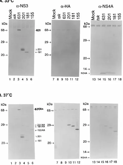
The results demonstrated that the NS3
631/4A-HA, NS3
201/4A-HA, NS3
181/4A-HA, and NS3
155/4A-HA constructs were
ex-pressed at different levels (Fig. 2, left blots). The NS3
155/
4A-HA protein was not detected at 33
8
C (Fig. 2A, lane 6) and
was produced only at a low level at 37
8
C (Fig. 2B, lane 6). The
expression level of NS3
181/4A-HA (Fig. 2A and B, lane 5) was
somewhat lower than that of NS3
631/4A-HA or NS3
201/4A-HA
(Fig. 2A and B, lanes 3 and 4). The NS3
201/4A-HA construct
produced two NS3
201products which might be due to N-linked
glycosylation, since there is a putative N-linked glycosylation
site at the C-terminal end of NS3
201. However, tunicamycin, a
potent inhibitor of N-linked glycosylation, failed to inhibit the
production of the larger NS3
201product. Thus, it is not clear
why more than one NS3
201product was produced. All of the
NS3/4A-HA products except for NS3
155/4A-HA are active in
processing the cis cleavage site at the NS3/4A junction,
releas-ing the NS4A protein shown at the bottom of the Western blot
(Fig. 2A, right blot). A product which is slightly larger than the
NS4A protein was also released from the NS3
201/4A-HA and
NS3
181/4A-HA constructs. This product is not NS4A-HA
be-cause it cannot be detected by the
a
-HA monoclonal antibody
(Fig. 2A, center blot). It is possible that this product results
from inaccurate cleavage at the NS3/4A site because of spatial
and conformational constraints created in the corresponding
constructs. The observation that no products could be detected
on the blot with
a
-HA monoclonal antibodies (Fig. 2A, lanes 9
[image:3.612.320.554.75.388.2]to 11) suggested that the HA epitope was quickly removed
because of trans cleavage at the NS4A/4B site. Interestingly, no
FIG. 1. Construction of the amplicon expression plasmids. The N-terminal protease domain (open bar) and the C-terminal RNA helicase domain (hatched bar) of NS3 are indicated. NS4A is indicated by a shaded bar. Amino acid sequences at both the cis (NS3/4A) and trans (NS4A/4B) cleavage sites are shown. Arrows, scissile bonds where the cleavages occur. The same cis- and
trans-cleavable sites are present in all of the constructs. The amplicon vector
pF19-CMV contains both HSV ori (DNA replication origin) and HSV pac (pack-aging signal). PIE, the cytomegalovirus (CMV) immediate-early promoter; SV40,
simian virus 40; aa, amino acids.
FIG. 2. Expression of NS3/4A-HA constructs in mammalian cells with the HSV amplicon system. (A and B) Expression at 33 and 378C, respectively. Western blotting analyses were carried out with three different antibodies:a-NS3 (lanes 1 to 6),a-HA (lanes 7 to 12), anda-NS4A (lanes 13 to 18). 631, 201, and 181, NS3631, NS3201, and NS3181, respectively; 631/4A, 201/4A, 181/4A, and
155/4A, the uncleaved precursors NS3631/4A-HA, NS3201/4A-HA, NS3181
/4A-HA, and NS3155/4A-HA, respectively.
on November 9, 2019 by guest
http://jvi.asm.org/
shown that the NS3 protease domain forms complexes with
NS4A (11, 34, 38). The results shown in Fig. 2 suggested that
the autoproteolytic processing of the NS3/4A-HA products
occurred rapidly, removing the HA epitope and the N-terminal
15 amino acids of NS4B. Thus, the product from each gene
construct was designated NS3/4A complex, such as NS3
631/4A
complex, NS3
201/4A complex, and NS3
181/4A complex. To
fur-ther characterize the protease activity as well as the
NS3-associated NTPase/RNA helicase activities, we attempted to
purify the NS3
631/4A complex expressed in mammalian cells.
The purification includes 25% ammonium sulfate precipitation
and then passage through Mono Q, Mono S, and poly(U)
affinity columns (Materials and Methods). This protocol was
utilized to purify both the NS3
631/4A and the NS3
201/4A
com-plexes expressed in mammalian cells. The purification was
monitored by Western blotting analysis with
a
-NS3 antibodies
(Fig. 3A) as well as by a protease assay with a substrate
con-taining the NS5A/5B site (Fig. 3B). Both the NS3
631/4A and
NS3
201/4A complexes exhibited the same chromatographic
be-havior up until the final poly(U) affinity column passage, which
was designed to purify the NS3
631/4A complex on the basis of
the RNA-binding activity of the C-terminal RNA helicase
do-main (46). The NS3
201protein was absent in the final poly(U)
fraction (Fig. 3A, lane 12) because it lacks the RNA helicase
domain and does not bind to the poly(U) column.
RNA-stim-ulated ATPase activity was also analyzed at each purification
step in the absence or presence of 1 mM poly(U)
homopoly-mers (Fig. 3C). RNA-stimulated ATPase activity was observed
in all of the NS3
631/4A fractions (lanes 2 to 11). In contrast,
only background ATPase activity was detected in the starting
lysate and the 25% ammonium sulfate precipitate of the
NS3
201/4A fractions. This suggested that the endogenous
ATPase activities were efficiently removed from the partially
purified NS3
631/4A fraction. Proteins in each fraction during
the purification were also analyzed on a 10 to 20%
polyacryl-amide gel stained with Coomassie brilliant blue. A 70-kDa
protein band is present in the final poly(U) fraction derived
from the NS3
631/4A lysate (Fig. 4A, lane 6) but not from the
NS3
201/4A lysate (Fig. 4A, lane 7). Western blotting analysis
confirmed that the 70-kDa protein is the full-length NS3
631protease (Fig. 4B, lane 2). Although the NS4A protein is
dif-ficult to detect on the gel stained with Coomassie brilliant blue
because of its small size (
;
6 kDa) (Fig. 4A, lane 6), it
copu-rified with the NS3
631protease as detected by
a
-NS4A
anti-bodies (Fig. 4B, lane 5). The purity of the NS3
631protein was
estimated at approximately 10 to 20% compared with the
amounts of other contaminating proteins, which were present
in the final poly(U) fractions of both NS3
631/4A and NS3
201/4A
(Fig. 4A, lane 6 and 7). The same protocol had been used to
purify the NS3
631/4A complex produced in insect cells with the
baculovirus system, which yielded a purity of approximately
90% (Fig. 4C, lane 1). The NS4A again copurified with the
full-length NS3 protein (Fig. 4C, lane 2). The levels of
expres-sion as well as the enzymatic activities of the NS3
631/4A
com-plexes from both expression systems (HSV amplicon versus
baculovirus) are comparable (21). However, for large-scale
production of the proteins, we prefer the baculovirus system.
Differential optimal temperatures for NS3 protease and
NS3-associated RNA-stimulated ATPase.
To further
charac-terize NS3 protease activity and the NS3-associated ATPase
activity, in vitro assays for both enzymes were established. To
confirm the observations that NS3 proteases were less active
when expressed at 37
8
C than at 33
8
C (Fig. 2), partially purified
NS3
631/4A and NS3
201/4A were used in an in vitro assay to
characterize the optimal temperatures for the protease activity.
The results revealed that while the proteases were most active
at lower temperatures ranging from 4 to 25
8
C, they were
ob-viously less active at 37
8
C than at 30
8
C and were completely
[image:4.612.317.555.72.395.2]inactive at 42
8
C (Fig. 5A). The decrease in protease activity at
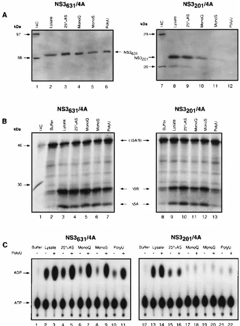
FIG. 3. Partial purification of the NS3631/4A complex in parallel with the
NS3201/4A complex. (A) Monitoring of the NS3631/4A complex (lanes 2 to 6) and
the NS3201/4A complex (lanes 8 to 12) during purification by Western blotting
analysis witha-NS3 antibodies. (B) Monitoring of the purification by in vitro
trans cleavage protease assaying with an in vitro-translated substrate containing
the NS5A/5B site.D5A/B, the uncleaved substrate labeled with [35S]methionine;
D5A andD5B, cleavage products. Substrate alone (buffer) without NS3 proteins is the negative control, as indicated in lanes 2 and 8. Lanes 3 to 7, samples from the NS3631/4A purification; lanes 9 to 13, samples from the NS3201/4A
purifica-tion. (C) Comparative analysis of NS3-associated ATPase activities during the purification of NS3631/4A and NS3201/4A. ATPase assaying was performed in the
absence (2) or in the presence (1) of 1 mM poly(U) nucleotides, respectively. The amounts of samples used in the above analyses were normalized between NS3631/4A and NS3201/4A fractions by protein concentration.
on November 9, 2019 by guest
higher temperatures is probably not due to any host-related
protease inactivation, because no degradation was detected by
Western blotting analysis before and after incubation at higher
temperatures (data not shown). To characterize the optimal
temperature for the NS3-associated RNA-stimulated ATPase
activity, the ATPase activities of the poly(U) fractions from
both NS3
631/4A and NS3
201/4A were compared. In contrast to
the protease, the NS3-associated RNA-stimulated ATPase has
a higher optimal temperature of 37 to 42
8
C (Fig. 5B). The
optimal temperatures for both enzyme activities were also
ver-ified with the 90% pure NS3
631/4A derived from the
baculovi-rus system (data not shown). One explanation for the
differ-ential optimal temperatures is that the NS3-associated
RNA-stimulated ATPase domain may be more thermostable than
the NS3 protease domain. To address the thermostability of
[image:5.612.131.456.73.240.2]both domains, heat inactivation of the NS3
631/4A complex was
FIG. 4. Purity of the NS3631/4A complex. (A) Protein analysis on an SDS-10 to 20% polyacrylamide gel stained with Coomassie brilliant blue. MW, molecular
weight. (B) Western blotting analysis of the total lysate (lanes 1 and 4) and the poly(U) fractions (lanes 2, 3, 5, and 6) witha-NS3 antibodies (lanes 1 to 3) anda-NS4A antibodies (lanes 4 to 6). (C) SDS-PAGE analysis of the NS3631/4A complex produced in a baculovirus expression system. Lanes: 1, silver stain; 2, Western blotting
with botha-NS3 anda-NS4A antibodies.
FIG. 5. Optimal temperatures for NS3 protease and RNA-stimulated ATPase. (A) Temperature optimization for protease activity with the in vitro trans cleavage assay. Sub, substrate alone; tsk, tsk helper virus-infected cell lysate; 631/4A and 201/4A, the corresponding Mono S fractions during the purification;D5A/B, the uncleaved substrate labeled with [35
S]methionine;D5A andD5B, the cleavage products. (B) Temperature (temp) optimization for RNA-stimulated ATPase activity in the presence (1) of 1 mM poly(U). The poly(U) fractions of NS3631/4A (lanes 2 to 9) and NS3201/4A (lanes 11 to 18) were used in the ATPase assay analyzed by
thin-layer chromatography.
on November 9, 2019 by guest
http://jvi.asm.org/
[image:5.612.127.485.417.680.2]carried out by preincubating the proteins at different
temper-atures. The results indicated that both enzyme activities were
inactivated at 50
8
C after preincubation (21). Furthermore, the
thermoinstability of the protease domain is probably not due to
the dissociation of NS4A, since adding excess amounts of
NS4A peptide (2
m
M) failed to prevent the NS3 protease from
being inactivated.
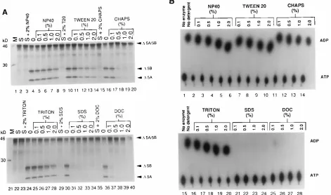
Detergent effects on the activities of NS3 protease and
NS3-associated RNA-stimulated ATPase.
It has been proposed that
a potential role of NS4A is to recruit NS3 to membranes where
the HCV virus assembly is believed to occur (19, 33, 44). Thus,
it is interesting to know how detergents affect the NS3
func-tions. The effect of detergents on NS3 protease activity has
been reported with somewhat contradictory results (3, 9, 33).
We investigated the effects of detergent on both protease
ac-tivity and RNA-stimulated ATPase acac-tivity. The results in Fig.
6 showed that while nonionic detergents such as Triton X-100,
Nonidet P-40, and Tween 20 did not affect both enzymatic
activities, anionic detergents such as SDS and deoxycholic acid
were inhibitory. Interestingly, D’Souza et al. (9) reported
stim-ulation of NS3 protease activity with a zwitterionic detergent,
3-[(3-cholamidopropyl)-dimethyl-ammonio]-1-propanesulfon-ate (CHAPS), at concentrations of up to 2.5% (40 mM); in our
hands, 0.5% (8.0 mM) CHAPS is inhibitory (Fig. 6A, lane 18).
However, RNA-stimulated ATPase activity was not affected
(Fig. 6B, lane 12). Our results regarding the effects of
deter-gent on protease activity are similar to those reported by
Bouf-fard et al. (3), with the exception that, in their hands, 1% SDS
was not inhibitory, whereas we observed complete inhibition
with 0.1% SDS (Fig. 6A, lane 31). Although we failed to
observe any significant stimulating effect on protease activity
with the addition of detergents using the in vitro cleavage
assay, in a more quantitative high-performance liquid
chroma-tography assay, stimulation with detergent such as 0.1%
Non-idet P-40 was detected (21).
The NS3
631/4A complex produced in mammalian cells has
RNA helicase activity with a 3
*
-to-5
*
directionality.
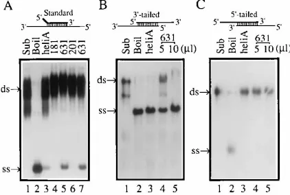
An in vitro
RNA-unwinding (strand displacement) assay for RNA helicase
activity was developed under conditions similar to those
de-scribed previously (30). Three types of RNA helicase
sub-strates were prepared to determine RNA helicase activity and
its directionality (31, 46). The poly(U) fractions were used to
characterize the NS3
631/4A-associated RNA helicase with both
NS3
181/4A and NS3
201/4A fractions as controls. As shown in
Fig. 7A, the NS3
631/4A complex unwound the standard duplex
RNA substrate (lanes 5 and 7), while the similarly purified
NS3
181/4A and NS3
201/4A fractions failed to unwind the
sub-strate (lanes 4 and 6). As a positive control, RNA helicase A
(31) also unwound the standard RNA duplexes (Fig. 7A, lane
3). In a test of directionality, the NS3
631/4A-associated RNA
helicase unwound the 3
9
-tailed RNA duplexes (Fig. 7B, lanes 4
and 5) but not the 5
9
-tailed RNA duplexes (Fig. 7C, lane 4 and
5), suggesting that the NS3 RNA helicase is a 3
9
-to-5
9
RNA
[image:6.612.72.543.75.353.2]helicase. The control RNA helicase A has the same
direction-ality as the NS3 RNA helicase (Fig. 7B, lane 3; Fig. 7C, lane 3).
Further characterization to optimize the NS3 RNA helicase
activity and to compare activities from different RNA helicase
constructs has been carried out and will be presented
else-where. RNA helicases from other positive-stranded RNA
vi-ruses such as the NS3 protein of bovine viral diarrhea virus
FIG. 6. The effect of detergents on NS3 protease and RNA-stimulated ATPase. (A) Effects of detergent on protease activity. S, substrate (D5A/B) alone without the NS3631/4A complex; T20, Tween 20;D5A/B, uncleaved substrate labeled with [35S]methionine;D5A andD5B, the cleavage products. (B) Effects of detergent on
RNA-stimulated ATPase activity. The concentration of each detergent can be converted to molar concentrations as follows: 1% Triton X-100, 16 mM; 1% Nonidet P-40, 17 mM; 1% Tween 20, 8 mM; 1% SDS, 35 mM; 1% deoxycholate (DOC), 24 mM; 1% CHAPS, 16 mM.
on November 9, 2019 by guest
(46) and the cylindrical inclusion (CI) protein of plum pox
viruses (29) have shown properties similar to those of the HCV
NS3 RNA helicase.
DISCUSSION
In this report, we have demonstrated three enzymatic
activ-ities that are associated with the HCV NS3 protein. NS3
pro-tease activity and NS3 RNA-stimulated NTPase activity have
been characterized previously (2, 14, 41, 45). Our results have
further extended the effort to characterize and optimize the
effects of temperature on protease activity in comparison with
NTPase activity. We have utilized the HSV amplicon system to
express NS3/4A proteins in mammalian cells. More
impor-tantly, for the first time, conventional purification of the native
NS3
631/4A complex without any fusion tag is presented, and we
have demonstrated RNA helicase activity, the third activity of
the NS3 protein.
An interesting finding in our report is the comparison of the
optimal temperatures and the detergent effects on both
pro-tease and RNA-stimulated ATPase activities (Fig. 5 and 6).
While the protease is most active at 4 to 25
8
C, the ATPase is
more active at 37 to 42
8
C. Although a similar observation for
the protease activity has been reported previously (3), it has
not been discussed systematically. The difference in the
opti-mal temperatures may suggest that the protease domain is less
thermostable than the ATPase domain (Fig. 5). However, heat
inactivation experiments have indicated that the
thermostabili-ties are similar between the protease domain and the NTPase
domain. Furthermore, inactivation of the protease may not be
due to the dissociation of NS4A (21). Detergents seem to affect
both activities similarly, although a slight difference in the
effect of the zwitterionic detergent CHAPS was observed (Fig.
6B). These results suggest that both the protease domain and
the RNA-stimulated NTPase domain have similar
require-ments for enzyme stability. In a recent report by Lin et al. (32),
the protease was shown to be more active at 30 or 37
8
C, and
the protease was inhibited by nonionic detergents at
concen-trations that did not affect the protease activity in our assays.
Several reasons may explain the contradictory observations: (i)
different systems were used to produce the enzymes for the in
vitro cleavage assays, (ii) different substrates were used
taining different cleavage sites, and (iii) different gene
con-structs were used. It is also quite possible that different HCV
isolates or cDNA clones encode proteases with different
tem-perature sensitivities. In our hands, the difference seems not to
result from the temperatures at which various NS3 enzymes
have been produced. We have generated our proteases at
different temperatures (33 versus 37
8
C), which show similar
requirements for lower optimal temperatures. Furthermore,
after incubation at 37
8
C for 30 min, the protease is equally
active compared with the protease incubated at 25
8
C (data not
shown). The slower autoproteolytic processing of the NS3
pro-tease when expressed at 37
8
C (Fig. 2) also confirms that the
NS3 protease is less active at higher temperatures. This result
seems to contradict the fact that HCV replicates in human
liver, where the temperature is approximately 37
8
C. It is
pos-sible that there is a specific cellular or viral factor which
sta-bilizes enzyme activity at 37
8
C. Alternatively, HCV may have
developed a suboptimal enzyme activity to regulate its growth
in humans, and this might reflect the observation that HCV
replicates poorly in cultured cells (39, 50).
One role proposed for NS4A is to recruit NS3 to membranes
for its activity (19, 33, 44) or to stabilize the active
conforma-tion of the protease domain. Indeed, it has been shown that
NS4A forms complexes with the N-terminal domain of NS3
(11, 38, 44). We have demonstrated that NS3 and NS4A
form stable complexes and are copurified during the entire
purification procedures. The complexes are very stable and
cannot be disrupted in the presence of 1% Nonidet P-40
detergent and 2 M salt (NaCl). Furthermore, the complexes
are eluted off a gel filtration column (FPLC Superose 12
column; Pharmacia) right after the void volume (molecular
weight,
'
2,000,000), suggesting that the NS3/4A complexes
form aggregates, possibly with detergents in the form of
micelles (21).
The RNA-stimulated ATPase activity in the poly(U)
frac-tion of NS3
631/4A showed requirements for optimal activity
similar to those previously reported (41), such as temperature,
MgCl
2, and stimulation by polynucleotides with a preference
for poly(U) (data not shown). This activity is also inhibited by
KCl. A slight difference was observed, i.e., that NS3
631/4A has
a slightly broader range of pH values for optimal ATPase
activity (data not shown), in contrast to the stringent optimal
pH (6.5) reported previously (41). The difference could be due
to the fact that a truncated C-terminal domain was used in the
previous characterization. The RNA helicase activity
demon-strated in this report also showed properties similar to those
reported for other positive-strand RNA viruses (29, 46). For
example, it unwinds RNA duplexes only with the 3
9
-unpaired
region and KCl is inhibitory. Further characterization of the
HCV NS3 RNA helicase is currently in progress to determine:
(i) if it binds to single-stranded RNA or DNA in the absence
of ATP; (ii) if it can displace DNA/RNA hybrid duplexes or
DNA/DNA duplexes; and (iii) what its requirements in the 3
9
unpaired region for optimal RNA-unwinding activity are.
ACKNOWLEDGMENTS
We thank Jerome Schwartz, W. Robert Bishop, and Gregory Reyes for support and helpful discussion. We also appreciate excellent technical assistance from Elizabeth B. Smith, Nancy Butkiewicz, and Ronald Jubin.
REFERENCES
1. Alter, H. J., H. S. Margolis, K. Krawczynski, F. N. Judson, A. Mares, W. J. Alexander, P. Y. Hu, J. K. Miller, M. A. Gerber, R. E. Sampliner, E. L. Meeks, and M. J. Beach.1992. The natural history of community-acquired hepatitis C in the United States. N. Engl. J. Med. 327:1899–1905. 2. Bartenschlager, R., L. Ahlborn-Laake, J. Mous, and H. Jacobsen. 1993.
Nonstructural protein 3 of the hepatitis C virus encodes a serine-type pro-FIG. 7. NS3 RNA helicase activity. (A) RNA-unwinding activity with the
standard substrate. Sub, RNA duplex; Boil, denatured single-stranded RNA molecules made by heating the RNA duplex at 958C for 30 min; heli A, RNA helicase A; 181, 201, and 631, the partially purified poly(U) fractions of NS3181/
4A, NS3201/4A, and NS3631/4A complexes, respectively. (B) RNA-unwinding
activity with the 39-tailed substrate. (C) RNA-unwinding assay with the 59-tailed substrate.
on November 9, 2019 by guest
http://jvi.asm.org/
[image:7.612.71.282.74.216.2]8. Choo, Q.-L., K. H. Richman, J. H. Han, K. Berger, C. Lee, C. Dong, C. Gallegos, D. Coit, A. Medina-Selby, P. J. Barr, A. J. Weiner, D. W. Bradley, G. Kuo, and M. Houghton.1991. Genetic organization and diversity of the hepatitis C virus. Proc. Natl. Acad. Sci. USA 88:2451–2455.
9. D’Souza, E. D. A., K. Grace, D. V. Sangar, D. J. Rowlands, and B. E. Clarke. 1995. In vitro cleavage of hepatitis C virus polyprotein substrates by purified recombinant NS3 protease. J. Gen. Virol. 76:1729–1736.
10. Failla, C., L. Tomei, and R. De Francesco. 1994. Both NS3 and NS4A are required for proteolytic processing of hepatitis C virus nonstructural pro-teins. J. Virol. 68:3753–3760.
11. Failla, C., L. Tomei, and R. De Francesco. 1995. An amino-terminal domain of the hepatitis C virus NS3 protease is essential for interaction with NS4A. J. Virol. 69:1769–1777.
12. Falgout, B., M. Pethel, Y.-M. Zhang, and C.-J. Lai. 1991. Both nonstructural proteins NS2B and NS3 are required for the proteolytic processing of den-gue virus nonstructural proteins. J. Virol. 65:2467–2475.
13. Gorbalenya, A. E., A. P. Donchenko, V. Koonin, and V. M. Blinov. 1988. A conserved NTP-motif in putative helicases. Nature (London) 333:22. 14. Grakoui, A., D. W. McCourt, C. Wychowski, S. M. Feinstone, and C. M. Rice.
1993. Characterization of the hepatitis C virus-encoded serine proteinase: determination of the proteinase-dependent polyprotein cleavage sites. J. Virol. 67:2832–2843.
15. Grakoui, A., D. W. McCourt, C. Wychowski, S. M. Feinstone, and C. M. Rice. 1993. A second hepatitis C virus-encoded proteinase. Proc. Natl. Acad. Sci. USA 90:10583–10587.
16. Grakoui, A., C. Wychowski, C. Lin, S. M. Feinstone, and C. M. Rice. 1993. Expression and identification of hepatitis C virus polyprotein cleavage prod-ucts. J. Virol. 67:1385–1395.
17. Hijikata, M., N. Kato, Y. Ootsuyama, M. Nakagawa, and K. Shimotohno. 1991. Gene mapping of the putative structural region of the hepatitis C virus genome by in vitro processing analysis. Proc. Natl. Acad. Sci. USA 88:5547– 5551.
18. Hijikata, M., H. Mizushima, T. Akagi, S. Mori, N. Kakiuchi, N. Kato, T. Tanaka, K. Kimura, and K. Shimotohno. 1993. Two distinct proteinase activities required for the processing of a putative nonstructural precursor protein of hepatitis C virus. J. Virol. 67:4665–4675.
19. Hijikata, M., H. Mizushima, Y. Tanji, Y. Komoda, Y. Hirowatari, T. Akagi, N. Kato, K. Kimura, and K. Shimotohno.1993. Proteolytic processing and membrane association of putative nonstructural proteins of hepatitis C virus. Proc. Natl. Acad. Sci. USA 90:10773–10777.
20. Hong, Z., M. Beaudet-Miller, J. Durkin, R. Zhang, and A. D. Kwong. 1996. Identification of a minimal hydrophobic domain in the herpes simplex virus type 1 scaffolding protein which is required for interaction with the major capsid protein. J. Virol. 70:533–540.
21. Hong, Z., and A. D. Kwong. Unpublished data.
22. Houghton, M. 1996. Hepatitis C viruses, p. 1035–1058. In B. N. Fields, D. M. Knipe, and P. M. Howley (ed.), Virology. Raven Press, New York. 23. Kaito, M., S. Watanabe, K. Tsukiyama-Kohara, K. Yamaguchi, Y.
Koba-yashi, M. Konishi, M. Yokoi, S. Ishida, S. Suzuki, and M. Kohara.1994. Hepatitis C virus particle detected by immunoelectron microscopic study. J. Gen. Virol. 75:1755–1760.
24. Kaplitt, M. G., A. D. Kwong, S. P. Kleopoulos, C. V. Mobbs, S. D. Rabkin, and D. W. Pfaff.1994. Preproenkephalin promoter yields region-specific and long-term expression in adult brain after direct in vivo gene transfer via a defective herpes simplex viral vector. Proc. Natl. Acad. Sci. USA 91:8979– 8983.
25. Kim, D. W., Y. Gwack, J. H. Han, and J. Choe. 1995. C-terminal domain of hepatitis C virus NS3 protein contains an RNA helicase activity. Biochem. Biophys. Res. Commun. 215:160–166.
26. Kuo, G., Q.-L. Choo, H. J. Alter, G. L. Gitnick, A. G. Redeker, R. H. Purcell, T. Miyamura, J. L. Dienstag, M. J. Alter, C. E. Stevens, G. E. Tegtmeier, F.
and two distinct E2-specific products with different C termini. J. Virol. 68:5063–5073.
33. Lin, C., and C. M. Rice. 1995. The hepatitis C virus NS3 serine proteinase and NS4A cofactor: establishment of a cell-free trans-processing assay. Proc. Natl. Acad. Sci. USA 92:7622–7626.
34. Lin, C., J. A. Thomson, and C. M. Rice. 1995. A central region in the hepatitis C virus NS4A protein allows formation of an active NS3-NS4A serine proteinase complex in vivo and in vitro. J. Virol. 69:4373–4380. 35. Miller, R. H., and R. H. Purcell. 1990. Hepatitis C virus shares amino acid
sequence similarity with pestiviruses and flaviviruses as well as members of two plant virus supergroups. Proc. Natl. Acad. Sci. USA 87:2057–2061. 36. Rice, C. M. 1996. Flaviviridae: the viruses and their replication, p. 931–960.
In B. N. Fields, D. M. Knipe, and P. M. Howley (ed.), Virology. Raven Press,
New York.
37. Saito, I., T. Miyamura, A. Ohbayashi, H. Harada, T. Katayama, S. Kikuchi, T. Y. Watanabe, S. Koi, M. Onji, Y. Ohta, Q.-L. Choo, M. Houghton, and G. Kuo.1990. Hepatitis C virus infection is associated with the development of hepatocellular carcinoma. Proc. Natl. Acad. Sci. USA 87:6547–6549. 38. Satoh, S., Y. Tanji, M. Hijikata, K. Kimura, and K. Shimotohno. 1995. The
N-terminal region of hepatitis C virus nonstructural protein 3 (NS3) is essential for stable complex formation with NS4A. J. Virol. 69:4255–4260. 39. Shimizu, Y. K., A. Iwamoto, M. Hijikata, R. H. Purcell, and H. Yoshikura.
1992. Evidence for in vitro replication of hepatitis C virus genome in a human T-cell line. Proc. Natl. Acad. Sci. USA 89:5477–5481.
40. Spaete, R. R., and N. Frenkel. 1982. The herpes simplex virus amplicon: a new eucaryotic defective-virus cloning-amplifying vector. Cell 30:295–304. 41. Suzich, J. A., J. K. Tamura, F. Palmer-Hill, P. Warrener, A. Grakoui, C. M.
Rice, S. M. Feinstone, and M. S. Collett.1993. Hepatitis C virus NS3 protein polynucleotide-stimulated nucleoside triphosphatase and comparison with the related pestivirus and flavivirus enzymes. J. Virol. 67:6152–6158. 42. Takamizawa, A., C. Mori, I. Fuke, S. Manabe, S. Murakami, J. Fujita, E.
Onishi, T. Andoh, I. Yoshida, and H. Okayama.1991. Structure and orga-nization of the hepatitis C virus genome isolated from human carrier. J. Virol. 65:1105–1113.
43. Tamura, J. K., P. Warrener, and M. S. Collett. 1993. RNA-stimulated NT-Pase activity associated with the p80 protein of the pestivirus bovine viral diarrhea virus. Virology 193:1–10.
44. Tanji, Y., M. Hijikata, S. Satoh, T. Kaneko, and K. Shimotohno. 1995. Hepatitis C virus-encoded nonstructural protein NS4A has versatile func-tions in viral protein processing. J. Virol. 69:1575–1581.
45. Tomei, L., C. Failla, E. Santolini, R. de Francesco, and N. La Monica. 1993. NS3 is a serine protease required for processing of hepatitis C virus polypro-tein. J. Virol. 67:4017–4026.
46. Warrener, P., and M. Collett. 1995. Pestivirus NS3 (p80) protein possesses RNA helicase activity. J. Virol. 69:1720–1726.
47. Warrener, P., J. K. Tamura, and M. S. Collett. 1993. RNA-stimulated NTPase activity associated with yellow fever virus NS3 protein expressed in bacteria. J. Virol. 67:989–996.
48. Wengler, G., and G. Wengler. 1991. The carboxyl-terminal part of the NS3 protein of the West Nile flavivirus can be isolated as a soluble protein after proteolytic cleavage and represents an RNA-stimulated NTPase. Virology 184:707–715.
49. Wiskerchen, M., and M. S. Collett. 1991. Pestivirus gene expression: protein p80 of bovine viral diarrhea virus is a serine proteinase involved in polypro-tein processing. Virology 184:341–350.
50. Yoo, B. J., M. J. Selby, J. Choe, B. S. Suh, S. H. Choi, J. S. Joh, G. J. Nuovo, H.-S. Lee, M. Houghton, and J. H. Han.1995. Transfection of a differenti-ated human hepatoma cell line (Huh7) with in vitro-transcribed hepatitis C virus (HCV) RNA and establishment of a long-term culture persistently infected with HCV. J. Virol. 69:32–38.