Abstract: Skin cancer is being classified among the mortal and exaggerating forms of cancer since decade. Notwithstanding, the early diagnosis of skin cancer is very important and it is an extravagant procedure. The presence of human skin is tough to examine and to model. This is because of its complex surface. The difficulty of the irregular edge, tone, appearance of thick hair and other alleviating features generate the skin tough to be analyzed. Human skin has some non-identical sorts of textures that diseased skin can characterize between the textures of the healthy one. In consequence, considerable achievements have not been brought into the evolution of diagnosis approach for skin cancer. On the other hand, skin cancer diagnosis in dermoscopic portrayals is an exceptionally troublesome chore. Notwithstanding, the early diagnosis of skin cancer is very important and it is an extravagant procedure. In consequence, considerable achievements have not been brought into the evolution of diagnosis approach for skin cancer. For precise diagnosis and categorization of skin cancer, particular features are recommended that one may categorize benign and malignant illustration. A midst analysts, governing the efficacious mechanism of skin cancer diagnosis is an all-important matter of contention. Ascertaining the more proficient methods of diagnosis to minimize the amount of errors is a crucial subject among analysts. Image processing is used to recognize the affected area by disease, its form and color etc. The reason behind it is to minimize the percentage of miscalculations. Computer vision can bring about influential contribution in skin cancer detection. Computer Aided Diagnosis provides support in the premature diagnosis of skin cancer. Programmed skin abrasion segregation is a stimulating chore because of the low contrast between abrasion and encompassing skin and asymmetrical periphery. In this paper we present an amalgamation of statistical features, GLCM features and GLRLM features. The recommended methodology is enacted on 100 images from PH2 database. In this work 2 segmentation tactics namely Otsu’s thresholding and Region growing have been explored. The performance of two systems is measured in terms of sensitivity, specificity, accuracy, precision, recall and f-measure
Index Terms: Biomedical image processing, Skin cancer, Gray-level Co-occurrence Matrix, Grey-level Run Length Matrix, Region Growing, Otsu thresholding, supervised classification
I. INTRODUCTION
Skin that accounts for 4-5 kg mass and surface area of about 1.2 m2 - 2.2 m2 is an eminent and gigantic organ of human. The very organ safeguards the body from outer environment. It overhauls the temperature of body and entitles
Revised Manuscript Received on June 23, 2019. .
Ashima Kalra, PhD Research Scholar, I.K. Gujral Punjab Technical University, Jalandhar(Punjab),India
Khushmeen Brar, Mtech Research Scholar, Electronics and Communication Engineering, Chandigarh Engineering College, Mohali, India
Piyush Samant, Chandigarh University, Ghauran, Mohali, India.
human to perceive sensations. Bearings that obstruct, modify skin appearance or deteriorate the skin can induce traits like inflammation, redness and rawness. Hypersensitivity, annoyance, and specific diseases can create diverse skin problems. But the most menacing disease, the skin can breed is cancer. Skin cancer is the utmost frequent and widespread sort of cancer that elucidates about 3.5 million cases. It is fatal if not diagnosed prematurely. Skin cancer can be tabulated as benign and the malignant. The malignant one is mortal form of cancer and in such a way it necessitates instantaneous diagnosis. It emerges from cancerous extension in pigmented abrasion. Meanwhile, benign skin abrasion is a usual skin and not menacing like malignant skin abrasion. Diagnosis of skin cancer is henceforth an important chore. Various methodologies have been implemented as of now to categorize the skin abrasion. The ABCD rule is by far considered as foremost dermoscopy innovation for the diagnosis of skin cancer [1],[2]. Neural network is considered as the efficient approach for classification purpose [3]. Amongst classifiers, the support vector machine(SVM) and k-means clustering can significantly perform well for classification or regression issues [4]-[5]. In the latest research, analysts employ snake’s algorithm for optimum results [6].Nevertheless untimely diagnosis of skin cancer is a complex job. Initially, the cast intra-class diversification of skin cancer concerning colour, appearance, form, magnitude in dermoscopy portrayals and resemblance among benign and malignant make it hard to differentiate malignant from non-malignant ones. Second, the respective low variation and hazy peripheries among skin abrasions and usual skin part make the programmed diagnosis function even complicated. Last, the appearance of clamour may un-focus the abrasions [7]. Numerous attempts have been devoted on resolving this complication. Premature evaluation aims to affect features to discriminate skin cancer from normal skin comprising colour [8], [9], texture [10] or both [11], [12]. Malignant melanoma cases have been notably exacerbating in decades, making it one of the tumours that has been experiencing consideration from medical management and tumour analysis domain. While numerous fresh instances of skin cancer are being detected every year, a programmed methodology to support the avoidance and premature diagnosis is distinctly desirable. With a view to refine the diagnostic pursuance of skin cancer, dermoscopy methodology was established. Dermoscopy is an approach that is utilized by dermatologists to detect skin cancer at the present time. The medical practitioner settles the gel on the abrasion and monitors it with an amplification tool that expands the abrasion.
This expansion entitles the detection of specific surface
Ashima Kalra, Khushmeen Brar, Piyush Samant
Improved Technique to Diagnose Skin Cancer
using Advanced Image Processing and Machine
that is out of sight to naked eye. This can then be employed to detect an abrasion utilizing specific symptomatic algorithms like ABCD rule [1], [13], menzies method, seven point checklist having alike initial step, that is, discerning the abrasion with reference to its emergence as melanocytic or non-melanocytic. Dermoscopy can literally intensify the vulnerability of melanoma diagnosis by 10-27%, only if practitioner has endured official guidance.
The detection of abrasion by medical practitioner is nevertheless instinctive as it is build upon human observation. Thus programmed dermoscopy image evaluation can be employed to pursue this difficulty. In this type of methodologies, a computer is employed as a diagnostic appliance to investigate doubtful abrasions. Automatically fragmenting the melanoma from adjoining skin is an indispensable step in programmed or automated interpretation of dermoscopic portrayals. The identical perspective for prematurely diagnosing skin cancer comprises divergent steps of pre-refining, subdivision, feature withdrawn and categorisation. The outcomes of each step pay vital contribution to avert misdiagnosis. A review on procedures and steps of CAD system on melanoma has been provided by [14]. Whilst the accomplishment of CAD methodologies relies upon Pre-refining, the foremost level of CAD methodology has solemnly consequences on misleading the outcomes. A contrast of various pre-refining approaches has been illustrated in [15]. Some analysts offered to engage feature selection algorithms and the amalgamation of features [16]. Few analysts offered to accomplish segmentation [17] and on the basis of this, classify the melanoma as benign and malignant. The latest research reveals that the smart phone application can be much beneficial for the diagnosis of skin cancer [18]..From literature, it may be noticed that an improved algorithm, in order to segment the skin abrasion is a prerequisite task. Also the literature reveals the use of statistical, structural and textural features. The propounded features lack on the subject of robustness and therefore the introduction of robust feature set is an essential condition. In order to reduce the complexity of categorization of various algorithms, the reduced feature set is a prerequisite. The contrast among different classifiers is required to be explored that one may be able to select the appropriate one. The comparison of various classifiers with reference to their accuracy and other performance metrics need to be developed.
In this paper the contrast amidst segmentation approaches for skin cancer diagnosis has been carried out.The first methodology uses the otsu’s segmentation that automatically implements clustering-based image thresholding. It reduces gray scale image into a binary one. Otsu’s methodology recapitulates uninterruptedly to every feasible threshold values, computing a measure of stretch for pixel magnitudes on adjoining side of threshold. This methodology picks the threshold to reduce the intra-class variation of threshold among the pixels. The second system uses region-growing methodology that explores adjoining pixels of preliminary seed points. It regulates whether or not the adjoining pixels should be attached to the region and the mechanism is recapitulated on. The major objective of this technique is to divide an image into regions. This segmentation governs the region precisely. This paper attempts to discover contrast amongst the two approaches and implement the results in skin
cancer detection.
II. PROPOSEDMETHODOLOGY
The variation in skin abrasion as malignant and non-malignant makes it inconvenient to diagnose the skin cancer. Moreover, the skin cancer is evolving haphazardly thus determining the optimum descriptor to separate benign and malignant skin cancer is a vital function. In this paper, we introduce a set of particular features acquired from distinct descriptors in order to spot among benign and malignant abrasions. The elaborated flowchart of the recommended feature extraction and its fusion procedure is encapsulated in fig.1.
Fig.1 Flowchart of Proposed Methodology
A.Dataset
The images have been acquired from the PH2 database [19]. A total of 100 images have been considered for the analysis.
B.Pre-image Processing
Pre-refining is an eminent practice of designating benign and malignant skin cancer. Pre-refining comprises converting the image into a meaningful form. The statistics might be uncertain, partial or might also comprise error. The method is required to evacuate clamor that causes uncertainty to the classifier. The dermoscopic portrayals are 8 bit RGB images. The portrayals of the abrasion are transformed into grey scale form.
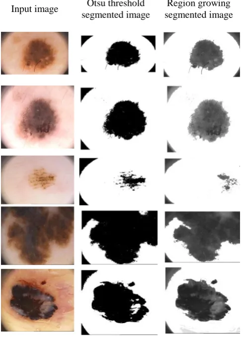
Segmentation is an approach of segregating digital portrayals into segments that measure related characteristics to facilitate the characterization and creating it better for the investigation and explanation. Image segmentation is a requisite part of image evaluation procedure. The methodology discriminates among the entity to be examined and the alternative entities. The paper uses otsu’s thresholding [20] and the region-growing technique for segmentation [21]. Fig. 2 shows the segmented sample images from both the methods.
Input image Otsu threshold segmented image
Region growing segmented image
Fig.2 Input images versus otsu threshold segmented and region growing segmented images
D.Feature Extraction
The subsequent step is the extraction of features of the respective portrayals for the successive classification.
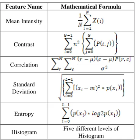
Features simply define the data extricated from images in numeral form and that can’t be analyzed by human being. Features are distinctive signatures of particular portrayal. In this paper, GLCM Features, GLRLM Features and the statistical features have been extracted.
GLCM Features
GLCM or gray-level spatial dependence matrix has demonstrated to be a prominent statistical approach of withdrawing features from the portrayals. A co-occurrence allocation is a matrix specifically defined above a representation to be the distribution of co-occurring grayscale standards by a prearranged offset. Graycomatrix utility is
made use of to generate GLCM by estimating how frequently a pixel amid intensity value i come about in an explicit spatial connection to pixel amid value j. Every factor (i, j) in the resultant GLCM is basically the summation of the amount of times the pixel through value i transpired in a particular spatial connection to a pixel amid value j in the input figure [18]. GLCM extricates second order dermographic textural features named as: Autocorrelation, Cluster Entropy prominence, Cluster shade, Contrast, Correlation, Difference entropy, Difference variance, Dissimilarity, Energy, Homogeneity, Information measure of correlation 1, Information measure of correlation 2, Inverse difference, Maximum probability, Sum average, Sum entropy, Sum of squares variance, Sum variance.
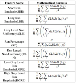
GLRLM Features
GLRLM is the set of running pixels with the identical grey level i and run length j for given direction. Grey level run length matrix is evaluated that one may withdraw textural features. The following GLRLM features have been extricated in this research (Table I):
Table 1 GLRLM Feature set with mathematical formulas
[image:3.595.50.295.195.534.2]Statistical Features
[image:4.595.41.266.146.375.2]The method identifies the texture laterally[22]. Statistical method is employed that one may examine the dimensional spread of grey value by enumerating features consistently in image and acquiring a set of data from spread of features [23-26]. Table II shows statistical features which have been extricated in this paper.
Table II Statistical Feature set with mathematical formulas
E.Classification
Six classification parameters of 20 different classifiers have been calculated and compared in this paper.
III. PERFORMANCEANALYSIS
The estimation of propounded methodology is computed maneuvering 6 performance measures that are Sensitivity, Specificity, Accuracy, Precision, Recall and F-measure:
Sensitivity= TP/TP+FN (1)
Specificity= TN/TN+FP (2)
Accuracy= TP+TN/TP+FN+TN+FP (3)
Precision=TP/TP+FP (4)
Recall=TP/TP+FN (5) F-measure=(2*Precision*Recall) / (Precision+Recall)(6)
IV. RESULTANDDISCUSSION A.Performance Analysis of Otsu Thresholding The performance of the classifiers is illustrated employing the table known as confusion matrix. The necessary terms related to the matrix may be stated as TP or True Positive, TN or True Negative, FP or False Positive and FN or False Negative. Figure 3 shows the accuracy of different classifiers for the segmented images by ostu thresholding method. Table III compared the segmentation Results obtained by otsu’s thresholding methodology.
Classif ier Sens itivit y Speci ficity Accur acy Precis ion Recall F-mea sure Compl ex Tree 92.8
0% 75% 90%
95.12 %
92.85
% 93%
Mediu m Tree
92.8
0% 75% 90%
95.12 %
92.85
% 93%
Simple Tree
92.8
0% 75% 90%
95.12 %
92.85
% 93%
Linear SVM
91.5 0%
64.70
% 87%
92.68 % 91.56 % 91.49 % Quadra tic SVM 91.7 6% 73.30
% 89%
95.12 % 91.76 % 92.95 % Cubic SVM 93.6 7% 61.90
% 87%
90.24 % 93.67 % 91.47 % Fine Gaussi an SVM 92.0 4% 91.66
% 92%
98.78 % 92.04 % 94.90 % Mediu m Gaussi an SVM 94.0 4% 81.25
% 92%
96.34 % 94.04 % 94.98 % Coarse Gaussi an SVM 82.8 2% 100
% 83% 100%
82.82 % 90.10 % Fine KNN 95.1 2% 77.77
% 92%
95.12 %
95.12
% 95%
Mediu m KNN
95% 70% 90% 92.68
% 95%
93.47 %
Coarse
KNN 82% 0/0 82% 100% 82%
90.10 % Cosine
KNN 95%
70.00
% 90%
92.68
% 95%
93.47 % Cubic KNN 93.8 2% 68.42
% 89%
92.68 % 93.82 % 92.49 % Weight ed KNN 95.1 8% 82.35
% 93%
96.34 % 95.18 % 95.49 % Booste d Trees 95.0 6% 73.68
% 91%
93.90 % 95.06 % 93.98 % Bagge d Trees 94.0 4% 81.25
% 92%
96.34 % 94.04 % 94.98 % Subspa ce Discri minant 92.6 8% 66.66
% 88%
92.68 %
92.68
% 92%
Subspa ce KNN 94.0 4% 81.25
% 92%
96.34 % 94.04 % 94.98 % RUS Booste d Trees 97.3 6% 72.72
% 90%
90.24 % 97.36 % 93.36 % Feature Name Mathematical Formula
Mean Intensity Contrast Correlation Standard Deviation Entropy
Table III. Performance evaluations of sensitivity, specificity, accuracy, precision, recall and F-measure for various classifiers under the otsu’s thresholding methodology
The table presents as to which classifier performs better. The results obtained by the table illustrates that from sensitivity point of view, RUS Boosted trees outperformed all other classifiers with 97.36%. The specificity of 100% is achieved by Coarse Gaussian SVM classifier. The accuracy of 93% by Weighted KNN, precision of 100% by Coarse KNN, recall of 97.36% by RUS Boosted Trees and F-measure of 95% was achieved by Fine KNN classifier. The table presents the examples of segmentation results obtained by applying Otsu’s thresholding segmentation method to original images. Fig.3 comprises the illustration results that analyze how efficaciously the abrasions were segmented by the otsu’s thresholding technique. The figure demonstrates the accuracy versus classifiers relationship. The evaluation of results achieved demonstrated that the otsu’s thresholding method is effective in diagnosis of skin abrasion and extracting their outlines from PH2 dataset figures.
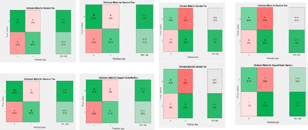
Figure 4 shows the examples of confusion matrices for the otsu threshold algorithm of some selective classifiers. Table 3 illustrates the performances achieved in the name of sensitivity, specificity, accuracy, precision, recall and F-measure for various classifiers under the otsu’s thresholding methodology. In order to measure the performance of otsu thresholding 6 performance measures, namely sensitivity, specificity, accuracy, precision, recall and F-measure have been evaluated. Then performances are evaluated by making use of the equations described above in section 3. Table III shows that from the sensitivity point of view, the best result is achieved maneuvering RUS Boosted Trees with sensitivity of 97.36%. If we consider specificity metrics, the best result is carried out by the Coarse Gaussian SVM classifier with 100% value. Weighted KNN reveals the
best accuracy result with 93%. Considering precision performance metric, the best result is given by the Coarse Gaussian SVM classifier with the 100% result. With regard to
recall metric, the best result is shown by RUS Boosted Tree with 97.36%. And best result for F-measure with 95.49% is given by the Weighted KNN classifier. The results have revolved that for Otsu thresholding Boosted tree, coarse gausian SVM and Weighted
KNN classifiers showing the best results.
Feature Name Mathematical Formula Short Run
Emphasis(SRE)
Long Run Emphasis(LRE)
Grey Level Non Uniformity(GLN) Run Percentage (RP) Run Length Non-Uniformity(RL N)
Low Grey Level Run Emphasis(LGRE)
High Grey Level Run Emphasis(HGRE) Classifi er Sensitiv ity Specif icity Accur acy Precis ion Reca ll F-mea sure Comple
x Tree 86.20%
89.15
% 83% 50%
66.66 %
53.45 % Mediu
m Tree 52.94%
89.15
% 83% 50%
52.94 % 53.45 % Simple Tree 60% 89.28
% 84% 50% 56% 53%
Linear
SVM 91.66%
92.04
% 92% 61%
91.66 %
73.03 % Quadrat
ic SVM 76.92%
90.80 % 89% 55.55 % 76.92 % 63.81 % Cubic SVM 62.50% 90.47 % 86% 55.55 % 62.50 % 58.29 % Fine Gaussia n SVM
0/0 82% 82% 0/0 0/0 0/0
Mediu m Gaussia n SVM 80% 88.88 % 88% 44.44 % 80.00 % 56.77 % Coarse Gaussia n SVM 83.30% 86.17 % 86% 27.77 % 83.33 % 40.74 % Fine KNN 84.60% 91.95 % 91% 61.11 % 84.60 % 70.67 % Mediu
m KNN 88.88% 89% 89%
44.44 % 88.88 % 58.66 % Coarse
KNN 0/0 82% 82% 0/0 0/0 0/0
Cosine
KNN 50% 88% 82%
44.44 % 50.00 % 22.68 % Cubic KNN 88.88% 89.01 % 89% 44.44 % 88.88 % 58.66 % Weight ed KNN
90% 90% 90% 50% 90.00
%
64.28 %
Booste
d Trees 76.92%
B.Performance analysis by Region growing
Performance evaluation of region growing segmentation algorithm has been shown in figure 5, 6 and table 4. Table IV demonstrates the performances realized in the name of sensitivity, specificity, accuracy, precision, recall and F-measure for a variety of classifiers under the region growing tactic.
Table IV reports the performance evaluations for various classifiers for region growing segmentation technique maneuvering Sensitivity, Specificity, Accuracy, Precision, Recall and F-measure. According to above table, the best result in terms of sensitivity is achieved by Linear SVM classifier with 91.66%. If we consider in terms of specificity, the best result is given by the RUS Boosted Tree with 92.68%. In terms of accuracy, the Linear SVM provides best result with 92%. The RUS Boosted Tree gives best result with just 66.66% in terms of precision metrics. If only recall is
Table IV Performance evaluations of sensitivity, specificity, accuracy, precision, recall and F-measure for various
classifiers under the region growing methodology
considered, the best result is given by Linear SVM with 91.66%. And in case, only F-measure is considered, the best result is provided again by Linear SVM. So, from this discussion it is clear that for region growing segmentation, Linear SVM shows best results. The comparison between table IV and table V show that the otsu thresholding segmentation technique shows better result than region growing in all aspects i.e., in terms of sensitivity, specificity, accuracy, recall, precision and F-measure.
V. CONCLUSION
Commencing the outcomes over, the associated conclusions can be determined employing neural system diagnosis of skin diseases. This is feasible and realizable all the way through extraction, division and classification methodology. This paper has compared two approaches namely otsu thresholding and region growing segmentation for the skin cancer diagnosis characterized by GLCM features, GLRLM features and the statistical features. Initially [27], the abrasion region is segmented using otsu thresholding and region growing segmentation. Later, the features are extracted in order to represent the abrasion region. And then the classification is performed maneuvering various classifiers. The two approaches were investigated using a database known as dermoscopy data-set. The experiments are carried out on 100 dermoscopic portrayals. The system parameters i.e., sensitivity (SE), specificity (SP), accuracy, precision, recall and F-measure were tested employing various classifiers. The results reveal that the otsu threshold technique outperforms the region growing segmentation in all performance metrics.
REFERENCES
[1] A. Rajesh, “Classification of malignant melanoma and Benign Skin Lesion by using back propagation neural network and ABCD rule,” Cluster Comput., pp. 1–8, 2018.
[2] S. Jain, V. Jagtap, and N. Pise, “Computer aided melanoma skin
cancer detection using image processing,” Procedia Comput. Sci., vol. 48, no. C, pp. 736–741, 2015.
[3] P. Dubai, S. Bhatt, C. Joglekar, and S. Patii, “Skin cancer detection and classification,” Proc. 2017 6th Int. Conf. Electr. Eng. Informatics Sustain. Soc. Through Digit. Innov. ICEEI 2017, vol. 2017–Novem, pp. 1–6, 2018.
[4] H. Alquran et al., “The melanoma skin cancer detection and classification using support vector machine,” 2017 IEEE Jordan Conf. Appl. Electr. Eng. Comput. Technol. AEECT 2017, vol. 2018–Janua, pp. 1–5, 2018.
[5] S. M. Jaisakthi, P. Mirunalini, and C. Aravindan, “Automated skin lesion segmentation of dermoscopic images using GrabCut and k-means algorithms,” IET Comput. Vis., vol. 12, no. 8, pp. 1088–1095, 2018.
[6] P. Bumrungkun, K. Chamnongthai, and W. Patchoo, “Detection
skin cancer using SVM and snake model,” 2018 Int. Work. Adv. Image Technol. IWAIT 2018, pp. 1–4, 2018.
[7] S. Joseph and J. R. Panicker, “Skin lesion analysis system for melanoma detection with an effective hair segmentation method,” in International Conference on Information Science, ICIS, 2016, pp. 91–96.
[8] N. Petrellis, “Using Color Signatures for the Classification of Skin Disorders,” 2018 7th Int. Conf. Mod. Circuits Syst. Technol., pp. 1–4, 2018.
[9] A. Fidalgo Barata, E. Celebi, and J. Marques, “Improving Dermoscopy Image Classification Using Color Constancy,” IEEE J. Biomed. Heal. Informatics, vol. 2194, no. c, pp. 1–1, 2014. [10] M. N. Islam, J. Gallardo-Alvarado, M. Abu, N. A. Salman, S. P.
Rengan, and S. Said, “Skin disease recognition using texture analysis,” 2017 IEEE 8th Control Syst. Grad. Res. Colloquium, ICSGRC 2017 - Proc., vol. 1, no. August, pp. 144–148, 2017. [11] C. Barata, M. Ruela, M. Francisco, T. Mendonca, and J. S.
Marques, “Two systems for the detection of melanomas in dermoscopy images using texture and color features,” IEEE Syst. J., vol. 8, no. 3, pp. 965–979, 2014.
[12] O. Abuzaghleh, B. D. Barkana, and M. Faezipour, “Automated skin lesion analysis based on color and shape geometry feature set for melanoma early detection and prevention,” 2014 IEEE Long Isl. Syst. Appl. Technol. Conf. LISAT 2014, 2014.
[13] N. F. M. Azmi, H. M. Sarkan, Y. Yahya, and S. Chuprat, “ABCD
rules segmentation on malignant tumor and Benign skin lesion images,” 2016 3rd Int. Conf. Comput. Inf. Sci. ICCOINS 2016 - Proc., pp. 66–70, 2016.
[14] A. N. Hoshyar, A. Al-Jumaily, and R. Sulaiman, “Review on automatic early skin cancer detection,” 2011 Int. Conf. Comput. Sci. Serv. Syst. CSSS 2011 - Proc., pp. 4036–4039, 2011. [15] A. N. Hoshyar, A. Al-Jumaily, and A. N. Hoshyar, “The beneficial
techniques in preprocessing step of skin cancer detection system comparing,” Procedia Comput. Sci., vol. 42, no. C, pp. 25–31, 2014.
[16] F. Adjed, S. J. Safdar Gardezi, F. Ababsa, I. Faye, and S. Chandra Dass, “Fusion of structural and textural features for melanoma recognition,” IET Comput. Vis., vol. 12, no. 2, pp. 185–195, 2018. [17] R. Sumithra, M. Suhil, and D. S. Guru, “Segmentation and classification of skin lesions for disease diagnosis,” Procedia Comput. Sci., vol. 45, no. C, pp. 76–85, 2015.
[18] R. Maurya, S. K. Singh, A. K. Maurya, and A. Kumar, “GLCM and Multi Class Support vector machine based automated skin cancer classification,” 2014 Int. Conf. Comput. Sustain. Glob. Dev., pp. 444–447, 2014.
[19] T. M. P. M. Ferreira1 and J. S. M. A. R. S. M. J. R. Abstract—The, “PH2 - A dermoscopic image database for research and benchmarking*,” J. ACM, vol. 14, no. 4, pp. 677–682, 1967. [20] C. Hima Bindu and K. Satya Prasad, “An Efficient Medical Image
Segmentation Using Conventional OTSU Method,” Int. J. Adv. Sci. Technol., vol. 38, pp. 67–74, 2012.
[21] S. Kamdi and R. K. Krishna, “Image Segmentation and Region Growing Algorithm,” Int. J. Comput. Technol. Electron. Eng., vol. 2, no. 1, pp. 2249–6343, 2012.
[22] J. Virmani and R. Agarwal, “ScienceDirect Effect of despeckle filtering on classification of breast tumors using ultrasound images,” Integr. Med. Res., pp. 1–21, 2019.
[23] P. Samant and R. Agarwal, “Machine learning techniques for
medical diagnosis of
diabetes using iris
images,” Comput.
Methods Programs
121–128, 2018.
[24] Kriti, Jitendra Virmani and Ravinder Agarwal, "Effect of despeckle filtering on classification of breast tumors using ultrasound images", Biocybernetics and Biomedical Engineering, Vol. 39, No. 2, pp. 536-560, 2019.
[25 Jitendra Virmani, Vinod Kumar, Naveen Kalra and Niranjan Khandelwal, "Neural network ensemble based CAD system for focal liver lesions using B-mode ultrasound", Journal of Digital Imaging, Vol. 27, No. 4. Pp. 520-537, 2014.
[26] Jitendra Virmani, Vinod Kumar, Naveen Kalra and Niranjan Khandelwal, "SVM-based characterization of liver cirrhosis by singular value decomposition of GLCM matrix", International Journal of Artificial Intelligence and Soft Computing, Vol. 3, No.3, 2013, pp. 276-296.
[27] P Samant, R Agarwal, "Comparative analysis of classification based algorithms for diabetes diagnosis using iris images", journal of Medical Engineering & Technology, vol. 42, 2018.
AUTHORS PROFILE
Ms. Ashima Kalra, Gold Medalist in B.Tech in Electronics from Kurukshetra in 2003. Received M tech degree from Punjab Technical University, Kapurthla (Punjab)in 2008 and pursuing PhD from Punjab Technical University, Kapurthala(Punjab) in the field of soft Computing.She has published more than 25 papers in reputed journals and 3 book chapters in Springer series. Her research activities include neural networks, fuzzy systems, supervised learning .machine learning, image processing. Lifetime IEEE member and has been serving as a fellow member of IEEE Delhi section, India. She organized 3 IEEE international conferences as a chairperson.
Khushmeen Brar is M Tech Research scholar in Chandiagrh Engineering College, Landran(Mohali). She received her B.Tech degree in electronics and communication from Punjab Technical University in 2017. Her research interests are in the field of image processing, biomedical image and dermoscopy.
Fig. 3 Accuracy of different classifiers for the segmented images by otsu thresholding method
Fig. 4 Confusion matrices for the otsu threshold algorithm of some selective classifiers
Fig. 5 Accuracy of different classifiers for the segmented images by region growing method
[image:8.595.48.553.259.472.2]