M E T H O D O L O G Y
Open Access
Development of standardized laboratory methods
and quality processes for a phase III study of the
RTS, S/AS01 candidate malaria vaccine
Christine Swysen
1*, Johan Vekemans
1, Myriam Bruls
1, Sunny Oyakhirome
2,3and Chris Drakeley
4and
Peter Kremsner
2,3and Brian Greenwood
4, Opokua Ofori-Anyinam
1and Brenda Okech
1, Tonya Villafana
5and
Terrell Carter
5, Barbara Savarese
5, Adriano Duse
6, Andrea Reijman
7, Charlotte Ingram
7, John Frean
8,
Bernhards Ogutu
9and the Clinical Trials Partnership Committee
Abstract
Background:A pivotal phase III study of the RTS,S/AS01 malaria candidate vaccine is ongoing in several research centres across Africa. The development and establishment of quality systems was a requirement for trial conduct to meet international regulatory standards, as well as providing an important capacity strengthening opportunity for study centres.
Methods:Standardized laboratory methods and quality assurance processes were implemented at each of the study centres, facilitated by funding partners.
Results:A robust protocol for determination of parasite density based on actual blood cell counts was set up in accordance with World Health Organization recommendations. Automated equipment including haematology and biochemistry analyzers were put in place with standard methods for bedside testing of glycaemia, base excess and lactacidaemia. Facilities for X-rays and basic microbiology testing were also provided or upgraded alongside health care infrastructure in some centres. External quality assurance assessment of all major laboratory methods was established and method qualification by each laboratory demonstrated. The resulting capacity strengthening has ensured laboratory evaluations are conducted locally to the high standards required in clinical trials.
Conclusion:Major efforts by study centres, together with support from collaborating parties, have allowed standardized methods and robust quality assurance processes to be put in place for the phase III evaluation of the RTS, S/AS01 malaria candidate vaccine. Extensive training programmes, coupled with continuous commitment from research centre staff, have been the key elements behind the successful implementation of quality processes. It is expected these activities will culminate in healthcare benefits for the subjects and communities participating in these trials.
Trial registration:Clinicaltrials.gov NCT00866619
Background
A candidate malaria vaccine, RTS,S/AS01, is in late-stage clinical trials in infants and children in sub-Saharan Africa. Earlier studies of the RTS,S/AS vaccine have demonstrated a promising safety profile and effi-cacy in this population [1-7]. Partners in the clinical development of this vaccine include the manufacturer
GlaxoSmithKline (GSK) Biologicals, the Program for Appropriate Technology in Health Malaria Vaccine Initiative (PATH-MVI) as the main funding partner, the Malaria Clinical Trials Alliance (MCTA) as the main capacity strengthening funding partner, and the Clinical Trial Partnership Committee (CTPC), consisting of representatives from the research centres where the trials are being conducted.
Although considerable expertise in biochemistry, hae-matology, microbiology and malaria-related laboratory * Correspondence: [email protected]
1GlaxoSmithKline Biologicals, Rixensart, Belgium
Full list of author information is available at the end of the article
methods already existed in some of the participating research centres, there were many centres which had yet to develop these capabilities. Therefore, implementation of quality processes during a pivotal phase III trial pre-sented an important opportunity for capacity strength-ening of laboratories across the centres conducting the trials. The extent of capacity strengthening depended on the existing scientific and technical knowledge and laboratory infrastructure at each site prior to trial pre-paration. Sharing best practice experience across labora-tories during the capacity strengthening exercise was valuable.
This paper describes the laboratory methods used in the pivotal phase III study of the candidate RTS, S/AS01 malaria vaccine, and highlights how the efforts of the study investigators and the study centre staff, together with support from collaborating partners, have effec-tively built laboratory capacity to support the conduct of the trial to good clinical practice (GCP) and good clini-cal laboratory practice (GCLP) regulatory standards [8] in all the 11 African centres participating in the study.
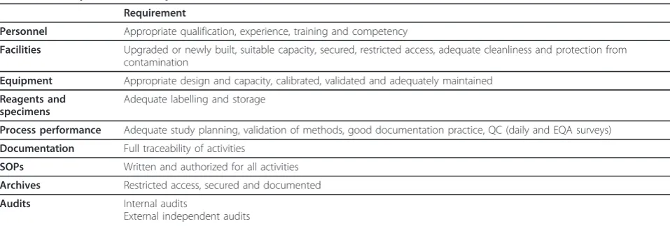
Quality assurance: implementation and challenge
The quality assurance (QA) programme is based on the criteria of GCLP from the British Association of Research Quality Assurance (BARQA) and the Interna-tional Committee on Harmonization guidelines on GCP that cover organization, personnel, facilities, equipment, specimens and reagents, performance and process con-duct, standard operating procedures (SOPs), documenta-tion, reporting, archiving, and the QA programme (Table 1). Implementation of the QA process during this large and complex trial has involved the services of Contract Laboratory Services (CLS - Johannesburg, South Africa), a highly experienced company in African projects. Presently CLS plays a key role in monitoring and evaluating participating laboratories, including
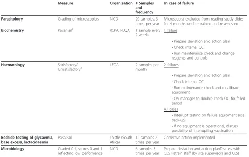
on-site laboratory appraisals, providing training, facilitating external quality assessment (EQA) and advising on equipment and processes. The EQA programme is sum-marized in Table 2.
Building infrastructure at some of the centres was an important part of the implementation of the overall QA process. Although some participating centres already possessed state-of-the-art facilities, others lacked some basic requirements such as an adequate power supply. Bringing a level of parity between the centres was, therefore, necessary. Provision of new infrastructure, such as the construction of laboratories and installation and maintenance of equipment, was made possible by financial support from MCTA and resource investments from GSK Biologicals and MVI.
Other practical challenges faced in the development and implementation of the laboratory strengthening pro-gram were high staff turnover, with consequent loss of experienced and trained personnel, lack of technical knowledge among equipment vendors, and delays in supply which hampered the installation of equipment. Logistical difficulties included limitations in available transport, compounded by long transportation distances and the challenging climatic conditions, which presented problems in the operation of equipment and preserva-tion of samples and reagents.
Slide reading for malaria parasites: methodology and quality assurance/quality control
Accurate measurement of the Plasmodium falciparum
[image:2.595.57.536.559.721.2]count in blood samples is a vital part of the phase III efficacy evaluation of the RTS,S/AS01 malaria candidate vaccine, as it is crucial for the diagnosis of clinical malaria and severe malaria, the key endpoints for eva-luation of vaccine efficacy. Different methods exist for counting parasite density; however, the World Health Organization (WHO) expert panel on the measurement
Table 1 Quality assurance requirements
Requirement
Personnel Appropriate qualification, experience, training and competency
Facilities Upgraded or newly built, suitable capacity, secured, restricted access, adequate cleanliness and protection from contamination
Equipment Appropriate design and capacity, calibrated, validated and adequately maintained
Reagents and specimens
Adequate labelling and storage
Process performance Adequate study planning, validation of methods, good documentation practice, QC (daily and EQA surveys)
Documentation Full traceability of activities
SOPs Written and authorized for all activities
Archives Restricted access, secured and documented
Audits Internal audits
External independent audits
of malaria vaccine efficacy in phase III clinical trials recommends a method based on reading linked to a defined number of white blood cells and use of indivi-dually calculated white cell counts [9].
The methods used for the trial were established by a CTPC working group, consisting of investigators, repre-sentatives of GSK Biologicals and MVI, as well as exter-nal experts from CLS and the South African Natioexter-nal Institute for Communicable Diseases (NICD). The work-ing group assessed different available methods for repro-ducibility, ease of use, and accuracy, and agreed on two
methods as acceptable. Method 1 countsP. falciparum
parasites against a known white blood cell concentration determined at the same time as the smear is performed, and follows the principles described by Greenwood and Armstrong [10]. Method 2 counts against an assumed known blood volume and follows the principles
described by Planche et al [11]. The consistency of
results from the two methods is established by perfor-mance assessment.
Parasite density counting methodology
Blood samples are collected by venipuncture or finger or heel prick and either transferred to the slide directly or
to a microcontainer containing ethylene diamine tetraa-cetic acid (EDTA). Two slides per subject are prepared. The asexual blood stage parasites of all parasite species are identified at 100× magnification.
Method 1: counting against known blood cell concentration
[image:3.595.55.542.101.407.2]One thin and one thick smear per slide are made, using 2 μL of blood for the thin smear and 6 μL of blood for the thick smear using a template. Slides are stained using Giemsa. A graphic representation of the metho-dology is shown in Figure 1. A minimum of 100 fields needs to be counted on the thick smear before a slide is recorded as negative. If the parasite density is less than 10 per 200 white blood cells on the thick smear, the number of parasites per 500 white blood cells is counted. If the parasite density is more than 10 but less than 100 in the first field of the thick smear, the num-ber of parasites is counted per 200 white blood cells. If 100 or more parasites are seen in the first field of the thick smear, the parasite density is assessed on the thin blood smear as the proportion of parasitized red blood cells in the total number of red blood cells counted. The red blood cells and parasitized red blood cells are counted on the thin blood smear until a minimum of 20 parasitized blood cells is counted. If 20 parasitized red
Table 2 Measures in External Quality Assessment
Measure Organization # Samples and frequency
In case of failure
Parasitology Grading of microscopists NICD 20 samples, 3 times per year
Microscopist excluded from reading study slides for 4 months until re-trained and re-assessed
Biochemistry Pass/Fail1 RCPA, I-EQA 1 sample every
2 weeks
1 failure
•Prepare deviation and action plan •Check internal QC
•Run maintenance check and change reagents and controls
Haematology Satisfactory/
Unsatisfactory2 I-EQA 2 samples permonth 2 failures
•Prepare deviation and action plan •Check internal QC
•Run maintenance check and recalibrate equipment
•QA manager to double check QC for failed period
All cases
•Interrupt testing on failure equipment (use back-up)
•If no equipment is operational, discuss possibility of interrupting vaccination
Bedside testing of glycaemia, base excess, lactacidaemia
Pass/Fail Thistle (South
Africa)
12 samples 2 times per year
Corrective action implemented
Microbiology Graded 0-4, scores 0 and 1 reflecting low performance
NICD 6 samples 3
times per year
Prepare deviation and action planDiscuss with CLS Retrain staff (by site supervisors and CLS)
blood cells are counted before all the red blood cells in the field are counted, counting is finished in that field. The subject’s true blood cell count is used in the calcu-lation of the number of parasites per microlitre of blood; complete blood counts are performed immedi-ately after collection using an automated method.
Method 2: counting against known blood volume
In this method, 10μL of blood is spread onto an area of 1 × 1.8 cm, using a template. Slides are stained using
Giemsa. One hundred fields must be parasite-free for a slide to be declared negative. If there are between one and nine parasites per field, 100 fields are counted; if there are 10-99 parasites per field, 10 fields are counted; if there are 100-999 parasites per field, one field is counted. The parasite count is calculated according to the formula: parasites/μL = parasites/field x microscope factor. When >1 field is counted, the mean number of parasites/field is calculated. The microscope factor
[image:4.595.55.541.89.226.2] [image:4.595.61.537.386.701.2]predetermined for each microscope - is the assumed blood volume per microscope high power field. To cal-culate the microscope factor, the number of white blood cells is counted across 10 fields, and the mean number per field is calculated. The number of white blood cells in 10μL of blood is counted immediately after collec-tion using an automated blood analyzer. The micro-scope factor is equal to the number of white blood cells in 10μL of blood (automated analyzer) divided by the mean number of white blood cells per high power field.
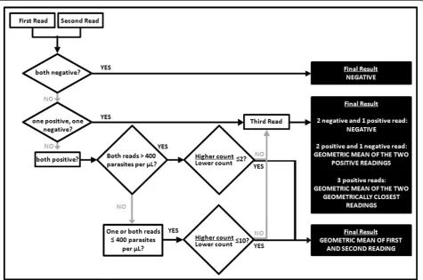
Criteria for concordance and determination of final result
All slides are read twice by two independent microsco-pists. A third independent microscopist also reads the slide if there are any of the following discrepancies between the first two readings: (1) a positive reading by one microscopist and a negative reading by the other; (2) both microscopists record a parasitaemia >400 para-sites/μL but the higher count divided by the lower count is >2; (3) at least one microscopist records a para-sitaemia ≤400 parasites/μL but the higher reading is more than 10 times the lower reading.
If the initial two readings give concordant results, the final parasite density is considered to be the geometric mean of the two readings. If the readings are discordant, then the following principles are applied: (1) where one reading is positive and the other negative, the majority decision obtained following the reading by the third microscopist is adopted - if positive, the final result is the geometric mean of the two positive results; (2) where all three readings are positive, the final result is the geo-metric mean of the two closest readings (Figure 2).
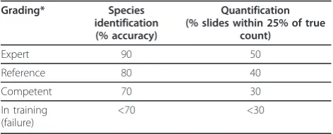
Slide reading performance measurement
After each preparation of a new batch of stain, internal quality control (QC) is performed on one negative and one positive slide. The external QA process for slide reading comprises species identification and parasite quantification. It is based on WHO recommendations [12] (Table 3).
Three assessments per year are planned and include 20 samples per microscopist. The samples in each assessment batch are provided by NICD (South Africa)
and present a variety of challenges, e.g. negative sam-ples, samples with very high counts, samples repeated to evaluate consistency (within the current survey and from previous surveys), samples with non-falciparum species and non-malaria pathogens. The true value of each sample parasite count is taken to be the median of the values obtained from the Parasitology Reference Unit of the NICD, two WHO reference laboratories and the laboratories of the study centres. Microscopists who fall below the level defined as competent are considered to be ‘in training’ and are not allowed to read study slides until retrained and re-assessed (Table 3).
Biochemistry and haematology: methodology and quality assurance/quality control
Before participating in the trial, many sites used manual methods for biochemical and haematological tests. How-ever, it was decided to introduce automated equipment for these tests in order to improve reliability, increase sample handling per day, reduce contamination risk, reduce blood volume requirement and enhance QA/QC assessment. Equipment for bedside measurements (blood gases, glucose and base excess), and other bio-chemical and haematological assays was chosen on the basis of performance, capacity, ease of use and availabil-ity of local technical support. The choice of equipment was made by individual centres, in collaboration with CLS. Duplicates of each major piece of equipment (main and back-up) were supplied. Two trained person-nel operate each piece of equipment. Significant efforts by study centre personnel were required in order to overcome the many challenges faced during the installa-tion of equipment, including delivery logistics and acces-sing reagents. Also, qualifying the methods before study start on the respective equipment proved a long and dif-ficult exercise due to lack of prior experience and avail-ability of samples.
[image:5.595.56.292.627.722.2]All biochemistry automated analyzers were initially enrolled with International EQA, formerly known as UKNEQAS (UK National External Quality Assessment Service) and then switched to the Royal College of Pathologists of Australasia (RCPA), because the latter was more appropriate for the study requirements at the time. All haematology automated analyzers are enrolled in EQA by UKNEQAS. Critical equipment such as ana-lyzers, autoclaves, fridges/freezers, incubators, laminar flow hoods and bedside equipment (I-STAT and Nova) were validated at installation by the vendor, who checked performance parameters and ran a calibration as needed. After this, a detailed installation qualifica-tion-operational qualification (IQ-OQ) certificate was provided. The users then performed some tests and ran QC samples to check the performance under routine conditions, reporting it in a performance quality (PQ)
Table 3 Grading of competency in parasite microscopy (species identification, and quantification)
Grading* Species identification (% accuracy)
Quantification (% slides within 25% of true
count)
Expert 90 50
Reference 80 40
Competent 70 30
In training (failure)
<70 <30
document. Thereafter, a yearly calibration is run by the vendors. For every piece of equipment, all interventions and incidents are recorded in a logbook. Each laboratory has needed to demonstrate method qualification for bio-chemistry and haematology. This includes analysis of repeatability and reproducibility on 10 samples covering the analysis range that is tested in duplicate on two to three independent runs. In addition, it includes analysis of linearity, tested by consecutive dilutions of five sam-ples, and accuracy or bridging of a panel of 60 samples between main and back-up analyzing equipment. For the latter, very low titered bilirubin samples are avoided, because bridging was proved to be difficult in the low ranges, while low ranges are of no concern for safety. Finally, stability of the QC samples is documented in the method qualification reports.
To show stability of the equipment, two or three QC samples are tested daily until the expiration date of the batch, and Levy-Jennings charts [13] of the test results are created to monitor whether they remain within the acceptable limits as provided by the supplier of the QC samples. One biochemistry QC sample (for bilirubin, creatinine and alanine aminotransferase) is sent to each laboratory every two weeks, and the criteria of accept-ability are defined by International EQA and the RCPA (results are pass or fail). Two haematology samples (for white blood cell count, red blood cell count, platelet count, and haemoglobin concentration) are sent to each laboratory every month; acceptability criteria are defined by International EQA (results are satisfactory or unsatis-factory). QC samples for EQA of bedside equipment to be analyzed over the subsequent six months are sent to each laboratory twice yearly by Thistle QA, South Africa (Table 2). Performance measurements by EQA are shown in Table 2. It is important to note that perfor-mance was better when QC reagents are provided by the vendor of the equipment.
Microbiology: methodology and quality assurance/quality control
Microbiological testing is essential for the diagnosis of malaria and associated co-morbidities. The primary case definition of severe malaria, adopted for evaluation of endpoints in this phase III trial, requires the exclusion of several co-morbidities including sepsis and meningi-tis, which are defined by a positive blood or cerebrosp-inal fluid culture [14].
Microbiology facilities in many participating centres had to be set up from the beginning, including installa-tion of dedicated laboratories, purchase of equipment, and staff training. A working group consisting of expert consultants and representatives from collaborating par-ties recommended standard microbiology methods using
automated Bactec™ incubators and paediatric bottles
(Bactec BD Diagnostic Systems, USA). The considera-tions that led to the choice of the automated system were standardization of methodology across sites, the
high sensitivity of the Bactec™ system with reduced
chances of contamination, and the availability of techni-cal support. Conventional systems are labour intensive and require skilled operators. This is an important con-sideration when skilled people are scarce and turnover is high.
Positive blood cultures are detected by carbon dioxide production, and positive automated cultures are sub-cul-tured using standard methods [15,16]. Further bacterial
identification relies on the API bioMerieux™and BBL
CRYSTAL™ methods supported by computerized
sys-tems that generate the name of the most likely organism based on the biochemical reactions obtained. Additional manual rapid tests used to differentiate between organ-isms and/or groups of organorgan-isms include oxidase, cata-lase and indole biochemical testing. Serotyping is used for identification of Salmonella and Shigella species. Rapid latex identification kits are used for confirmation of staphylococci, streptococci and pneumococci.
For the purpose of the trial analysis, as opposed to clinical care, there was an effort to standardize pathogen classification, according to a previously published algo-rithm [16] that was endorsed by a microbiology working group composed of representatives of the investigator groups and international experts in microbiology in Africa. A blood culture is considered positive if a defi-nite pathogen is isolated (e.g.Streptococcus pneumoniae,
Streptococcus agalactiae,Streptococcus pyogenes, Haemo-philus influenzae, Salmonellaspecies) or if a bacterium that could be either a pathogen or a contaminant is iso-lated within 48 hours of incubation (e.g.Escherichia coli,
Klebsiella pneumoniae,Staphylococcus aureus, Entero-coccus faecalis). A blood culture is considered to be con-taminated if a likely contaminant is isolated or if a bacterium that could be either a pathogen or a contami-nant is isolated after more than 48 hours of incubation.
CSF is collected by lumbar puncture, biochemical (glucose and protein) analyses and a white blood cell count is performed. Gram-stained direct smears are examined microscopically for the presence of bacteria, CSF is cultured using internationally accepted standar-dized microbiological methods, and bacterial growth is checked for after incubation for 24 and 48 hours. Bac-terial identification is performed according to methods described previously, as well as latex antigen agglutina-tion tests for S. pneumoniae, H. influenzaandNeisseria meningitidis.
Microbiology performance measurement
Microbiology EQA covers evaluation of microscopy,
antimicrobial for particular isolates, and antimicrobial susceptibility testing (Table 2). Six samples (with at least two meningeal and two enteric organisms) are sent to each laboratory three times per year as part of the NICD microbiology EQA, and the criteria of acceptabil-ity are defined by NICD (Table 2). Internal qualacceptabil-ity con-trol using American Type Culture Collection concon-trol strains for species identification testing is performed weekly, when a new batch of reagent is received or when discordant results occur. Internal QC is carried out on all antimicrobial discs available for antimicrobial susceptibility testing for use in the lab. The contamina-tion rate of the clinical specimens is evaluated monthly by internal assessment. Continuous assessment allows re-training programmes for both hospital and laboratory staff and more intense quality evaluation in case of high contamination rates.
Conclusions
Development of standardized laboratory methods and quality assurance and control processes in the ongoing phase III study of the RTS,S/AS01 candidate malaria vaccine is an example of a successful collaboration between research centres, trial sponsors and funding partners. Despite major challenges, study centres made tremendous achievements in leading this process, along with the sponsor and funding partners who provided support, training and coordination.
The focus has been to develop and maintain standar-dized laboratory facilities and procedures that meet cur-rent criteria for the conduct of clinical trials. High importance has been given to QC and QA in this study. External QA assessment of all laboratory methods (hae-matology, biochemistry, parasitology, microbiology) and bedside measurements are key aspects of this process. Automated equipment has replaced manual methods previously used by some centres. Through implementa-tion of the QA systems and standard operaimplementa-tion proce-dures covering all activities, the study centres have been able to effect considerable improvements in staff knowl-edge and competency.
Whilst this paper demonstrates what is possible, there remain challenges for the sustainability of the developed capacity described in this paper [17-19]. Although only equipment that could be serviced in country was selected, expensive consumables and maintenance contracts will be required after the trial. Many staff have been trained and assessed continuously in laboratory techniques and these capable and experienced staff must be retained. However, the research centres now have greater capabil-ities and the potential to diversify their research portfo-lios beyond malaria. A number of centres have already leveraged this capacity to successfully attract research grants. Given the limited microbiology services in some
areas, particularly rural areas, another opportunity that is being explored by some centres is to act as a national reference laboratory for microbiology or as a site for sen-tinel surveillance. All centres together with MCTA are looking at ways for the capacity to be sustained and to contribute to research and health care provision.
In conclusion, huge progress has been made in putting standardized methods into place and developing QA/QC processes in various research laboratories in Africa. There is adequate capacity to perform laboratory evalua-tions required in the conduct of the pivotal phase III study of the RTS,S/AS01 paediatric malaria candidate vaccine. Therefore, partners in the clinical development of this advanced malaria vaccine candidate are confident that the laboratory evaluations can be performed to the high standards required for clinical trials. Crucially, the capacity strengthening now in place will ensure a posi-tive, significant effect on the potential to conduct other research and healthcare benefits for the communities participating in this trial on its conclusion.
Acknowledgements
The authors would particularly like to acknowledge the significant contributions towards the parasite reading and microbiology working group of Richard Adegbola, Belinda Ngongo, Safi Traore, Sibongile Xaba, Leigh Dini, Rita van Deventer and Bhavani Poonsamy. Supportive computer programs for the slide reading process were developed and shared by James Beard. Statistical analyses of method qualification were provided by Cédric Bievelet. Special thanks go to the staff of each of the participating research laboratories for their dedication. Editorial assistance and manuscript coordination were provided by Julia Donnelly, Jarno Jansen, and Sarah Benns on behalf of GlaxoSmithKline Biologicals.
Members of the CTPC during the period of protocol development (2005-2009) included: Kintampo Health Research Centre, Ghana: Seth Owusu-Agyei, Kwaku Poku Asante; Ifakara Health Institute, Tanzania: Salim Abdulla; Centro de Investigação em Saude de Manhiça, Mozambique: Eusebio Macete, Jahit Sacarlal; KEMRI-Wellcome Trust Research Programme, Kenya: Philip Bejon, Trudie Lang, Kevin Marsh, Patricia Njugana, Ally Olotu; Institut de Recherche en Science de la Santé, Burkina Faso: Tinto Halidou; Albert Schweitzer Hospital, Gabon: Selidji Todagbe Agnandji, Bertrand Lell; Kumasi Centre for Collaborative Research, Ghana: Jennifer Evans; School of Medical Sciences, Kwame Nkrumah University of Science and Technology, Ghana: Tsiri Agbenyega, Daniel Ansong; KEMRI/CDC Research and Public Health Collaboration, Kenya: Mary Hamel, Simon Kariuki; KEMRI-Walter Reed Project, Kenya: David Jones, Walter Otieno; University of North Carolina Project, Malawi: Francis Martinson; Institut de Recherche pour le Development, Senegal: Cheikh Sadibou-Sokhna, Jean-François Trape; National Institute of Medical Research, Tanzania: Samwel Gesase, Martha Lemenge, John Lusingu; Prince Leopold Institute of Tropical Medicine, Belgium: Umberto
D’Allessandro; University of Tübingen, Germany: Peter Kremsner, University of Barcelona, Spain: John Aponte; Swiss Tropical Institute, Switzerland: Marcel Tanner; London School of Hygiene and Tropical Medicine, UK: Brian Greenwood, Lorenz von-Seidlein; Centers for Disease Control and Prevention, USA: Laurence Slutsker; University of North Carolina at Chapel Hill, USA: Irving Hoffman; laxoSmithKline Biologicals, Belgium: W. Ripley Ballou, Alice Grasset, Didier Lapierre, Amanda Leach, Johan Vekemans, Laurence Vigneron, Anne Walsh; Malaria Vaccine Initiative, USA: Alan Brooks, Filip Dubovsky, Santiago Ferro Carol Hooks, Christian Loucq, Melinda Moree, John McNeil, Carolyn Petersen, David Poland, Patricia Atkinson, Barbara Savarese, Marla Sillman, Tonya Villafana
Author details
1GlaxoSmithKline Biologicals, Rixensart, Belgium.2Institute of Tropical
Medicine, University of Tübingen, Tübingen, Germany.3Albert Schweitzer Hospital, Kumasi, Gabon.4London School of Hygiene and Tropical Medicine,
University of London, London, UK.5PATH Malaria Vaccine Initiative,
Washington, USA.6University of the Witwatersrand Medical School,
Johannesburg, South Africa.7Contract Laboratory Services, Johannesburg,
South Africa.8National Health Laboratory Service, National Institute for Communicable Diseases, Johannesburg, South Africa.9Malaria Clinical Trials
Alliance (MCTA), INDEPTH Network, Accra, Ghana.
Authors’contributions
All authors contributed to the development of this manuscript through discussions and document review. CS led the writing of the manuscript coordinating the incorporation of all reviewer comments. JV and BS coordinated the input of the MVI team. All authors critically contributed to the discussions on laboratory methodology. All authors read and approved the final manuscript.
Competing interests
CS, JV, MB, OOA, and BOk are employees of GSK Biologicals or were so during the writing of this manuscript. JV holds stock options in GSK Biologicals. CS, JV, MB, PK, BG, OOA, Bok, AR, CI, and JF declare their institution received a grant from MVI for the clinical trial described in this manuscript. JV, MB, and OOA declare their institution has received grants from MVI for previous clinical trials. BG, AD, AR, CI, and JF declare receiving travel funds from GSK Biologicals for travel related to this clinical trial. AR and CI declare their institution receiving financial support for lab support from GSK Biologicals. During the development of this manuscript, TV, TC, and BS were employed by MVI. SO and BOg declare no potential conflicts of interest.
Received: 3 March 2011 Accepted: 4 August 2011 Published: 4 August 2011
References
1. Abdulla S, Oberholzer R, Juma O, Kubhoja S, Machera F, Membi C, Omari S, Urassa A, Mshinda H, Jumanne A, Salim N, Shomari M, Aebi T,
Schellenberg DM, Carter T, Villafana T, Demoitié MA, Dubois MC, Leach A, Lievens M, Vekemans J, Cohen J, Ballou WR, Tanner M:Safety and immunogenicity of RTS,S/AS02D malaria vaccine in infants.N Engl J Med
2008,359:2533-2544.
2. Agnandji ST, Asante KP, Lyimo J, Vekemans J, Soulanoudjingar SS, Owusu R, Shomari M, Leach A, Fernandes J, Dosoo D, Chikawe M, Issifou S, Osei-Kwakye K, Lievens M, Paricek M, Apanga S, Mwangoka G, Okissi B, Kwara E, Minja R, Lange J, Boahen O, Kayan K, Adjei G, Chandramohan D, Jongert E, Demoitié MA, Dubois MC, Carter T, Vansadia P, Villafana T, Sillman M, Savarese B, Lapierre D, Ballou WR, Greenwood B, Tanner M, Cohen J, Kremsner PG, Lell B, Owusu-Agyei S, Abdulla S:Evaluation of the safety and immunogenicity of the RTS,S/AS01 malaria candidate vaccine when integrated in the Expanded Program of Immunization.J Infect Dis2010,
202:1076-1087.
3. Alonso PL, Sacarlal J, Aponte JJ, Leach A, Macete E, Aide P, Sigauque B, Milman J, Mandomando I, Bassat Q, Guinovart C, Espasa M, Corachan S, Lievens M, Navia MM, Dubois MC, Menendez C, Dubovsky F, Cohen J, Thompson R, Ballou WR:Duration of protection with RTS,S/AS02A malaria vaccine in prevention ofPlasmodium falciparumdisease in Mozambican children: single-blind extended follow-up of a randomised controlled trial.Lancet2005,366:2012-2018.
4. Aponte JJ, Aide P, Renom M, Mandomando I, Bassat Q, Sacarlal J, Manaca MN, Lafuente S, Barbosa A, Leach A, Lievens M, Vekemans J, Sigauque B, Dubois MC, Demoitié MA, Sillman M, Savarese B, McNeil JG, Macete E, Ballou WR, Cohen J, Alonso PL:Safety of the RTS,S/AS02D candidate malaria vaccine in infants living in a highly endemic area of Mozambique: a double blind randomised controlled phase I/IIb trial. Lancet2007,370:1543-1551.
5. Bejon P, Lusingu J, Olotu A, Leach A, Lievens M, Vekemans J, Mshamu S, Lang T, Gould J, Dubois MC, Demoitié MA, Stallaert JF, Vansadia P, Carter T, Njuguna P, Awuondo KO, Malabeja A, Abdul O, Gesase S, Mturi N, Drakeley CJ, Savarese B, Villafana T, Ballou WR, Cohen J, Riley EM, Lemnge MM, Marsh K, von Seidlein L:Efficacy of RTS,S/AS01 vaccine
against malaria in children 5 to 17 months of age.N Engl J Med2008,
359:2521-2532.
6. Asante KP, Abdulla S, Agnandji S, Lyimo J, Vekemans J, Soulanoudjingar S, Owusu R, Shomari M, Leach A, Jongert E, Salim N, Fernandes J, Dosoo D, Chikawe M, Saadou I, Osei-Kwakye K, Lievens M, Paricek M, Möller T, Apanga S, Mwangoka G, Dubois M-C, Tigani M, Kwara E, Minja R, Houkpatin A, Boahen O, Kayan K, Adjei G, Chandramohan D, Carter T, Vansadia P, Sillman M, Savarese B, Loucq C, Lapierre D, Greenwood B, Cohen J, Kremsner P, Owusu Agyei S, Tanner M, Lell B:Safety and efficacy over 19 months of the RTS,S/AS01E candidate malaria vaccine co-administered with Expanded Program of Immunization vaccines: an open randomized controlled trial.Lancet Infect Dis.
7. Sacarlal J, Aide P, Aponte JJ, Renom M, Leach A, Mandomando I, Lievens M, Bassat Q, Lafuente S, Macete E, Vekemans J, Guinovart C, Sigaúque B, Sillman M, Milman J, Dubois MC, Demoitié MA, Thonnard J, Menéndez C, Ballou WR, Cohen J, Alonso PL:Long-term safety and efficacy of the RTS, S/AS02A malaria vaccine in Mozambican children.J Infect Dis2009,
200:329-336.
8. Schluger N, Karunakara U, Lienhardt C, Nyirenda T, Chaisson R:Building clinical trial capacity for tuberculosis drugs in high-burden countries. PLoS Med2007,4:e302.
9. Moorthy V, Reed Z, Smith PG, on behalf of the WHO Study Group on Measures of Malaria Vaccine Efficacy:Measurement of malaria vaccine efficacy in phase III trials: Report of a WHO consultation.Vaccine2007,
25:5115-5123.
10. Greenwood BM, Armstrong JRM:Comparison of two simple methods for determining malaria parasite density.Trans R Soc Trop Med Hyg1991,
85:186-188.
11. Planche T, Krishna S, Kombila M, Engel K, Faucher JF, Ngou-Milama E, Kremsner PG:Comparison of methods for the rapid laboratory assessment of children with malaria.Am J Trop Med Hygiene2001,
65:599-602.
12. World Health Organization:Informal consultation on quality control of
malaria microscopyGeneva: WHO; 2006.
13. Westgard JO:Basic QC Practices: Training in Statistical Quality Control for Medical Laboratories.Madison WI: Westgard Quality Corporation;, 3 2010. 14. Vekemans J, Marsh K, Greenwood B, Leach A, Kabore W, Soulanoudjingar S,
Asante KP, Ansong D, Evans J, Sacarlal J, Bejon P, Kamthunzi P, Salim N, Njuguna P, Hamel M, Otieno W, Gesase S, Schellenberg D:Assessment of severe malaria in a multicentre, phase III, RTS,S/AS01 malaria candidate vaccine trial: case definition, standardization of data collection and patient care.Malar J.
15. Mahon CR, Manuselis G:Textbook of Diagnostic Microbiology.2 edition. Philadelphia: Saunders; 2000.
16. Winn W, Allen S, Janda W, Koneman E, Procop G, Schreckenberger P, Woods G:Koneman’s Color Atlas and Textbook of Diagnostic Microbiology.6 edition. Philadelphia: Lippincott Williams and Wilkins; 2006.
17. Participants in the 2001 Conference on Ethical Aspects of Research in Developing Countries [Participants]. Fair benefits for research in developing countries. Science2002,298:2133-2134.
18. Participants in the 2001 Conference on Ethical Aspects of Research in Developing Countries [Participants]. Moral standards for research in developing countries. From“reasonable availability”to“fair benefits”. Hastings Cent Rep2004,34:17-27.
19. Schulz-Baldes A, Vayena E, Biller-Andorno K:Sharing benefits in international research.EMBO Reports2007,8:8-13.
doi:10.1186/1475-2875-10-223