This is a repository copy of
A dual function for Pex3p in peroxisome formation and
inheritance
.
White Rose Research Online URL for this paper:
http://eprints.whiterose.ac.uk/10225/
Article:
Munck, J.M., Motley, A.M., Nuttall, J.M. et al. (1 more author) (2009) A dual function for
Pex3p in peroxisome formation and inheritance. Journal of Cell Biology, 187 (4). pp.
463-471. ISSN 0021-9525
https://doi.org/10.1083/jcb.200906161
[email protected] https://eprints.whiterose.ac.uk/ Reuse
Unless indicated otherwise, fulltext items are protected by copyright with all rights reserved. The copyright exception in section 29 of the Copyright, Designs and Patents Act 1988 allows the making of a single copy solely for the purpose of non-commercial research or private study within the limits of fair dealing. The publisher or other rights-holder may allow further reproduction and re-use of this version - refer to the White Rose Research Online record for this item. Where records identify the publisher as the copyright holder, users can verify any specific terms of use on the publisher’s website.
Takedown
If you consider content in White Rose Research Online to be in breach of UK law, please notify us by
The Rockefeller University Press $30.00
Correspondence to Ewald H. Hettema: [email protected]
Abbreviations used in this paper: PMP, peroxisomal membrane protein; WT, wild type.
Introduction
Eficient functioning of individual organelles depends on their presence in a characteristic copy number, size, and position within different cell types. Therefore, eukaryotic cells have evolved molecular mechanisms to ensure the accurate segregation of organelles during cell division. As Saccharomyces cerevisiae cells grow asymmetrically, forming a bud that is initially much smaller than the mother cell, organelles must be actively trans-ported from the mother cell to the bud (Yaffe, 1991).
Yeast peroxisomes multiply by growth and division of preexisting peroxisomes (Motley and Hettema, 2007; Nagotu et al., 2008). Therefore, movement of peroxisomes during cell division is regulated tightly to ensure accurate segregation between mother and daughter cells (Hoepfner et al., 2001; Fagarasanu et al., 2005, 2006). Segregation of peroxisomes is achieved by two opposing processes: transport and retention. Approximately half of the total population of peroxisomes is transported from the mother cell into the bud. This transport occurs along actin cables and is driven by the class V myosin Myo2p (Hoepfner et al., 2001), which is recruited to peroxi-somes by the integral peroxisomal membrane protein Inp2p (Fagarasanu et al., 2006). Transport to the bud is balanced by the second process, which involves retention of the remaining peroxisomes within the mother cell. This retention is depen-dent on the peripheral peroxisomal membrane protein Inp1p (Fagarasanu et al., 2005). As peroxisomes that are retained within the mother cell have a predominantly peripheral localization,
Inp1p has been suggested to provide an anchor between the per-oxisome and the cell periphery (Fagarasanu et al., 2005).
Actin/myosin-dependent transport to the bud is required also for inheritance of vacuoles, cortical ER, and late Golgi ele-ments, and mechanisms for retention have thus far been sug-gested also for late Golgi, cortical ER, and mitochondria (Yang et al., 1999; Rossanese et al., 2001; Wiederkehr et al., 2003; Boldogh et al., 2004; Cerveny et al., 2007).
Peroxisome biogenesis depends on a large set of proteins called peroxins (abbreviated as pex) (Distel et al., 1996). Most peroxins are required for the post-translational import of lume-nal proteins; in mutants lacking these factors, matrix proteins are mislocalized to the cytosol, whereas peroxisomal mem-brane proteins (PMPs) assemble into peroxisomal memmem-brane “ghosts” (Gould and Valle, 2000). Pex3p, Pex16p, and Pex19p have been shown to be responsible for peroxisomal membrane biogenesis in mammalian cells, and two models for PMP im-port have been suggested. According to the irst model, PMPs are inserted post-translationally into peroxisomes in a Pex3-, Pex16-, Pex19-dependent process. In the second model, at least some PMPs are inserted irst into the ER, and from here they are sorted to peroxisomes. There is evidence in support of both of these mechanisms (Tabak et al., 2003; Fang et al., 2004; Jones et al., 2004; Kim et al., 2006; Matsuzaki and Fujiki, 2008; Toro et al., 2009).
S
accharomyces cerevisiae Pex3p has been shown toact at the ER during de novo peroxisome formation. However, its steady state is at the peroxisomal mem-brane, where its role is debated. Here we show that Pex3p has a dual function: one in peroxisome formation and one in peroxisome segregation. We show that the peroxisome retention factor Inp1p interacts physically with Pex3p in vitro and in vivo, and split-GFP analysis shows that the site
of interaction is the peroxisomal membrane. Furthermore, we have generated PEX3 alleles that support peroxisome formation but fail to support recruitment of Inp1p to per-oxisomes, and as a consequence are affected in peroxi-some segregation. We conclude that Pex3p functions as an anchor for Inp1p at the peroxisomal membrane, and that this function is independent of its role at the ER in per-oxisome biogenesis.
A dual function for Pex3p in peroxisome formation
and inheritance
Joanne M. Munck, Alison M. Motley, James M. Nuttall, and Ewald H. Hettema
Department of Molecular Biology and Biotechnology, University of Sheffield, Sheffield S10 2TN, England, UK
© 2009 Munck et al. This article is distributed under the terms of an Attribution– Noncommercial–Share Alike–No Mirror Sites license for the first six months after the publica-tion date (see http://www.jcb.org/misc/terms.shtml). After six months it is available under a Creative Commons License (Attribution–Noncommercial–Share Alike 3.0 Unported license, as described at http://creativecommons.org/licenses/by-nc-sa/3.0/).
THE
JOURNAL
OF
CELL
BIOLOGY
on December 2, 2009
jcb.rupress.org
Downloaded from
JCB • VOLUME 187 • NUMBER 4 • 2009 464
importance of Pex3 for traficking from ER to peroxisomes. More and more PMPs have now been shown to be able to trafic from ER to peroxisomes (Ma and Subramani, 2009). There is no evidence for direct import of membrane proteins into yeast peroxisomes, and it has been suggested that all S. cerevisiae PMPs trafic to peroxisomes via the ER in a Pex3-dependent manner (Tabak et al., 2008).
However, the steady-state localization of Pex3p in WT cells is at the peroxisomal membrane: only newly synthesized Pex3p has been detected in the ER. This raises the question of whether the role of Pex3p at the ER is different to its role at the peroxisomal membrane.
Here we show that in addition to its role in peroxisome formation, Pex3p is also required for peroxisome segregation. We have pinpointed the role of Pex3p in this process to provid-ing the anchor for Inp1p at the peroxisomal membrane. Further-more, we demonstrate that the roles of Pex3p in peroxisome formation and segregation can be separated genetically. S. cerevisiae cells lacking Pex3p or Pex19p are devoid of
any peroxisomal structures and rapidly degrade most PMPs (Hettema et al., 2000). Pex16p is not present in the S. cerevisiae genome. An S. cerevisiae strain conditionally expressing Pex3p-GFP as the sole copy of Pex3p can form peroxisomes de novo under permissive conditions. Careful time-lapse microscopy analysis shows that Pex3p-GFP appears irst in the ER, where it concentrates in punctate structures that subsequently lose their association with the ER and mature over several hours into per-oxisomes containing matrix proteins. Pex19p is required for the exit of Pex3p from the ER during de novo peroxisome forma-tion (Hoepfner et al., 2005); Pex3 is able to follow the same pathway in wild-type (WT) cells (Hoepfner et al., 2005). We re-cently proposed that in WT cells this pathway supplies existing peroxisomes with membrane constituents, thus allowing growth and subsequent division (Motley and Hettema, 2007). A non-functional, truncated Pex3p-GFP can exit the ER only in the presence of WT Pex3p (Tam et al., 2005), showing the crucial
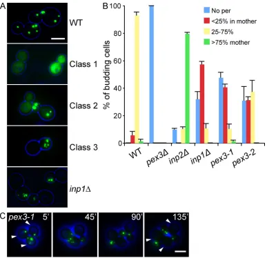
Figure 1. Isolation of a new class of pex3 mutants. (A) pex3 cells expressing the lumenal peroxisomal marker GFP-PTS1 were transformed with
plasmids containing a WT PEX3 allele or a representative member of each pex3 mutant class. inp1 cells are included for comparison. (B) Quantitative
description of peroxisome distribution in WT, pex3, inp1, inp2, and the class III mutants pex3-1 and pex3-2. Overnight cultures were diluted and
grown for 6 h in selective glucose medium, examined by epifluorescence and phase contrast, and scored for peroxisome distribution. More than 100
budding cells were analyzed for each strain. Three independent experiments were performed. Error bars represent SEM. (C) pex3-1 cells expressing
GFP-PTS1 were spotted on an agarose pad and peroxisome distribution was followed with time. A and C show merged brightfield (blue) and fluorescent images (green). Bar, 5 µm.
on December 2, 2009
jcb.rupress.org
Downloaded from
[image:3.612.121.508.56.430.2](Fagarasanu et al., 2005; Hettema and Motley, 2009). We have shown previously that cells that fail to inherit peroxisomes will form them de novo. However, this is a slow process, taking lon-ger than the duration of the cell cycle, hence the large propor-tion of peroxisome-deicient cells (Motley and Hettema, 2007; Hettema and Motley, 2009). We conclude that Pex3p has a dual function in peroxisome formation and segregation.
Inp1p is mislocalized to the cytosol in cells lacking Pex3p
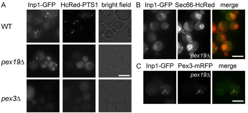
Inp1p is peripherally associated with the peroxisomal membrane, where it is required for anchoring peroxisomes to the cell pe-riphery (Fagarasanu et al., 2005). When expressed at endogenous levels, Inp1p-GFP labeled peroxisomes (Fig. 2 A). Subsequently, we controlled the expression of Inp1-GFP with the conditional GAL1/10 promoter. When cells are induced to express Inp1-GFP for 3 h by growth on galactose medium, the level of Inp1-GFP is comparable to endogenous levels (Fig. S1 A). Upon continued growth on galactose medium, Inp1-GFP is overexpressed and labeling of peroxisomes becomes initially more intense before a cytoplasmic pool of Inp1p-GFP becomes evident. This demon-strates that the association of Inp1p with peroxisomes de-pends on a saturable factor (Fig. S1 B). We hypothesized that Pex3p may be this factor, or may be required for the activity of this factor.
Indeed, in pex3∆ cells, Inp1p-GFP displayed a cytosolic labeling pattern: no peripheral or punctate labeling was ob-served (Fig. 2 A). This implies that Pex3p is required for associ-ation of Inp1p with membranes. However, because many PMPs are unstable in pex3∆ cells (Hettema et al., 2000) we analyzed the localization of Inp1p-GFP in a range of peroxisomal mu-tants: Inp1-GFP was localized in punctae in all mutants exam-ined (Fig. S1 C). We conclude that none of these proteins are essential for the recruitment of Inp1 to membranes. One inter-pretation of these data is that Pex3p is responsible for the asso-ciation of Inp1p with the peroxisomal membrane. However,
Results and discussion
A new class of pex3 mutants
Deciphering the function of Pex3p is crucial if we are to under-stand the process of peroxisome biogenesis. To this end, we gen-erated a plasmid library of random pex3 mutants by error-prone PCR. We were able to distinguish three classes of mutants; of the
1,000 strains analyzed (see Materials and methods), 720 failed to import the peroxisomal marker GFP-PTS1 (class I) (Fig. 1 A); class II mutants (258 in total) had a mild pex phenotype, in which cells partially mislocalized GFP-PTS1 to the cytosol; class III mutants comprised 15 strains which displayed an unequal distri-bution of peroxisomes between mother cell and bud. We recov-ered the PEX3 plasmids from class III mutants and reintroduced them into a pex3∆ strain; these new transformants (1) reformed peroxisomes (i.e., were able to support de novo peroxisome for-mation) and (2) displayed the segregation defect, conirming the phenotype is plasmid linked. Several mutations were found in each pex3 allele, although no mutation hot spots were observed. We focused our studies on pex3-1 and pex3-2 cells, as these dis-played the strongest phenotype. The pex3-1 allele had six amino acid substitutions (V81E, N178D, N188I, N242D, N247Y, and F353I) and the pex3-2 allele had ten amino acid substitutions (F29L, Y44N, F55Y, N158S, F186Y, Q217R, N242Y, S307T, N326K, and K369E). Further phenotypic analysis of pex3-1 and pex3-2 cells (Fig. 1 B) reveals they have a peroxisome retention defect. In 40% of budding pex3-1 cells, the mother cell contained less than 25% of the number of peroxisomes present in the bud. In more than half of these, the mother cell was completely devoid of peroxisomes, something hardly ever (<1%) observed in WT cells. A similar trend was observed in pex3-2 cells, although the segregation defect was less pronounced. Time-lapse microscopy shows the unequal distribution is due to a retention defect in the mother (Fig. 1 C).
[image:4.612.87.514.56.250.2]A large proportion of cells in both strains were completely devoid of peroxisomes. This phenotype is found in inp1 cells
Figure 2. Inp1p localization in peroxisome-deficient cells. A plasmid that expresses Inp1p-GFP under the control of its endogenous promoter was
trans-formed into WT (A), pex19 (A–C), and pex3 (A) cells expressing HcRed-PTS1 (A), Sec66p-HcRed (B), and Pex3p-mRFP (C). The strains were grown on
selective medium and examined by epifluorescence and phase contrast. For A and C, multiple epifluorescence images were acquired in the z axis and flattened into a single image. For B, a single focal plane was taken. Bar, 5 µm.
on December 2, 2009
jcb.rupress.org
JCB • VOLUME 187 • NUMBER 4 • 2009 466
Pex3p in punctae close to the ER (Fig. 2 C). Combined with the observation that Inp1p is mislocalized to the cytosol in the ab-sence of Pex3p (Fig. 2 A), we conclude that Pex3p is involved in the membrane association of Inp1p.
Inp1p binds Pex3p in vitro
To test whether Inp1p and Pex3p interact, we performed an in vitro–binding assay with an Escherichia coli–expressed GST fusion of the cytosolic domain of Pex3p (amino acids 40–441) and a yeast lysate of Inp1p-HA–expressing cells (Fig. 3 A). We found that Inp1p-HA binds speciically to Pex3p, as the sorting nexin Mvp1p, an unrelated peripheral membrane protein of the endosomal system, does not bind Pex3p. The observation that Inp1p and Pex3p interact in vitro is consistent with the hypoth-esis that Pex3p is responsible for the membrane association of Inp1p. However, this experiment does not distinguish between direct or indirect binding. We expressed both 6xHis-Pex3p 40–441 and GST-Inp1p in E. coli and found a speciic interaction between Inp1p and Pex3p (Fig. 3 B). This result indicates that no additional yeast proteins are required for Inp1p to bind Pex3p, and that the binding is therefore direct.
Inp1p and Pex3p interact in vivo
Because Pex3p and Inp1p colocalize at the peroxisomal mem-brane, and because they interact directly in vitro, we tested whether they interact at the peroxisomal membrane in vivo. We performed an inducible bimolecular luorescence complemen-tation assay or split-GFP experiment. Inp1 was tagged at its C terminus with the N-terminal part of GFP (aa 2–156), and Pex3p was tagged at its C terminus with the C-terminal part of GFP because pex3 cells are completely devoid of peroxisomal
structures (Hettema et al., 2000), the cytosolic localization of Inp1p-GFP may simply be attributable to the lack of peroxi-somal membranes.
Inp1p is localized to a subdomain of the ER
in pex19 cells
To discriminate between these two interpretations, the localiza-tion of Inp1p-GFP was analyzed in a pex19 strain. Pex19p and Pex3p act together at an early stage of peroxisomal membrane formation and like pex3∆ cells, pex19∆ cells lack peroxisomal membrane structures. However, in contrast to pex3∆ cells, in pex19 cells Inp1p-GFP showed a punctate labeling pattern (Fig. 2 A). We conclude that the lack of peroxisomal mem-branes, by itself, is not causing Inp1 to be mislocalized to the cytosol.
A major difference between pex3∆ and pex19∆ cells is that in pex19∆ cells, Pex3p is mislocalized to punctate struc-tures associated with the ER membrane (Hoepfner et al., 2005; Tam et al., 2005; Motley and Hettema, 2007). Because Inp1p localization appears to be dependent on Pex3p, we hypothesized that Inp1p might also be localized to these structures. To test this, Inp1p-GFP was coexpressed alongside a red ER membrane marker (Sec66p-HcRed) in pex19 cells. Inp1p-GFP was ob-served in luorescent punctae that were localized close to the ER (Fig. 2 B). Subsequently, Inp1p-GFP was coexpressed along-side Pex3p-RFP in pex19 cells. As shown in Fig. 2 C, Inp1p-GFP completely colocalized with Pex3p-RFP.
In summary, Inp1p is associated with peroxisomes in WT cells (Fig. 2 A), and in pex19 cells it colocalizes with
Figure 3. Inp1p binds directly to the cytosolic domain
of Pex3p in vitro. GST-Pex3p (40–441) and GST were bound to glutathione Sepharose beads and incubated with a detergent lysate of spheroplasts expressing HA-tagged Inp1p and Mvp1p at endogenous levels (A). After extensive washing, the bound fraction and lysate were analyzed by SDS-PAGE and immunoblotting using the HA monoclonal 12CA5. Yeast lysates (YL) represent 5% of the lysate added to the beads and analyzed by blot-ting. Because the signal of Inp1p-HA was too low in the YL, 5 times more lysate was reloaded on a separate gel and compared with the GST- and GST-Pex3–bound frac-tion (right-hand panel). Bottom panel shows Coomassie staining. (B) GST-Inp1p and GST were bound to
gluta-thione Sepharose and incubated with a lysate of E. coli
expressing either 6xtagged Pex3p (40–441) or
HIS-tag only, or with lysis buffer only (). After extensive
wash-ing, bound fractions were analyzed by SDS-PAGE and Coomassie staining of the gel. A lane was included with partially purified 6xHIS-Pex3p as control. M, molecular weight marker. Arrow indicates 6xHIS-Pex3p. Asterisks
indicate multiple GST-Inp1p fragments. EL, E. coli lysate
of 6xHIS-Pex3p–expressing cells.
on December 2, 2009
jcb.rupress.org
Downloaded from
[image:5.612.262.561.57.366.2]somes to the bud (Fagarasanu et al., 2005). The luorescence signal seen using split-GFP takes at least 1 h longer to appear than when expressing full-length GFP fusions. This is most likely because Pex3p and Inp1p must interact before GFP can reassemble and the chromophore can mature. A combination of positive and negative controls were performed to test the speci-icity of the interaction (Fig. 4, A and B). From these data it is clear that Inp1p and Pex3p interact on the peroxisomal mem-brane in vivo.
We subsequently redirected Pex3p to a nonnative local-ization within the cell by fusing the cytosolic domain of Pex3p to the mitochondrial outer membrane protein Tom70p and (aa 157–end). The fusion proteins were expressed from the
GAL1 promoter. The GFP fragments do not luoresce (Fig. 4 A) unless GFP is reconstituted by interaction between the bait and target proteins (Wilson et al., 2004; Park et al., 2007). After 4 h of induction on galactose medium, a punctate luorescent sig-nal was observed in mother cells, whereas the buds remained empty (Fig. 4 B). These punctae represent peroxisomes as con-irmed by their ability to import HcRed-SKL (Fig. 4 C). The effect on peroxisome segregation is comparable to previous ob-servations after expressing Inp1p-GFP under control of the GAL1 promoter (Fig. S1 B), and is in accordance with the ind-ing that overexpression of Inp1p prevents transport of
peroxi-Figure 4. Inp1p interacts with cytosolic domain
of Pex3p in vivo. (A–C) Split-GFP analysis be-tween Inp1p and Pex3p in WT cells. Tagged
pro-teins were expressed under control of the GAL1
promoter for 4 h (short) or 8 h (long) and scored for the presence and intensity of fluorescence
(A). , no signal; +, faint; +++, strong. (B) Selected
images of WT cells induced for 4 or 8 h. (C) WT cells were induced to express Inp1p-GFP-N and Pex3p-GFP-C for 4 h, followed by mating with
pex3 cells expressing HcRed-PTS1, and imaging
2 h after mating. GFP and HcRed signals overlap in mated cell (arrow). (D) The expression of a chi-meric protein consisting of the cytosolic domain of Pex3p fused at its N terminus to Tom70p and tagged at its C terminus with mRFP
(mito-Pex3p-mRFP) was induced on galactose for 3 h in pex3∆
cells expressing Inp1p-GFP under control of its endogenous promoter. Image shows two budding cells, one of which is expressing mito-Pex3p-RFP that recruits Inp1p-GFP. The strains were grown on selective medium and examined by epifluores-cence and phase contrast. Multiple epifluoresepifluores-cence images were acquired in the z axis and flattened into a single image. The brightfield image is blue in the merged pictures. Bar, 5 µm.
on December 2, 2009
jcb.rupress.org
[image:6.612.47.374.55.565.2]JCB • VOLUME 187 • NUMBER 4 • 2009 468
redirected to mitochondria when expressed with the Tom70-Pex3p chimera in pex3 cells. From these data we conclude that Inp1p and Pex3p interact in vivo, with the localization of Inp1p being determined by that of Pex3p. For reasons that are not clear, we were unable to coimmunoprecipitate Inp1p with Pex3p from yeast lysates.
[image:7.612.114.507.56.599.2]tagging the construct at its C terminus with mRFP. When expressed in pex3 cells, the chimera is present in a pattern characteristic of mitochondria. (Fig. 4 D). The Tom70-Pex3p chimera does not restore peroxisome biogenesis after expres-sion in pex3 cells, as indicated by the continuing absence of peroxisomes (unpublished data). Strikingly, Inp1p-GFP is
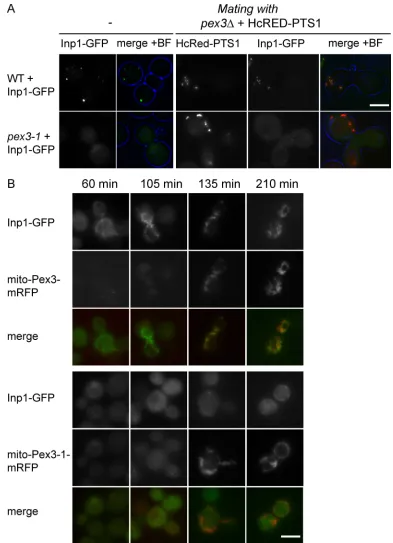
Figure 5. Pex3-1p fails to recruit Inp1p-GFP to peroxisomes. (A) WT and pex3-1 cells expressing Inp1p-GFP at endogenous levels were mated with pex3∆
cells expressing HcRed-PTS1 and imaged after 2 h. (B) The cytosolic domain of Pex3p and Pex3-1p was redirected to mitochondria by fusion to Tom70p in pex3∆ cells expressing Inp1p-GFP at endogenous levels. Mito-Pex3-mRFP and mito-Pex3-1-mRFP expression was induced on galactose medium for times
indicated. Signals of mito-Pex3p and mito-Pex3-1p are directly comparable; Inp1p-GFP signals are more enhanced in pex3-1 cells. The strains were grown
on selective medium and examined by epifluorescence and phase contrast. Bar, 5 µm.
on December 2, 2009
jcb.rupress.org
Downloaded from
used are pex28/pex29 (Vizeacoumar et al., 2003), pex11/ pex25/pex27 (Vizeacoumar et al., 2003), pex30/pex31/pex32
(Vizeacoumar et al., 2004), and c13-ABYS-86 (Heinemeyer et al., 1991). The Mvp1p-HA strain is a derivative of c13-ABYS-86. Mvp1p was genomically tagged with the C-terminal triple HA cassette using homologous recombination.
Plasmids
All yeast expression plasmids were based on the parental plasmids ycplac33 and ycplac111 (Gietz and Sugino, 1988). The majority of constructs used in this study were generated by homologous recombination in yeast (Uetz et al., 2000). The open reading frame (ORF) of interest was amplified by
PCR. The 5 ends of the primers included 18 nucleotide extensions
homolo-gous to plasmid sequences flanking the intended insertion site, to enable repair of gapped plasmids by homologous recombination. For expression,
constructs of INP1 under control of its own promoter 600 bp upstream from
the ORF were included. Galactose-inducible constructs contained the GAL1
and GAL10 intragenic region and MFA2 terminator. Other constructs
con-tain the PGK1 terminator. For constitutive expression of the peroxisomal
lumenal markers HcRed-PTS1 and GFP-PTS1, the Tpi1 promoter was used (Motley and Hettema, 2007). Tom70-Pex3p fusions were constructed by appending full-length Tom70p with the cytosolic domain of Pex3p (amino acid 49–441) or Pex3-1 (49–441) and mRFP. We used GFPS65T and triple-HA tag for tagging. Split-GFP constructs were based on the plasmids designed by Barnard et al. (2008). However, we introduced the split-GFP
fragments behind the GAL1/10 promoter into centromeric plasmids to
gen-erate a conditional split-GFP system. Sec66-HcRed marker was provided by Alistair Goldman (University of Sheffield, Sheffield, UK).
For E. coli expression PEX3 (a.a.40-441) was cloned into pET42a
and pET30a and full-length INP1 in pET42a.
Growth conditions
For all experiments, cells were grown overnight in selective glucose me-dium. For analysis of phenotypes by microscopy, cells were subsequently
diluted to OD600 = 0.1 in fresh selective glucose medium and grown for two
to three cell divisions (4–6 h) before imaging. Where the induction of a re-porter protein was required, cells were transferred to selective galactose
medium at OD600 = 0.1 and grown for the time indicated in the figures and
text. Growth media components are as follows: minimal glucose/galactose media for the selection of uracil and tryptophan prototrophic markers, 2% glucose/galactose, 0.17% yeast nitrogen base (without amino acids and ammonium sulfate), 0.5% ammonium sulfate, 1% casamino acids. Minimal glucose/galactose media for the selection of all prototrophic markers, 2% glucose/galactose, 0.17% yeast nitrogen base (without amino acids and ammonium sulfate), 0.5% ammonium sulfate. The appropriate amino acid stocks were added to minimal media as required.
Mating assay
Overnight cultures of cells were diluted to OD600 = 0.1 in fresh selective
glucose medium and grown for 2–3 h. The cells were collected by filtration onto a 0.22-µm nitrocellulose filter (type GS, 25-mm diameter; Millipore), and the filter was incubated cell-side up on a prewarmed YPD plate at
30°C. 107 cells of each strain were collected per 25-mm filter. After 2 h,
cells were harvested by vortexing the filter in selective glucose medium.
Image acquisition
Live cells were analyzed with a microscope (Axiovert 200M; Carl Zeiss, Inc.) equipped with Exfo X-cite 120 excitation light source, band-pass filters (Carl Zeiss, Inc. and Chroma), and a Plan-Fluar 100x/1.45 NA or A-Plan 40x/0.65 NA Ph2 objective lens (Carl Zeiss, Inc.) and a digital camera (Orca ER; Hamamatsu). Image acquisition was performed using Openlab software (PerkinElmer). Fluorescence images were routinely collected as 0.3-µm z-stacks and merged into one plane after contrast enhancing in Openlab, and processed further in Photoshop where only levels adjustment was used. On occasion (as indicated in text) images were collected as single-plane images. Brightfield images were collected in one plane. Blue color was applied to the brightfield image using Photoshop. The level of the brightfield images were modified, and the image was blurred, sharpened, and blurred again before one more round of level adjustment so that only the circumference of the cell was visible.
In vitro–binding assay
The GST-Pex3p, 6xHIS-Pex3p, and GST-Inp1p fusion proteins were
ex-pressed in E. coli BL21 DE3. Cells were grown to OD600 = 0.6 in 2TY
media with 75 µg/ml ampicillin at 30°C. After 3 h of IPTG-induced
Pex3-1p is unable to recruit Inp1p to peroxisomes
Our data show that Inp1p binds Pex3p on peroxisomal membranes in WT cells. As mentioned above, budding pex3-1 cells are un-able to retain peroxisomes within the mother cells. We sought to determine the reason for this retention defect. Expression of Inp1p-GFP in pex3-1 cells labeled the cytosol. Because many pex3-1 cells lack peroxisomes, we introduced (by mating) a red peroxisomal lumenal marker (HcRed-PTS1): the pex3-1 cells shown in Fig. 5 A contain peroxisomes but mislocalize Inp1p-GFP to the cytosol. We conclude that pex3-1 cells are unable to recruit Inp1p, and that this inability to bind Inp1p gives rise to the retention defect in pex3-1 cells.
We also used the mitochondrial redirection assay to test the Pex3-1p interaction with Inp1p. The cytosolic domain of the pex3-1 allele was fused to Tom70p and tagged at its C terminus with mRFP. Expression was induced with galactose. As was the case for WT Pex3p chimera, the Pex3-1p chimera was success-fully targeted to mitochondria (Fig. 5 B). At early time points after induction, Inp1p-GFP was recruited to the mitochondrial membrane even before the chimeric Tom70-Pex3p-mRFP pro-tein was detectable. This is in contrast to the timing of Inp1p-GFP recruitment in the cells expressing the Pex3-1 chimeric protein: only after prolonged induction of Tom70-Pex3-1p-mRFP was a small amount of Inp1p-GFP recruited to mitochondria, and a pool of Inp1p-GFP remained in the cytosol. This shows that the ability of Pex3-1p to mediate recruitment of Inp1p to mem-branes is severely affected.
In summary, Pex3p is required for the recruitment of Inp1p to peroxisomes, where it acts as its anchor. Disruption of Inp1p recruitment by Pex3p results in a peroxisome retention defect and prevents maintenance of peroxisomes in mother cells. Because de novo peroxisome formation takes longer than the duration of one cell cycle, subsequent division of these mother cells results in daughter cells without peroxisomes. This illustrates that Pex3p is required not only for de novo peroxisome formation from the ER, but also for the subsequent maintenance of peroxisomes. This lat-ter role is performed at the peroxisomal membrane.
Although our results show distinct functions for Pex3p on the ER and the peroxisomal membrane, we cannot rule out an additional role for Pex3p on peroxisomes, as has been suggested for mammalian Pex3p (Fujiki et al., 2006; Matsuzaki and Fujiki, 2008).
Peroxisomes are not the only organelles that use a single factor for distinct processes. For instance, yeast vacuole inheri-tance relies on Vac8p, a factor that is also involved in homotypic vacuole fusion, cytoplasm-to-vacuole targeting pathway (Cvt), and microautophagy (Weisman, 2003). A theme is emerging whereby a single factor is used for segregation as well as for other pro-cesses speciic to that organelle, which may allow spatial and temporal coordination of these processes.
Materials and methods
Strains
The yeast strains used in this study were derivatives of BY4741 (MATA
his3-1 leu2-0 met15-0 ura3-0) or BY4742 (MAT a his3-1 leu2-0 lys2-0 ura3-0) obtained from the EUROSCARF consortium. Additional strains
on December 2, 2009
jcb.rupress.org
JCB • VOLUME 187 • NUMBER 4 • 2009 470
References
Barnard, E., N.V. McFerran, A. Trudgett, J. Nelson, and D.J. Timson. 2008. Development and implementation of split-GFP-based bimolecular luor-escence complementation (BiFC) assays in yeast. Biochem. Soc. Trans.
36:479–482. doi:10.1042/BST0360479
Boldogh, I.R., S.L. Ramcharan, H.C. Yang, and L.A. Pon. 2004. A type V myosin (Myo2p) and a Rab-like G-protein (Ypt11p) are required for retention of newly inherited mitochondria in yeast cells during cell division. Mol. Biol. Cell. 15:3994–4002. doi:10.1091/mbc.E04-01-0053
Cerveny, K.L., S.L. Studer, R.E. Jensen, and H. Sesaki. 2007. Yeast mitochon-drial division and distribution require the cortical num1 protein. Dev. Cell. 12:363–375. doi:10.1016/j.devcel.2007.01.017
Distel, B., R. Erdmann, S.J. Gould, G. Blobel, D.I. Crane, J.M. Cregg, G. Dodt, Y. Fujiki, J.M. Goodman, W.W. Just, et al. 1996. A uniied nomenclature for peroxisome biogenesis factors. J. Cell Biol. 135:1–3. doi:10.1083/ jcb.135.1.1
Fagarasanu, M., A. Fagarasanu, Y.Y. Tam, J.D. Aitchison, and R.A. Rachubinski. 2005. Inp1p is a peroxisomal membrane protein required for peroxisome inheritance in Saccharomyces cerevisiae. J. Cell Biol. 169:765–775. doi:10.1083/jcb.200503083
Fagarasanu, A., M. Fagarasanu, G.A. Eitzen, J.D. Aitchison, and R.A. Rachubinski. 2006. The peroxisomal membrane protein Inp2p is the peroxisome-speciic receptor for the myosin V motor Myo2p of Saccharomyces cere-visiae. Dev. Cell. 10:587–600. doi:10.1016/j.devcel.2006.04.012 Fang, Y., J.C. Morrell, J.M. Jones, and S.J. Gould. 2004. PEX3 functions as a
PEX19 docking factor in the import of class I peroxisomal membrane proteins. J. Cell Biol. 164:863–875. doi:10.1083/jcb.200311131 Fujiki, Y., Y. Matsuzono, T. Matsuzaki, and M. Fransen. 2006. Import of
peroxi-somal membrane proteins: the interplay of Pex3p- and Pex19p-mediated interactions. Biochim. Biophys. Acta. 1763:1639–1646. doi:10.1016/ j.bbamcr.2006.09.030
Gietz, R.D., and A. Sugino. 1988. New yeast-Escherichia coli shuttle vectors constructed with in vitro mutagenized yeast genes lacking six-base pair restriction sites. Gene. 74:527–534. doi:10.1016/0378-1119(88)90185-0 Gould, S.J., and D. Valle. 2000. Peroxisome biogenesis disorders: genetics and cell
biology. Trends Genet. 16:340–345. doi:10.1016/S0168-9525(00)02056-4 Heinemeyer, W., J.A. Kleinschmidt, J. Saidowsky, C. Escher, and D.H. Wolf.
1991. Proteinase yscE, the yeast proteasome/multicatalytic-multifunctional proteinase: mutants unravel its function in stress induced proteolysis and uncover its necessity for cell survival. EMBO J. 10:555–562.
Hettema, E.H., and A.M. Motley. 2009. How peroxisomes multiply. J. Cell Sci.
122:2331–2336. doi:10.1242/jcs.034363
Hettema, E.H., W. Girzalsky, M. van Den Berg, R. Erdmann, and B. Distel. 2000.
Saccharomyces cerevisiae Pex3p and Pex19p are required for proper localization and stability of peroxisomal membrane proteins. EMBO J.
19:223–233. doi:10.1093/emboj/19.2.223
Hoepfner, D., M. van den Berg, P. Philippsen, H.F. Tabak, and E.H. Hettema. 2001. A role for Vps1p, actin, and the Myo2p motor in peroxisome abun-dance and inheritance in Saccharomyces cerevisiae. J. Cell Biol. 155:979– 990. doi:10.1083/jcb.200107028
Hoepfner, D., D. Schildknegt, I. Braakman, P. Philippsen, and H.F. Tabak. 2005. Contribution of the endoplasmic reticulum to peroxisome formation.
Cell. 122:85–95. doi:10.1016/j.cell.2005.04.025
Jones, J.M., J.C. Morrell, and S.J. Gould. 2004. PEX19 is a predominantly cyto-solic chaperone and import receptor for class 1 peroxisomal membrane proteins. J. Cell Biol. 164:57–67. doi:10.1083/jcb.200304111
Kim, P.K., R.T. Mullen, U. Schumann, and J. Lippincott-Schwartz. 2006. The origin and maintenance of mammalian peroxisomes involves a de novo PEX16-dependent pathway from the ER. J. Cell Biol. 173:521–532. doi:10.1083/jcb.200601036
Ma, C., and S. Subramani. 2009. Peroxisome matrix and membrane protein bio-genesis. IUBMB Life. 61:713–722. doi:10.1002/iub.196
Matsuzaki, T., and Y. Fujiki. 2008. The peroxisomal membrane protein import receptor Pex3p is directly transported to peroxisomes by a novel Pex19p- and Pex16p-dependent pathway. J. Cell Biol. 183:1275–1286. doi:10.1083/jcb.200806062
Motley, A.M., and E.H. Hettema. 2007. Yeast peroxisomes multiply by growth and division. J. Cell Biol. 178:399–410. doi:10.1083/jcb.200702167 Nagotu, S., R. Saraya, M. Otzen, M. Veenhuis, and I.J. van der Klei. 2008.
Peroxisome proliferation in Hansenula polymorpha requires Dnm1p which mediates ission but not de novo formation. Biochim. Biophys. Acta. 1783:760–769. doi:10.1016/j.bbamcr.2007.10.018
Park, K., S.Y. Yi, C.S. Lee, K.E. Kim, H.S. Pai, D.W. Seol, B.H. Chung, and M. Kim. 2007. A split enhanced green luorescent protein-based re-porter in yeast two-hybrid system. Protein J. 26:107–116. doi:10.1007/ s10930-006-9051-2
expression at 30°C, cells were harvested and the pellet resuspended in 15 ml PBS, 1 mM PMSF including a protease inhibitor cocktail (Roche). The cells were subjected to 5x 15 s of sonication at an amplitude of 16 µm. 1% Triton X-100 was added to the lysate and incubated at 4°C
for 30 min. The lysate was subsequently centrifuged at 20,000 g for 5 min
and the supernatant retained. Glutathione Sepharose 4B beads (GE Health-care), prewashed in PBS, were added to GST-fusion protein lysates and incubated at 4°C for 1 h. The beads were subsequently washed three times in PBS, 1 mM PMSF. GST- and GST-Pex3p beads were incubated with yeast spheroplast lysate. Spheroplasts were prepared from yeast strains expressing C-terminally HA–tagged proteins. Whole cell lysates were generated by dounce homogenization of the spheroplasts in lysis
buffer (150 mM KCl, 20 mM Tris-Cl, pH 8.0, 5 mM MgCl2, 0.1% Triton
X-100, and 1 protease inhibitor tablet per 25 ml). Loaded glutathione Sepharose 4B beads were added to each yeast cell lysate and incubated for 4 h at 4°C. The beads were washed extensively with yeast lysis buffer, followed by a final wash with 0.1% Triton X-100/PBS. The bound mate-rial was eluted with SDS sample buffer. The eluted matemate-rial was resolved by SDS-PAGE and HA-tagged proteins were detected by Western blot-ting. Western blots were blocked in 2% (wt/vol) fat-free Marvel milk/PBS. HA-tagged proteins were detected using the monoclonal HA anti-body 12CA5. Antianti-body binding was visualized using antibodies conju-gated to HRP (Roche) and chemiluminescence.
For direct binding, GST-Inp1p and GST beads were incubated
with a total E. coli lysate expressing 6xHIS-Pex3p. Post-binding washes
were performed as described above. Washes with salt concentrations up to 500 mM KCL did not affect the binding. Subsequent analysis was done with SDS-PAGE and Coomassie staining of the gel. As a control, 6xHIS-Pex3p was partially purified on Ni-NTA beads according to the manufacturer’s instructions.
Random mutagenesis of PEX3
To generate random mutants of Pex3p, pex3 cells (MAT ) were
cotrans-formed with (1) a LEU2-containing PEX3 gap repair plasmid (pJM26),
which contained the PEX3 promoter region and 3 flanking region (but
lacked the PEX3 ORF), and (2) a PEX3-coding DNA fragment generated by
error-prone PCR. Taq PCR reaction included 0.4 mM MnCl2 and a biased
ratio of dNTPs (dTTP/dCTP/dATP/dGTP = 5:5:1:1). The PCR fragment
in-cluded flanking regions that were identical to the PEX3 promoter region
and 3 flanking region, in order to enable homologous recombination.
1,000 recombinants were transferred onto selective glucose plates in a
96-array format. Using robotics, the library was mated with a pex3 strain
(MAT A) constitutively expressing the peroxisomal marker GFP-PTS1 from a
URA3-containing plasmid. Diploids were selected by repinning the mated strains onto selective glucose media. Subsequently, the diploids were grown in liquid culture and their peroxisomal morphology analyzed by fluor-escence microscopy.
For the mating of the mutant library, all replications and inocula-tions were performed using the 96-pin replicator of a Biomek 2000 Labo-ratory Automation Workstation (Beckman Coulter), with movements programmed using the BioWorks Version software (Beckman Coulter).
The class III PEX3 mutant plasmids and a representative one of each other
class were recovered from the appropriate haploid yeast strains,
ampli-fied in E. coli, and reintroduced into pex3 cells (constitutively
express-ing GFP-PTS1). These strains were used for further analysis and for the images in Fig. 1.
Online supplemental material
In Fig. S1 the membrane association of Inp1p-GFP is analyzed. Using a conditional expression system it is shown that overexpression ini-tially results in an increased level of Inp1-GFP on peroxisomes and sub-sequently accumulation of Inp1p-GFP in the cytosol and a segregation defect are observed. Additionally, an array of gene deletion mutants was analyzed for the localization of Inp1p-GFP localization. Online supplemental material is available at http://www.jcb.org/cgi/content/ full/jcb.200906161/DC1.
The authors thank Stefan Millson and Peter Piper for assistance with robotic handling of the mutagenesis screen. We thank Alistair Goldman for the ER marker, and Rick Rachubinski and Ralf Erdmann for strains.
This work was funded by a Wellcome Trust Senior Research Fellowship in Basic Biomedical Science (awarded to E.H. Hettema).
Submitted: 25 June 2009 Accepted: 8 October 2009
on December 2, 2009
jcb.rupress.org
Downloaded from
Rossanese, O.W., C.A. Reinke, B.J. Bevis, A.T. Hammond, I.B. Sears, J. O’Connor, and B.S. Glick. 2001. A role for actin, Cdc1p, and Myo2p in the inheritance of late Golgi elements in Saccharomyces cerevisiae.
J. Cell Biol. 153:47–62. doi:10.1083/jcb.153.1.47
Tabak, H.F., J.L. Murk, I. Braakman, and H.J. Geuze. 2003. Peroxisomes start their life in the endoplasmic reticulum. Trafic. 4:512–518.
Tabak, H.F., A. van der Zand, and I. Braakman. 2008. Peroxisomes: minted by the ER. Curr. Opin. Cell Biol. 20:393–400. doi:10.1016/j.ceb.2008.05.008 Tam, Y.Y., A. Fagarasanu, M. Fagarasanu, and R.A. Rachubinski. 2005. Pex3p
initiates the formation of a preperoxisomal compartment from a sub-domain of the endoplasmic reticulum in Saccharomyces cerevisiae. J. Biol. Chem. 280:34933–34939. doi:10.1074/jbc.M506208200
Toro, A.A., C.A. Araya, G.J. Córdova, C.A. Arredondo, H.G. Cárdenas, R.E. Moreno, A. Venegas, C.S. Koenig, J. Cancino, A. Gonzalez, and M.J. Santos. 2009. Pex3p-dependent peroxisomal biogenesis initiates in the endoplasmic reticulum of human ibroblasts. J. Cell. Biochem. 107:1083– 1096. doi:10.1002/jcb.22210
Uetz, P., L. Giot, G. Cagney, T.A. Mansield, R.S. Judson, J.R. Knight, D. Lockshon, V. Narayan, M. Srinivasan, P. Pochart, et al. 2000. A compre-hensive analysis of protein-protein interactions in Saccharomyces cerevi-siae. Nature. 403:623–627. doi:10.1038/35001009
Vizeacoumar, F.J., J.C. Torres-Guzman, Y.Y. Tam, J.D. Aitchison, and R.A. Rachubinski. 2003. YHR150w and YDR479c encode peroxisomal inte-gral membrane proteins involved in the regulation of peroxisome number, size, and distribution in Saccharomyces cerevisiae. J. Cell Biol. 161:321– 332. doi:10.1083/jcb.200210130
Vizeacoumar, F.J., J.C. Torres-Guzman, D. Bouard, J.D. Aitchison, and R.A. Rachubinski. 2004. Pex30p, Pex31p, and Pex32p form a family of peroxi-somal integral membrane proteins regulating peroxisome size and number in Saccharomyces cerevisiae. Mol. Biol. Cell. 15:665–677. doi:10.1091/ mbc.E03-09-0681
Weisman, L.S. 2003. Yeast vacuole inheritance and dynamics. Annu. Rev. Genet.
37:435–460. doi:10.1146/annurev.genet.37.050203.103207
Wiederkehr, A., Y. Du, M. Pypaert, S. Ferro-Novick, and P. Novick. 2003. Sec3p is needed for the spatial regulation of secretion and for the inheritance of the cortical endoplasmic reticulum. Mol. Biol. Cell. 14:4770–4782. doi:10.1091/mbc.E03-04-0229
Wilson, C.G., T.J. Magliery, and L. Regan. 2004. Detecting protein-protein inter-actions with GFP-fragment reassembly. Nat. Methods. 1:255–262. doi:10.1038/nmeth1204-255
Yaffe, M.P. 1991. Organelle inheritance in the yeast cell cycle. Trends Cell Biol.
1:160–164. doi:10.1016/0962-8924(91)90017-4
Yang, H.C., A. Palazzo, T.C. Swayne, and L.A. Pon. 1999. A retention mecha-nism for distribution of mitochondria during cell division in budding yeast. Curr. Biol. 9:1111–1114. doi:10.1016/S0960-9822(99)80480-1
on December 2, 2009
jcb.rupress.org