0022-538X/87/113373-08$02.00/0
Copyright© 1987,American Society for Microbiology
Removal of Serine Phosphates from Simian Virus 40 Large
Antigen Increases
Its
Ability To Stimulate DNA
Replication
In
but Has No Effect
on
ATPase and DNA
Binding
FRIEDRICH A.
GRASSER,'
KRISTINE MANN,2 ANDGERNOTWALTER'*
Department of Pathology, University of California, San Diego, LaJolla, California
92093,1
and BiologyDepartment, University of Alaska, Anchorage, Alaska 995082Received 10 April 1987/Accepted 14 July 1987
Theeffect of phosphorylation onthe abilityofsimian virus 40 large T antigentostimulate DNAsynthesis in vitrowastested. Treatment of affinity-purifiedlarge T antigenwith calf intestinalalkaline phosphatase resulted in the removalof 70to80% of the phosphate residues. Only serine-boundphosphate residueswere affected. Phosphatase-treated large T antigen stimulated in vitro DNA synthesis fourfoldoverthe untreated control.The stimulationwasstrongest atearly timesof DNA replication. At later times,DNAreplication proceededatequal rateswith dephosphorylated and untreated large T antigen. The ATPaseactivity oflargeT antigenwas not affectedby phosphatasetreatment.Theorigin-binding activity of largeTantigenwastestedover awiderange oflarge Tantigen to DNAratios, including DNAexcess, and in the presence and absenceof carrier DNA. Undernoconditionwas aneffect ofdephosphorylation of large T antigenonitsDNA-binding activity observed. These findings might indicate that phosphorylation at serine residues modulates the interaction of large T antigen with cellular factors. During DNA synthesis largeT antigen was substantially rephosphorylated by kinases in the HeLa cellextract.Asshown by two-dimensional peptide mapping,thisphosphorylationoccurred atallknown in vivo sites. No phosphatase andproteaseactivitiesweredetectable in the HeLa cellextract.
Simian virus40(SV40) large T antigen is phosphorylated atserine and threonine residues clustered intworegions of the polypeptide chain. One region is located between Ser-106 and Thr-124 intheamino-terminalpartof the mole-cule; the other is located between Ser-639 and Thr-701 toward the carboxy terminus (17, 33). The phosphate resi-duesareaddedtolarge T antigen inastepwise fashion,some inthecytoplasm and others after itstransport tothe nucleus (19, 25). They differ markedly in turnover rates; serine phosphates turn overfaster thanthreonine phosphates (16, 34). Itappearsthatseinephosphates areattachedtolarge T antigen by cytoplasmic kinases and that threonine phos-phates are attachedby nuclear protein kinases.
The amino-terminal phosphate residues are not located within the DNA-binding domain, which maps between amino acids 139 and 223 on thepolypeptide (15, 24).
How-ever,phosphorylationmightexertanindirect effectonDNA
binding. The role of phosphorylation in the binding of large Tantigentothe origin of DNAreplication has been studied by several investigators, but the results from these studies are conflicting. Whereas Shaw and Tegtmeyer (22) found thatremoval of serine phosphates with alkaline phosphatase hadnoeffectonbinding, Simmonsetal. (26) reporteda
1.5-to2-fold increase in origin-binding activity after removal of phosphates withthe same enzyme. Baumann, on the other hand, observednoeffect of alkalinephosphatase but founda significant decrease inorigin-specific binding after removal ofserine and threonine phosphates with potato acid phos-phatase (1). These studies supportthe previous notionthat phosphorylationatThr-124mightbeaprerequisitefor DNA binding asmeasured by theMcKay assay (13, 18). Genetic studies do not support the idea that phosphorylation is important in controlling origin binding oflarge T antigen. Removal oftheamino-terminal serineand threonine residues
*Correspondingauthor.
by site-directed mutagenesis hadno effecton DNAbinding in vitro, although it severelyhampered DNAreplication in vivo (7).
There is no evidence that phosphorylation at serine or threonine residues plays a role in the ATPase activity of large T antigen (1), and its transforming properties are not affected by the phosphorylation of Ser-111, Ser-112, Ser-123, andThr-124,asshownby site-directed mutagenesis of these
potentialphosphorylation sites. However,adecreased trans-formationactivity was observed afterremoval of the phos-phorylation site Ser-106 (7).
The recent development ofa cell-free system for SV40 DNA replication made it possible to search for new func-tions of large T antigen and to study the potential role of phosphorylation in such functions (9, 10, 29, 35). Stillman and collaborators isolateda mutantform oflarge T antigen which behaved like the wildtypewithrespecttoATPase and
origin-bindingactivity but failedto supportDNAreplication in vitro (30). These findings indicated that an additional function besides ATPase and origin-binding activity is re-quired forreplication. Smale and Tjian reportedthatin vitro SV40DNAreplication wasinhibitedby certainmonoclonal antibodies against large T antigen which had no effect on ATPase andorigin binding (27). However, these antibodies prevented the formation of a complex between large T antigenandDNApolymerasea, suggestingthat theabilityof
large T antigen to interact with polymerase a might be
essentialfor theinitiation ofSV40DNAreplication. We studied the effect of dephosphorylation of large T antigenonitsabilitytosupport DNAreplicationinvitro. We foundthatdephosphorylationoflargeTantigenwithalkaline phosphatase, which removed phosphate from serine but notthreonineresidues,hadapronounced stimulatoryeffect
on DNA replication, whereas ATPase and origin-binding activity were not altered by this treatment. The effectwas strongest at early times of the replication reaction and 3373
T
Vitro
on November 10, 2019 by guest
http://jvi.asm.org/
seemed to shorten the
lag
thatprecedes
maximal DNAsynthesis.
MATERIALS AND METHODS
Cell
lines
andviruses. Celllines weregrown in Dulbeccomodified
Eagle
mediumsupplemented
with 10% fetal calf serum(Irvine Scientific). Large
Tantigen
wasprepared
fromconfluent
layers
of 293 cells infected with a recombinantadenovirus type 5-SV40
hybrid
virus(AdSVR111)
supplied
by
Y.Gluzman,
ColdSpring
HarborLaboratory,
ColdSpring Harbor,
N.Y. Inthe AdSVR111virus,
theearly (El)
region
of the adenovirus type 5 wasreplaced by
theearly
region
ofSV40
virus fused tothe
adenovirus type 5major
late promoter
(5).
Confluentmonolayers
of 293 cells wereinfected at a
multiplicity
of about 5 PFU per cell. The PAb419cell lineproduces
monoclonalantibodiesagainst
anepitope
inthe
N-terminalregion
ofSV40
large
Tantigen
(6).
Extraction, purification,
andmetabolip
labeling
oflarge
Tantigen.
Cell extracts from 293 cells infected with theAdSVR111 virus were
prepared
24 to 28 hpostinfection.
Approximately
109cells
werelysed
with20mloflysis
buffercontaining
20mMNa2HPO4,
(pH 9.0),
200 mMNaCl,
1mMEDTA,
1mMdithiothreitol,
1%(vol/vol)
NonidetP-40,
10%(vol/vol) glycerol,
50,uM
Leupeptin (Sigma
ChemicalCo.),
and 10 ,ugof
phenylmethylsulfonyl
fluorideper ml.Large
Tantigen
from109
cells was bound to 0.5 ml ofprotein
A-Sepharose (Pharmacia
FineChemicals)
that containedcovalently
bound PAb419(21).
Theprotein A-Sepharose
containing
bound PAb419 is referred to herein as PAb419 resin.The PAb419 resinwaswashed withbuffers ofincreas-ing ionic strength
andpH (3, 4, 23). Large
Tantigen
waseluted at
pH
10.7 and 750 mM NaCl. The eluate wasneutralizedwith 0.1 M
glycine
hydrochloride (pH 2.4) and
concentrated
in an Amicon ultrafiltration unit to aprotein
concentration
of0.4 to 1.2mg/ml.
Protein concentrationswere
determined
by
the method of Bradford with bovineserum albumin
(Sigma)
as a standard(2).
The final storagebufferconsisted of20 mM Tris
hydrochloride
(pH 8.0),
100 mMNaCl,
1 mMdithiothreitol,
1 mMEDTA,
and 50%glycerol.
Thepreparations
oflarge
Tantigen
wereusually
>95% pureas
judged
from Coomassie blue-stained sodiumdodecyl
sulfate(SDS)-polyacrylamide
gels (8)
butcontained variousamountsofanN-terminalfragment
ofapproximately
19 to20 kilodaltons. This
fragment
waspresumably
gener-atedthrough
incorrectsplicing
andprematuretermination
of translation ofthelarge-T-antigen
mRNA(14).
Samples
of2 x 107 293 cells infected with the AdSVR111 virus weremetabolically
labeled 24to28 hpostinfection
with 1 mCiof
H332P04
(ICN
PharmaceuticalsInc.)
in 3 ml ofphosphate-free Dulbecco modified
Eagle
mediumsupplemented
with2%
phosphate-free
fetal calfserum.Dephosphorylation
oflarge T antigen with calfintestinal alkalinephosphatase.
Dephosphorylation
was carriedoutonlarge
Tantigen
boundto PAb419resin. A 0.5-ml sample ofPAb419
resinwas washed withphosphatase
buffercontain-ing
20 mM Trishydrochloride
(pH8.5),
100 mMNaCl,
and 5 mMMgCl2.
The reactionwasstarted by
the additionof 0.5 ml ofphosphatase
buffercontaining
500 U of calfintestinal alkalinephosphatase
(Sigma)
followedby
a 15-minincuba-tion at
37°C
underagitation
of the reaction vessel. Thereaction was
stopped by
the addition of 10 ml ofice-coldlysis
buffer. After several washes with lysis buffer, large Tantigen
waseluted from thePAb419resin and concentratedas described above. To determine the extent of
de-phosphorylation,
in vivo 32P-labeledlarge
T antigen wastreated with calf intestinal alkalinephosphataseasdescribed andanalyzed on SDS-polyacrylamide gels. LargeTantigen
not treated with the enzyme was used as a control. The removal of radioactive label wasdeterminedby liquid scin-tillation spectrometry of the excised and solubilized gel
bands corresponding tolarge T antigen. The
dephosphoryla-tionof specific phosphorylation sites oflarge Tantigenwas determinedby two-dimensional phosphopeptide analysis of metabolically 32P-labeled large Tantigen.
Two-dimensional phosphopeptide analysis.Phosphopeptide analysis was carried out as described previously
(18).
Briefly, immunopurified large T antigen was analyzed on 12.5 or 15% SDS-polyacrylamide gels and eluted from the unfixed, dried gels with a buffer containing 50 mM
NH4HC03and 0.1% SDS. Aftertrichloroacetic acid precip-itation and oxidation with 3% performic acid, large Tantigen
was sequentially digestedwith trypsin and pronase E. The digestion mixture was applied to the middle of thin-layer
celluloseplates (Merck &Co., Inc.) and analyzed by
elec-trophoresis at pH 1.9 (6% [vol/vol] formic acid, 1.25%
[vol/vol]acetic acid,0.25% [vol/vol]pyridine) at 1,300 V for 25 min. Ascending chromatography was in isobutyric acid buffer (isobutyric acid-pyridine-acetic acid-butanol-water, 65:5:3:2:29) for 6 h. The phosphopeptides were visualizedby autoradiographywithintensifyingscreens. To determinethe content ofphosphoserine andphosphothreonineof dephos-phorylated large T antigen, the above digestion mixturewas
incubatedwith 6 N HCIat 110°C for 90min. The products were lyophilized over NaOH pelletsand analyzed by
elec-trophoresisonthin-layercelluloseplates in pH1.9buffer for 40min at1,000V. Thephosphoaminoacids were visualized
by autoradiography.
ATPaseactivity. ATPase assays were carried out in 25 mM HEPES (N-2-hydroxyethylpiperazine-N'-2-ethanesulfonic acid) (pH 7.2)-100mM NaCI-5 mM
MgCI.20.01%
Nonidet P-40-20 ,uM [a-32P]ATP (2 to 5 jiCi; ICN) at 37°C. Conver-sion of ATP to ADP was determined by spotting 1-,usamples of the reaction mix on phosphoethyleneimine-cellulose plates (Merck) that were developed in 0.75 M
KH2PO4 (pH 3.5).Thereaction products were-visualizedby
autoradiography, and the amount of ATP and ADP was
determinedby liquid scintillationspectrometry.Thespecific
activitywas expressedas conversionofnanomoles ofATP per hour permilligramoflargeTantigen.
DNAbinding.DNA-binding experimentswerecarriedout
by the method ofMcKay (13). SV40 DNA was agift from BenTseng.A1-jg sample ofSV40 DNA wasdigestedwith 8 U of restriction endonuclease AvaIl (Boehringer
Mann-heim Biochemicals) in avolume of30
RI
for 4 h at 37°C as recommended by the supplier. The fragments were end labeled with 5 U of the Klenowfragment ofDNA polymer-ase(New EnglandBioLabs,Inc.) and 10 ,uCiof[a-32P]dGTP as described by Maniatis et al. (12). Before the DNAbinding,0.7
jig
ofpurifiedlarge T antigen was dephosphoryl-ated with 3 U of calfintestinal alkalinephosphatase for 15 minat37°Cin 50RI
ofphosphatase buffer. Asa control, the enzymewasomittedfromthereaction. Various amounts of untreated ordephosphorylatedlarge T antigen were added to the DNA-binding assays. The binding buffer contained 20 mMNaH2PO4 (pH 7.0), 0.1 mM EDTA, 2 mM dithiothreitol,0.05% Nonidet P-40, 0.01% bovine serum albumin, 4 ng of
32P-labeled
SV40fragments, and 800 ng of sheared salmon sperm DNA, when present. The binding reactions were allowed to proceed at room temperature for 60 min. ThelargeT antigen-DNAcomplex was removed from the mix-tureby sequential additionof 20 jil ofPAb419 and 40jil of
on November 10, 2019 by guest
http://jvi.asm.org/
10% (wt/vol) formaldehyde-fixed Staphylococcus aureus
(Calbiochem-Behring).
Theimmunocomplexes
werewashed twice with10 mMTrishydrochloride (pH 8.0) containing150 mM NaCl and0.5%
Nonidet P-40. The DNA wasreleasedfrom the bacteria with50 mMTrishydrochloride (pH
6.8)-5
mMEDTA-2.5% SDS-10mMdithiothreitol-20%
,-mercap-toethanol-5% glycerol, heated at 65°C for 15 min, and
analyzed
on1.5%agarosegelsasdescribed previously(18).Theamountof
origin fragment
wasdeterminedby scintilla-tion spectrometryof the excised gel bands.DNA replication. HeLa cell extract and plasmid pKHSO
weregenerously providedto us by Joachim Liand Thomas
Kelly,
Jr. Plasmid pKHSO contains the complete SV40origin
of DNAreplication
clonedinto plasmid pKP55,orig-inally
designed by KeithPeden (11). The standard reaction mixture contained 30mMHEPES (pH 7.5), 7mMMgC92,
4 mMATP, 40mMphosphocreatine,0.1 mgof creatine kinase(Boehringer)
per ml, 15 mMNa2HPO4
(pH 7.7), 25 ,uM[a-32P]dCTP
(2.5 ,uCi; ICN), 100 ,uM (each) ribonucleoside anddeoxyribonucleoside
triphosphates, 30 ng of plasmidpKHSO,
75jig
ofHeLacellextract,andvariousamountsof untreated ordephosphorylated large
Tantigen
in a total volumeof 25,ll.
Thereactionswerestartedby adding largeT
antigen
andraising
the temperature to37°C. The specificincorporation
ofdeoxyribonucleoside
triphosphates intoac-id-precipitable
material was determined by liquid scintilla-tion spectrometry. The reactions were stopped at various timesby
the addition of SDS, EDTA, and proteinase K(Boehringer)
tofinal concentrations-of0.4%,
15mM,and 0.2mg/ml,
respectively. The purified DNA was analyzed on1.4%agarose
gels
and visualizedby autoradiography ofthedried
gels.
Rephosphorylationof largeTantigen duringDNA replica-tion.
Rephosphorylation
oflarge
Tantigen during
therepli-cation reactionwas
analyzed by
addition of[_y-32P]ATP
(5
x104
cpm/pmol;
AmershamCorp.)
tothe standardreplication
assay
c^-+-ining
0.8,ugofdephosphorylated large
Tantigen.
Since the in vitro system containeda
high
concentration ofATP,
itwas necessary toaddalarge
amountof radioactiveATP to get
large
Tantigen
sufficiently
labeled forpeptide
mapping.
Thereactionwasstopped
after2 hby
theaddition of 0.5 ml oflysis
buffer.Large
Tantigen
was reisolatedby
immunoprecipitation
andsubjected
tophosphopeptideanal-ysis.
The half-life ofy-32P-labeled
ATP in thereplication
mixture
containing
10,uCi
of[_y-32P]ATP
per 25 ,ul wasdetermined
by spotting
1-pI samples
of the mixture atvarious times on phosphoethyleneimine-cellulose plates.
Analysis
of the reactionproducts
was as described above.To test for the presence of
phosphatase
activity
in thereplication mixture, large
Tantigen metabolically
labeled with32p
was bound to PAb419 resin and incubated for2 hwiththe
replication
mixture. Asacontrol,
HeLa cellextract wasomitted from the reaction.LargeTantigen
wasreleased from theSepharose by boiling
withgel
buffer andanalyzed
on a
15%
SDS-polyacrylamide gel.
Theamountof radioac-tive label present in the bandscorresponding
tolarge
Tantigen
was determined asdescribed above.RESULTS
Removalofphosphatefrom serine residuesinSV40
large
Tantigen
has noeffectonitsATPase andDNA-binding
activity.
The
primary goal
of thisinvestigation
was tostudy
thepossible
role ofphosphorylation
ofSV40large
Tantigen
in SV40 DNAreplication.
This was doneby
treating large
Tantigen
with alkalinephosphatase
andassaying
it in an ina
913
Pi
12a
12
# 9
la
b
9 13.*. 12a
12
[image:3.612.324.539.64.445.2]0+
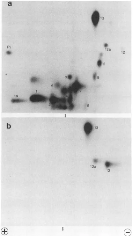
FIG. 1. Phosphopeptide analysis of dephosphorylated large T antigen. Large T antigen, metabolically labeled with
32p,
was dephosphorylated with calf intestinal alkaline phosphatase and analyzed by two-dimensional peptide mapping as described in Materials and Methods. Sampleswere appliedtothe middle of the thin-layercellulose plates as indicated by the vertical dashes. (a) Untreated; (b) dephosphorylated.vitro replication system. Inparallel, the effect of
phospha-tase treatment on ATPase and
origin-binding
activity wasalso measured. The
dephosphorylation
reactionwascarriedout with T
antigen
bound to monoclonal antibodiescova-lently
attached toprotein A-Sepharose.
The ATPase and DNAreplication experiments
were conducted with large Tantigen
released fromthemonoclonalantibodiesby high pHtreatment as describedin Materialsand Methods.
Figure
1 shows a two-dimensionalphosphopeptide
map of alkalinephosphatase-treated large
Tantigen
isolated from32p_
labeled cells infected with the adenovirus type 5-SV40 hybrid virus. Only threephosphopeptides, designated with numbers
12, 12a,
and13,
wereresistanttophosphatase
(Fig.lb).
Themap of untreatedlarge
Tantigen
is shown in Fig.la. As demonstrated
previously (19),
thephosphatase-resistant
peptides
12 and 13 contain Thr-124 and Thr-701,respectively.
We assumethat 12 and 12a representoverlap-ping
peptides
bothcontaining phosphothreonine
124.Pep-tide 12aismoreintenserelativeto12inthe map of untreated
on November 10, 2019 by guest
http://jvi.asm.org/
TABLE 1. Phosphorylation sitesoflarge Tantigen representedinphosphopeptides"
Peptide Phosphorylated residue
Amino-terminal region
1 Ser-106
3 Ser-106,Ser-111,orboth
4,7, 11 Ser-123,Thr-124
5 ?
4,6 Ser-111
12 Thr-124
Carboxy-terminal region
2,4 Ser-639
8,9,10 Ser-676, Ser-677, Ser-679
13 Thr-701
aData aretakenfromreference 18.
B than in thatofdephosphorylated large Tantigen. It is likely
thatthis shift in theratio of 12to 12aiscaused byachange inthespecificity ofpronaseafterdephosphorylation of large T antigen. The in vivo phosphorylation sites of large T antigen and the numbering of their corresponding phospho-peptides are shown in Table 1.
To test for ATPase activity, dephosphorylated large T antigenwas incubated with [at-32P]ATP, and the conversion
toADPwasdetermined by thin-layer chromatography (Fig. 2). Wefound nodifference between the activities of dephos-phorylated and untreated large T antigen. The specific activity of 1,200 nmol h-1 mg-1wascomparabletopublished values (1, 31, 32).
DNA binding was measured by incubation of
dephos-phorylatedanduntreated large T antigen with radioactively labeled AvaIl fragments of SV40 DNA and subsequent
precipitationof thelarge Tantigen with PAb419 monoclonal antibody. The DNA bound tolarge T antigen was analyzed
by agarose gel electrophoresis (18). The experiments were
carriedout atvarious large Tantigen-to-DNA ratios and in
thepresence orabsence of carrier DNA(Fig. 3, Table 2). At
a large Tantigen-to-DNA ratio of 30:1 (wt/wt) in the pres-enceofcarrierDNA, about 50% of the origin fragment was
a b c
d
e f 9
0-FIG. 2. ATPase activity of dephosphorylated large T antigen. Samples(0.8
jig)
ofdephosphorylatedand untreated largeTantigenwere assayed for their ability to hydrolyze ATP as described in
MaterialsandMethods.Samples(1,ul) of the reaction mixtureswere
spottedonphosphoethyleneimine-cellulose platesatvarious times ofthereaction. The plateswere developed in 0.5 M KH2PO4 (pH
3.5). Reactionproductswerevisualized by autoradiography.Lanes:
a, control (no large T antigen); b, d, and f, hydrolysis by
dephosphorylated large T antigen for 15, 30, and 60 min,
respec-tively;c, e,andg,hydrolysis by untreated largeTantigen for 15, 30, and60min, respectively. 0,Origin.
-CARRIER
phos
dephos-n
°O) Uc)Octou
M_ooVoo
A
+CARRIER
phos- dephos
-s°Lo C4N, LO °-Cl) M
phos-deph-U) uL
cN uL c4U)
U) e U)
-I
FIG. 3. DNA binding oflargeT antigendephosphorylated with calf intestinal alkaline phosphatase. Samples (4 ng) of32P-labeled Avall-cleaved SV40DNA wereincubated with variousamountsof purified large T antigen. For example, the number 30 indicates a
ratio of120 ng oflarge T antigen to 4 ngof labeled SV40 DNA fragments. Carrier DNA (800 ng) was added as indicated. The sampleson theright inpanel B representalongerexposureof the correspondingsamplesontheleft.Misthemarkertrack represent-ing one-halfof the labeled fragments added to each assay.
bound to large T antigen. When the ratio was lowered to
1.25:1, the amount of bound origin fragment dropped to
0.5%, indicating that at this ratio the amount of origin fragmentwasinlarge excess over thatof largeTantigen. At all large T antigen-to-SV40 DNA ratios tested in the
pres-TABLE 2. Origin binding by untreated anddephosphorylated largeTantigen'
T/DNAratioh Phosphatase Carrier' fragmentBoundorigin
(%)
30 - + 49
30 + + 46
10 - + 17
10 + + 19
5 - + 5
5 + + 4
2.5 - + 2
2.5 + + 1
1.25 - + 0.5
1.25 + + 0.5
10 - - 64
10 + - 76
1 - - 44
1 + - 36
0.5 - - 28
0.5 + - 28
0.1 - - 6
0.1 + - 3
0.05 - - 1.5
0.05 + - 1.5
aData wereobtained from theexperiment shownin Fig. 3. Theamountsof
orifragmentweredetermined by scintillation spectrometryof the excisedgel
bands andby scanningof theautoradiogram.
'DNAwasconstantat 4ngof32P-labeledAvaIl-cleavedSV40DNA, and
the amountoflargeTantigenwasvaried.
Carrier was 800 ng of salmon sperm DNA.
" Percent bound of32P-labeledorigin fragmentin the assay.
olp
.04 f.44
on November 10, 2019 by guest
http://jvi.asm.org/
[image:4.612.70.307.82.213.2] [image:4.612.326.562.452.658.2] [image:4.612.118.257.482.615.2]
hi80-E
0
30 60 90 12
CL
0 0.
20
30 60 90 120
[image:5.612.73.268.62.264.2]t(min)
FIG. 4. Stimulation of in vitro DNA replication by dephos-phorylation ofSV40large Tantigen. InvitroDNAreplicationwas
startedbytheadditionof variousamountsof dephosphorylated or
untreated large T antigen. Incorporation of deoxynucleotide triphosphateswasdetermined by liquidscintillationspectrometryof acid-insoluble material collectedat the times indicated. Symbols: (- *)0.7,ugofdephosphorylated largeTantigen,(0-4)0.3 pugofdephosphorylated largeTantigen,(E---E) 0.7,Lgof untreated largeTantigen,(L---L)0.3 ,ug of untreated largeTantigen.
ence of carrier DNA, no significant difference in origin binding between untreated and dephosphorylated large T antigen was observed. In the absence ofcarrier DNA, the
protein-to-DNAratiowasvaried from 10:1to0.05:1. Again, there was no difference between untreated and dephos-phorylated large T antigen in the ability to bind origin fragment. The levelof origin bindingwasgenerally higher in theabsencethan in thepresence ofcarrier. For example,at
aratio of10:1, about 65% of the origin fragmentwasbound withoutcarrier and only 17%was bound with added carrier DNA.Thisresultindicates that carrier DNAcompeteswith theorigin for bindingtolarge T antigen. We also noticed that intheabsence ofcarrier and athigh protein-to-DNA ratios, highamountsof the nonorigin fragmentswereboundtolarge Tantigen(Fig. 3), and that decreasing theamountof large T antigen resulted inmore specific binding of the origin
frag-ment. This result may be explained by assuming that the nonorigin fragments compete with the origin fragment in bindingtolargeTantigen.
Enhanced initiation ofin vitro SV40 DNA replication by
dephosphorylated large T antigen. After finding that the origin binding andATPaseactivitiesoflargeTantigenwere independent of its phosphorylation state, we asked if its abilitytosupportDNAreplicationinthe invitroreplication system described by Li and Kelly (9) was affected by phosphatase treatment. It seemed possible that an interac-tion oflarge T antigenwithsomecellularfactorsorenzymes required for SV40 DNA replicationwasinfluencedby phos-phorylation (27).AtimecourseofDNAreplicationwithtwo
concentrations ofdephosphorylated and untreated large T antigen is shown in Fig. 4. The data clearly demonstrate a stimulatory effect ofdephosphorylation which was two- to fourfoldafter15-and30-minreactions andwashigheratthe lower concentration oflarge T antigen. At later times, the ratesofDNAsynthesiswere similar withdephosphorylated and untreated large T antigen, as illustrated by the nearly
abc
def
high MW
~ii~
E
DNA
tw
"
Monomers
FIG. 5. Product analysis of DNA replication stimulated by dephosphorylation of SV40 large Tantigen. Purified dephosphory-lated oruntreated largeTantigen wasused in the in vitro replication assay. DNAsynthesis was terminated after various times of incu-bation, and the reaction productswereanalyzed ona1.4% agarose gel. The ladder of bands correspondstomonomericplasmid mole-cules with different degrees of supercoiling. The high-molecular-weight DNAs represent undissolved, catenated molecules. Lanes: a,b, and c, reaction products with dephosphorylated largeTantigen at30, 60,and120 min,respectively; d, e, and f, controls at 30, 60, and 120min,respectively.
parallel timecourses. The stimulationofDNAreplicationat
early times was also apparent when the reaction products
were analyzed on a 1.4% agarose gel (Fig. 5). The depen-denceof the stimulatory effecton theconcentrationoflarge Tantigenwasfurtherdemonstrated by the results shown in
Fig. 6. When the concentration of large T antigen was
decreased from0.8 to 0.1
jxg,
the stimulationofreplication causedby thedephosphorylation increased fromtwofold tofourfold, respectively.
Phosphorylation of large T antigen by HeLa cell extract.
One would have expected that dephosphorylated large T
antigen might have an advantage at each initiation event
throughout the entire reaction. This should have led to a
relatively higher amount of replicated DNA molecules
5-0 E
04
:
.22 4 .6 .
0. 3-0 0 0.
I- 2
z
.2 .4 .6 .8
pIg
TantigenFIG. 6. Difference in stimulation ofin vitro DNA replication betweendephosphorylatedand untreatedSV40largeTantigenas a function oflarge-T-antigenconcentration. Variousamounts of un-treated ordephosphorylated largeTantigen were added tothe in vitroDNAreplication reaction. Incorporationofdeoxynucleotides intoacid-insoluble materialaftera15-minreactionwasdetermined by liquid scintillation spectrometry. Symbols:
(-@*)
dephos-phorylated largeTantigen,
(@---@)
control.on November 10, 2019 by guest
http://jvi.asm.org/
[image:5.612.361.503.64.145.2] [image:5.612.331.533.430.641.2]a
9
PJ.
6 :
1. 7
MPb3sz...
b
c
O+>
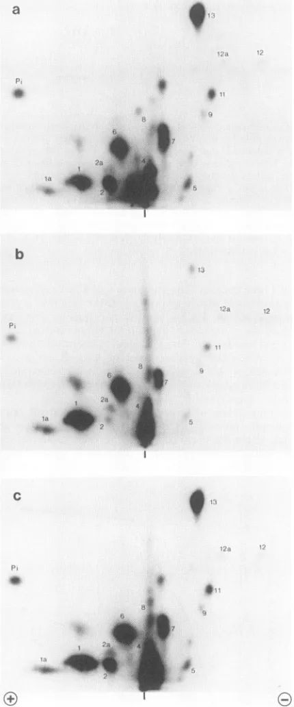
r----FIG. 7. Rephosphorylation of large T antigen by HeLa cell
extract. Dephosphorylated largeTantigen was added to the
repli-cation reactionsupplementedwith [y-32P]ATP. Large Tantigenwas
reisolated after 2 h and analyzed by two-dimensional
phospho-peptide mapping as described in Materials and Methods. In
vivo-32P-labeled large T antigen was used for a mixing experiment.
Samples were applied to the thin-layer chromatography plates as
indicatedby the verticaldashes.(a) Invivo-32P-labeledlarge T antigen;
(b) in vitro-phosphorylatedlargeTantigen;(c)mixtureofaand b.
towardthe end of the replication reactioninthepresenceof
dephosphorylated large T antigen. It was surprising that
after an initial lag phase, dephosphorylation had no further
effectonDNAreplication.However, itseemed possiblethat
dephosphorylated large Tantigen becamequicklymodified,
that is, rephosphorylated by kinases in the HeLa cell extract and,
therefore,
lost the ability to initiate replication faster than untreated large T antigen. In preliminary experiments we found, indeed, that large T antigen became quickly rephosphorylated in vitro. Dephosphorylated large T antigen was incubated for 2 h with HeLa cell extract under the same conditions used for DNAreplication, except that[,y-32P]ATP
was added. After the reaction, large T antigen was isolated by immunoprecipitation and subjected to peptide mapping.
Figure 7b shows a phosphopeptide map of the in vitro-labeled large T antigen. The pattern is similar to the map of the in vivo-labeled protein (Fig. 7a). All thephosphopeptides found in vivo were present in the map of the in vitro-phosphorylated T antigen, although the incorporation into specific sites differed considerably. Ser-106 (peptides 1 and la),
Ser-111
(peptides 4 and 6), and Ser-123(peptide 7) were themajor
phosphate acceptors. Thr-124 (peptides 11, 12, and 12a), Thr-701 (peptide 13), Ser-639 (peptide 2), and Ser-676, -677, and -679 (peptides 8 and 9) were phosphorylated to a small extent. One reason for the low phosphorylation of Thr-124 and Thr-701 could be that the phosphate bound to these residues was not removed by the alkaline phosphatase (Fig. 1). We calculated that the amount of phosphate incor-porated during the in vitro reaction was approximately 0.4 mol/mol of large T antigen. This may be an underestimation of the total amount incorporated, since the radioactive ATP became quickly degraded. The[.y-32P]ATP
added to the extract had a half-life of about 8min
(data not shown). If the higher activity of dephosphorylated large T antigen was lost because it became rephosphorylated during the lag phase of the in vitro replication reaction, then incubation of dephosphorylated large T antigen with HeLa cell extract before the start of DNA replication should reverse the effect of dephosphorylation. However, alkaline phosphatase-treated large T antigen retained its higher activity after preincubation with HeLacell
extract (data not shown). The reason might be that the extent of rephosphorylation of Ser-123 was not sufficient. The studies by Scheidtmann et al. (18, 19) and Baumann (1) indicated that phosphorylation at Ser-123 and Thr-124 might be crucial for the interaction of large T antigen with DNA. Although phosphorylation at Thr-124 could be the prerequisite for DNA binding, addi-tional phosphorylation at Ser-123 (and possibly other resi-dues) could downregulate DNA-binding activity in the pres-ence of cellular factors (1, 18).It was of interest to find out whether the HeLa cell extract contained active phosphatases in addition to protein kinases. This was determined by incubating in
vivo-32P-labeled
large T antigen with HeLa cell extract for 2 h and measuring the removal of32P
after isolation of the large T antigen by immunoprecipitation. There was no phosphatase activity in the extract under the conditions of DNA replication. Iden-tical amounts of radioactivity were recovered from large T antigen incubated with extract and from the control sample that was not incubated with extract (data not shown). Since no peptide maps were determined for the32P-labeled
large T antigen after the incubation with HeLa extract, we cannot exclude a small amount of phosphatase activity.DISCUSSION
Our studies demonstrate that the removal of serine-bound phosphates from SV40 large T antigen enhances its ability to stimulate in vitroSV40 DNA replication but has no effect on its ATPase and origin-specific DNA-binding activities.
i.11
6
V, Mk.
:I -,. 7
2a .i,
I
it i.
2
U
I
on November 10, 2019 by guest
http://jvi.asm.org/
[image:6.612.80.295.65.581.2]Thesefindingsareinagreementwithareportfrom Shaw and Tegtmeyer, who showed by DNA footprinting and DNase protection experiments that alkaline phosphatase treatment of large T antigen had no effect on DNA binding (22). Similarly, Baumann found that DNA bindingwasunaltered by alkalinephosphatase (1). On the other hand, Simmonset
al. reported a 1.5- to 2-fold stimulation of binding to the purified SV40 origin-containing fragment caused by alkaline phosphatase (26). They suggested that dephosphorylation produced more DNA-binding sites on large T antigen and also increased the rate of DNA binding. These authors carried out the DNA binding under DNA excess. Our DNA-binding studies were conducted under a variety of differentconditions, including considerable variation of the large Tantigen-to-SV40 DNA ratio (over 200-fold), and in the absence and presence of carrier DNA. We did not
observeaneffect ofdephosphorylationonbinding of large T antigentothe origin fragment underanyconditions.
Whileourstudieswere inprogress, we were informed of the recently published results of Mohr et al. (14). These authors found no effect of dephosphorylation of large T antigen with alkaline phosphatase on ATPase activity and only a "slight" stimulatory effect on DNA binding when using origin fragments containing large-T-antigen binding sites I and II. Interestingly, dephosphorylation caused a large increase in DNA bindingtothe isolated binding site II butnottotheisolated binding site I.As in thepresentstudy,
Mohretal. founda significant stimulation of in vitro DNA replication resulting from dephosphorylation of large T an-tigen.
In aprevious report by Scheidtmann et al. it was found thatthe DNA-binding activity of large T antigencorrelated with a distinct phosphorylation state (18). Newly synthe-sized large T antigen was phosphorylated to a low degree andexistedprimarily inamonomericform sedimenting with 5S. It had a high affinity for SV40 DNA as measured by bindingtoSV40 DNA cellulose. Bycontrast, "old" large T antigen was highly phosphorylated and predominantly ex-isted in an oligomeric form with a lower affinity for SV40 DNA. Two-dimensional phosphopeptide analysis revealed distinct phosphopeptide patterns for new and old large T antigen. Basedonthese studies thefollowingscheme for the life cycle of large T antigen was proposed: large T antigen becomes phosphorylated in the cytoplasm at certain sites (Ser-111, Ser-112, Thr-124, Thr-701). It is transportedtothe nucleus, where it binds to SV40 DNA while still in a low
phosphorylation state. Subsequentphosphorylation at addi-tional sites(Ser-123, Ser-106) then modulates the interaction with DNA, resulting in dissociation and oligomerization. According to this model, phosphorylation atThr-124 might be aprerequisite for DNA binding (18, 20). Furthermore, removal ofphosphate from Ser-106 and Ser-123withalkaline phosphatase might activate the DNA-binding activity of large Tantigen. To reconcile the findings of Scheidtmannet al. with the present data,one shouldconsider the possibili-tiesthatSV40 largeTantigeninteracts withcellularfactors which enhance itsDNA-binding ability and that phosphory-lation at Ser-123 and/or Ser-106 might interfere with the binding of these factors. In this case, removal of serine
phosphateswith alkalinephosphatase would have noeffect onDNAbindingin theabsence of these factors. Thismight explain why we and others did not observe an effect of alkalinephosphataseonDNAbindingsince purified largeT antigen was usedin the experiments (1, 22). On the other hand,the DNA-binding studies of Scheidtmann etal. were carried out with crude extracts containing cellular factors,
andthismaybe thereasonwhyin thiscasetheDNA-binding activity wasdependent onthe state of phosphorylation.
Stillmann et al. described a mutant of large T antigen (Lys-224 to Glu) which has wild-type levels of ATPase
activityandbinds to thereplicationoriginbutfails to support
SV40DNAreplicationinvitro(30). Theysuggestedthat the
binding of largeTantigento ahostfactor isanessentialstep in the initiation ofDNAreplication, and that this mutantis
defective in carrying out this interaction. Smale and Tjian reported that large T antigen binds to DNA polymerase a
andthat monoclonalantibodies that inhibitthis bindingalso
inhibit in vitro DNA replication (27). They suggested that this interaction might precedethebinding oflarge Tantigen
to the origin of
replication.
Itis possible that phosphoryla-tion of large Tantigen inhibits bindingtopolymerase a.A large number of mutations have been introduced into
the gene of large T antigen to localize regions in the
polypeptide responsible
forDNAreplicationand DNAbind-ing. Interestingly, mutants affecting the phosphorylation
sites Ser-106, Ser-123,andThr-124weredefective forSV40 DNA replication in vivo. However, the large T antigens
encoded by these mutants showed normal origin binding activity in vitro (7). These findingssuggest that phosphory-lationatthesesitesdoes notdirectlyinfluence DNAbinding butinterfereswith anotherpropertyof largeTantigen such
as interaction with host factors. This
interpretation
iscom-patible
withthefinding
that theDNA-binding
domainonthelarge-T-antigen polypeptide
maps betweenamino acids 139and 223, that is, downstream of the amino-terminal
phos-phorylation
sites.Recently, Woldetal. studiedthe events whichtakeplace during the lag phase preceding maximal DNA synthesis in the in vitro DNA
replication
system (36). They found that the lag phase could be almost completely eliminated bypreincubating
large Tantigen with SV40origin DNA andacellular factor,
presumably
asingle-stranded DNA-binding
protein,
in thepresenceofATP.Thefindings
suggestedthat the helicaseactivity of large Tantigen
mediatesunwinding andmelting oftheSV40origin (28). Again, itis conceivablethat
phosphorylation
interfereswith theinteraction between largeTantigen and the cellular factor.The elucidation of the role of phosphorylation in the
function oflarge T
antigen
is hampered by the finding that HeLacell extract contains high protein kinase activity. In the future,experiments
with a DNAreplication
systemconsisting
only ofpurified
components will be carried out.Hopefully,
theseexperiments
will allowustoobtainabetterunderstanding
of the role ofphosphorylation
in DNArepli-cationin vitro and in vivo.
ACKNOWLEDGMENTS
Wethank Lois C. Tack for adviceonthe ATPase assay,Arlene Carbone-Wiley and UteStober for excellent technical assistance, and Gail Parish fortypingthemanuscript.We thankJoachimJ. Li and ThomasJ. Kellyfor criticalreadingofthemanuscript.
Thisstudywassupported byPublic HealthService researchgrant CA36111 from theNational Cancer Institute toG.W.
LITERATURECITED
1. Baumann, E. A. 1985. DNA-binding propertiesof
phosphoryl-atedand dephosphorylated D2-Tantigen, asimianvirus 40 T antigenrelatedprotein. Eur. J. Biochem. 147:495-501. 2. Bradford, M. M. 1976. A rapidand sensitive method for the
quantitation of microgram quantities of protein utilizing the principle ofprotein-dye binding.Anal. Biochem.72:248-254. 3. Clark, R., M. J. Tevethia, and R. Tjian. 1984. The ATPase
activity ofSV40large T antigen, p. 363-368. InG. F. Vande
on November 10, 2019 by guest
http://jvi.asm.org/
Woude, A. T. Levine, W. C. Topp, and J. D. Watson (ed.), Cancer cells 2: oncogenes and viral genes. Cold Spring Harbor Laboratory, Cold Spring Harbor, N.Y.
4. Dixon, R. A. F., and D. Nathans. 1985. Purification of simian virus 40 large Tantigen by immunoaffinitychromatography. J. Virol.53:1001-1004.
5. Gluzman, Y., H. Reichl, and D. Solnick. 1982. Helper-free adenovirus type-5 vectors, p. 187-192. In Y. Gluzman (ed.), Eukaryotic viral vectors. ColdSpring Harbor Laboratory, Cold Spring Harbor, N.Y.
6. Harlow, E.,L. V.Crawford,D.C. Pim,and N. W.Williamson. 1981.Monoclonalantibodies specific for simian virus 40 tumor antigens. J. Virol. 39:861-869.
7. Kalderon,D.,and A. E. Smith. 1984. Invitromutagenesis ofa
putative DNA-binding domain of SV40 large T. Virology 139:109-137.
8. Laemmli, U.K.1970.Cleavage of structural proteins duringthe assembly ofthe head of bacteriophage T4. Nature (London) 227:680-685.
9. Li, J. J., and T. J. Kelly.1984. Simian virus 40 DNA replication invitro.Proc. Natl. Acad. Sci. USA 81:6973-6977.
10. Li, J. J., andT.J. Kelly.1985.Simian virus 40 DNA replication in vitro: specificity of initiation and evidence for bidirectional replication. Mol. Cell. Biol. 5:1238-1246.
11. Li, J. J., K. W. C. Peden,R. A. F.Dixon, and T. Kelly. 1986. Functional organization of the simian virus 40 origin ofDNA replication. Mol. Cell. Biol. 6:1117-1128.
12. Maniatas, T., E. F. Fritsch, and J. Sambrook. 1982. Molecular cloning:alaboratorymanual. Cold Spring Harbor Laboratory, ColdSpring Harbor, N.Y.
13. McKay,R.1981. Binding of a simian virus 40 T antigen related proteintoDNA. J. Mol. Biol. 145:471-488.
14. Mohr,I.J., B.Stilimann, and Y. Gluzman. 1987. Regulationof SV40 DNAreplication by phosporylation ofTantigen. EMBO J.6:153-160.
15. Paucha, E.,D.Kalderon,R. W.Harvey,and A. E. Smith. 1986. Simianvirus40origin DNA-binding domain on largeTantigen. J.Virol. 57:50-64.
16. Scheidtmann, K. H. 1986. Phosphorylation of simian virus 40 largeTantigen: cytoplasmic and nuclear phosphorylation sites differ in their metabolicstability. Virology 150:85-95. 17. Scheidtmann, K. H., B.Echle, and G. Walter. 1982. Simian virus
40largeTantigen isphosphorylatedatmultiple sites clustered intwoseparate regions.J. Virol. 44:116-133.
18. Scheidtmann, K. H., M. Hardung, B. Echle, and G. Walter. 1984. DNA-binding activity of simian virus 40 large T antigen correlateswithadistinctphosphorylationstate.J.Virol. 50:1-12. 19. Scheidtmann,K.H.,J.Schickedanz, G. Walter,R. E.Lanford, andJ. Butel. 1984. Differential phosphorylation of cytoplasmic and nuclear variantsof simian virus40largeTantigen encoded by simian virus 40-adenovirus 7 hybrid viruses. J. Virol. 50:
636-640.
20. Schickedanz, J., K. H. Scheidtmann, and G. Walter. 1986. Kinetics of nuclear transport and oligomerization of simian virus 40 large Tantigen. Virology 148:47-57.
21. Schneider, C.,R. A.Newmann,D. R.Sutherland,U.Asser,and M. F. Greaves. 1982. A one-step purification of membrane proteins using ahigh efficiency immunomatrix. J. Biol. Chem. 257:10766-10769.
22. Shaw,S. B.,and P. Tegtmeyer. 1981.Bindingof dephosphory-latedAproteintosimian virus 40DNA.Virology 115:88-96. 23. Simanis, V., and D.P. Lane. 1985.Animmunoaffinity
purifica-tionprocedure for SV40largeTantigen. Virology 144:88-150. 24. Simmons,D.T.1986.DNA-binding region of the simian virus 40
tumor antigen. J. Virol. 57:776-785.
25. Simmons, D. T. 1984. Stepwise phosphorylation ofthe NH2-terminalregion of the simian virus40large Tantigen. J. Biol. Chem. 259:8633-8644.
26. Simmons,D.T.,W. Chou,and K.Rodgers. 1986. Phosphoryla-tiondownregulatestheDNA-bindingactivity of simian virus40 Tantigen. J. Virol.60:888-894.
27. Smale, S. T.,and R.Tjian. 1986.T-antigen-DNA polymerasea complex implicated in simian virus 40 DNAreplication. Mol. Cell. Biol. 6:4077-4087.
28. Stahl, H., P. Droge, and R. Knippers. 1986. DNA helicase activity of SV40 largetumorantigen. EMBO J. 35:1939-1944. 29. Stillman,B.W.,andY. Gluzman. 1985.Replication and
super-coiling of simian virus40 DNAin cellextractsfrom human cells. Mol.Cell Biol. 5:2051-2060.
30. Stillmann, B., R. D. Gerard, R. A. Guggenheimer, and Y. Gluzman. 1985.Tantigen and templaterequirements forSV40 DNAreplicationinvitro.EMBO J. 4:2933-2939.
31. Tjian, R.,andA.Robbins. 1979.Enzymatic activities associated with apurified simian virus 40 Tantigen-related protein. Proc. Natl. Acad. Sci. USA76:610-614.
32. Tjian, R.,A. Robbins, andR.Clark. 1979. Catalyticproperties of the SV40 largeTantigen. ColdSpring Harbor Symp. Quant. Biol. 44:103-111.
33. VanRoy, F.,L. Fransen, and W. Fiers. 1983. Improved local-ization of phosphorylation sites in simian virus 40 large T antigen.J. Virol. 45:315-331.
34. VanRoy, F., L. Fransen, and W. Fiers. 1983. Metabolic turn-overofphosphorylationsitesinsimian virus40 largeTantigen. J. Virol. 45:442-446.
35. Wobbe, C. R., F. Dean,L.Weissbach, and J. Hurwitz. 1985.In vitro replication of duplex circular DNAcontaining the simian virus 40DNAorigin site. Proc. Natl. Acad. Sci. USA 82:5710-5714.
36. Wold,M.S., J. J.Li, and T.J. Kelly.1987. Initiation of simian virus 40 DNA replication in vitro: large-tumor-antigen- and origin-dependent unwinding of the template. Proc. Natl. Acad. Sci. USA 84:3643-3647.