0022-538X/85/060825-08$02.00/0
Copyright C 1985,American SocietyforMicrobiology
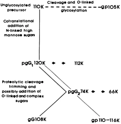
Synthesis
and
Processing of
Glycoprotein gG of
Herpes Simplex
Virus Type 2
N. BALACHANDRAN* AND L. M. HUTT-FLETCHER
DepartmentofComparative andExperimentalPathology, University of Florida, Gainesville, Florida 32610
Received10October1984/Accepted25 February 1985
Monoclonal antibody 13caC5-1-A11 immunoprecipitated two major polypeptides of molecular weights 108,000 and 120,000 from extracts of herpes simplex virus type 2-infected BHK-21 cells labeled with [35S]methionine or [3H]glucosamine. In pulse-chase experiments, both labels were chased from the 120,000-molecular-weight peptide (120K peptide) into the 108K molecule. Endoglycosidase H (endo H) reduced the 120K peptide to a 112K peptide but did not affect the 108K peptide. Similar profiles were obtained with monoclonalantibody AP-1 which reacts with a 92K glycoprotein, gG,whichmaps to the shortuniqueregion of thegenome. Cross-absorption experimentsindicated that both antibodies reacted with the same peptides, suggesting that the 120K peptide is a partially glycosylated high-mannose-type precursor of gG
(pgG,).
Immunoprecipitationfrommonensin-treated cells indicated thatpgG1(120K)may undergopeptide cleavage to
form a 74Khigh-mannose-typepeptide (pgG2) and that this 74K peptide may be further processed into an endo H-resistant 110K to 116Kpeptide.Inthepresenceoftunicamycin,gG(108K)wasreplacedby 110K and 105K peptides which were resistant to both endo H and endoglycosidase F. The 105K peptide was the only molecule labeled by [3H]galactose or [3H]glucosamine in the presence oftunicamycin, and none of the peptides were labeled with
[3H]mannose,
indicating theprobable presence of 0-linked sugars in the 105Kpeptide.Our resultsimplythatcotranslational glycosylationof theunglycosylated precursor 110K peptide results in the
high-man-nose-type
pgGl(120K),
whichprobably undergoes peptide cleavage. Thisputativecleavageproductmay then mature intogG(108K) by the trimming of sugars and the addition of complex and probably 0-linked sugars;thehigh-mannose-type pgG2(74K) isprobably an intermediate peptide formed in this process.
Inthe courseofproductive infection with herpes simplex
virus type 1 (HSV-1) and type 2 (HSV-2), at least four
virus-specified glycoproteins, designated gC, gB, gD, and
gE, aresynthesized and become incorporated intoboth the
infected cell membrane and the virion envelope (19). The
glycoproteins gB, gE, andgD possessbothtype-specificand
type-commonantigenic sites (7, 8,14),andthe genes
encod-ing them map colinearly in HSV-1 and HSV-2 genomes
(12-14). ThegCof HSV-1 wasoriginally thought tocontain
exclusively
type-specific
sites, and a counterpart in HSV-2was notknown(7). However, using monoclonal antibodies
we described two additional antigenically and structurally distinctHSV-2-specific glycoproteins (3). Oneofthese new
glycoproteins was originally designated gF (2, 3) but has
sincebeen showntobeencodedbyanHSV-2genemapping
colinearly with the gC gene of HSV-1. It shares antigenic
determinants with gC of HSV-1 (15, 25, 26, 27, 28)and is
nowreferredto asgC2. Thesecondapparentlytype-specific
glycoprotein is immunoprecipitated by monoclonal antibody
13aC5-1-A11
(13aC5). Wehereidentifythisglycoproteinas gG, thepreviously
described 92K glycoprotein of HSV-2whichmaps in theuniqueshort region ofHSV-2 DNA (11).
We describe the biosynthesis of the mature glycosylated
molecule and present evidence for probable major
proteo-lytic cleavage ofapartiallyglycosylatedprecursorduring its
maturation.
MATERIALSANDMETHODS
Cells and virus. Monolayers of baby hamster kidney
(BHK-21)andVero cellsweregrowninmodifiedDulbecco
medium(GIBCOLaboratories)supplementedwith antibiot-ics and heat-inactivated fetal bovine serum. Stock virus
*Correspondingauthor.
preparation and plaque assay of HSV-2
(strain
333) weredonein Verocells (18).
Monoclonal antibodies. Preparation and characterization
of ascitic fluid of monoclone 13aC5 have been described
previously (1-3). Monoclonal antibody AP-1 (ascitic fluid)
was a gift from A. C. Minson, University of Cambridge,
England(11).
Radiolabeling procedures. Confluent monolayers of BHK-21cellswere infected with HSV-2 at amultiplicity of
infection of10PFUpercellandallowedtoadsorb for2 to3
h. Forlong-term labeling (3to20h), cellswereincubated in
Hanks balanced salt solution containing 20
,uCi
per ml ofL-[35S]methionine (specific
activity, 1,380,uCi/mmol;
Amer-sham Corp.),
5%
dialyzed fetal bovine serum, and 1/10 thenormalconcentration of methionine. Cellswerealso labeled
for 3 to 20 h with
[3H]mannose
(specific activity, 27.2,Ci/mmol;NewEngland Nuclear Corp.),[3H]galactose
(spe-cific activity, 51.7
pCi/mmol;
New England Nuclear), and[3H]glucosamine
(specific activity, 15 ,uCi/mmol; ICN Phar-maceuticalsInc.) atconcentrations of100,uCi/ml
in mediumwith 1/10 the normal concentration of glucose. In
pulse-chase experiments, cells were labeled for 10 min with
[35S]methionine
(100,uCi/ml)
or for 30 min with[3H]
glucosamine (100 ,uCi/ml) at 5 h postinfection (p.i.) and
chased in medium containing 100times the normal
concen-tration of unlabeled methionine or glucose and
cyclohex-imide
(50
,ug/ml).
Inexperiments withtunicamycinormonensin, cells were
washed and preincubated for 3 h with medium containing 3 ,ug of tunicamycin (Sigma Chemical Co.) per ml or 1 ,uM monensin
(Sigma).
Bothdrugs were thenpresent throughouttheremainingcourse ofeach experiment.
Immunoprecipitation and electrophoresis.
Immunoprecipi-tation was carried out as previously described (2). Briefly,
825
on November 10, 2019 by guest
http://jvi.asm.org/
205K
1 16K( _ 97K_
66K.
45K.
1 2 3 4 5 6
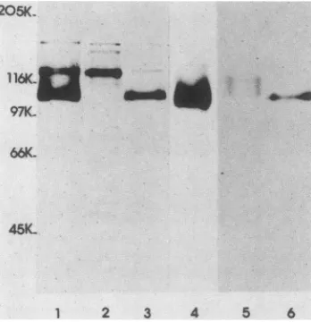
FIG. 1. Autoradiograms of peptides immunoprecipitated from HSV-2-infected BHK-21 cells by monoclonal antibody 13aC5. Lanes showinfected cellslabeled with: 1,[35S]methioninebetween 3to20 h p.i.; 2, [35S]methionine pulse for 10 min at 5 h p.i.; 3,
[35S]methioninepulsefor 10 minat 5 h p.i. followed by chase for5
h innonradioactive medium-cycloheximide-100x cold methionine; 4,[3H]glucosamine between 3 to 20 h p.i.; 5,[3H]glucosaminepulse for30minat 5hp.i.; 6,[3H]glucosaminepulse for 30 minat 5hp.i. followed by chase for 5 h in nonradioactive medium-cyclo-heximide-cold glucose. SDS-PAGE was carried out on 9% acrylam-ide cross-linked with N,N'-diallyltartardiamacrylam-ide.
cells were solubilized with RIPAbuffer containing 0.1 mM
phenylmethylsulfonylfluoride and 100 U of aprotinin per ml
(2)and mixedwith antibodyandproteinA-SepharoseCL-4B
beads (Sigma). The precipitates were washed, dissociated by boiling, and analyzed by sodium dodecyl
sulphate-polyacrylamide gel electrophoresis (SDS-PAGE) in 9%
acrylamide cross-linked with 0.24%
N,N'-diallyltartar-diamide. Molecular weightmarkers(Sigma)were
electropho-resed in parallel channels. Gels were stained, destained,
infused with 2,5-diphenyloxazole, dried onfilter paper,and
placed in contact with Kodak XAR-5 film at -70°C for
fluorography (4).
Enzyme digestion. Immunoprecipitated proteins boundto
theprotein A-Sepharose beadswereeluted byincubatingthe
pellets for20min at37°C in50 ,u ofsodium citrate buffer (50
mM,
pH 5.5)containing0.2% SDSandthenboiling for30 s.Endoglycosidase H (50,lI,endo H)in citrate buffer(25 mU;
Miles
Laboratories,
Inc.) was added to the samples andincubated for24h at 37°C. For digestion by endoF, beads
were suspended in 100 ,lIof sodiumphosphate buffer(100
mM,pH 6.1) containing 0.1% SDS,0.5% Nonidet P-40, and
50 mM EDTA. Five microliters of endo F (440
U/ml;
NewEngland Nuclear)wasaddedtothesample and incubated
for
24 hat 37°C. Control samples withoutadditional endo Hor
endo Fwere treated in the same way, and after incubation 100 ,ul ofsample buffer (2 x) containing 10% SDS and 5%
mercaptoethanolwas added and boiled for 3 min. Samples
containing equalamountsofradioactivity wereanalyzedby
SDS-PAGE andautoradiography. RESULTS
Characterization of glycoprotein
immunoprecipitated
by monoclonal antibodyl3aC5.
BHK-21 cells were infectedwithHSV-2 and labeled with
[35S]methionine
for3 to 20 h,and the
RIPA-solubilized
lysate was immunoprecipitated.Autoradiograms ofthe electrophoresed immunoprecipitate
showedtwomajor bands,one anintensely labeledpeptide of
approximate molecular weight 108,000 and the other a
peptide of about molecular weight120,000 (Fig. 1, lane 1).
Similar size peptides were also precipitated from infected
Vero cells and from BHK-21 cells in the absenceof
proteo-lytic inhibitors, and no peptides were precipitated from
uninfected cells (data not shown). In addition to the two
major peptides,two tothree high-molecular-weightpeptides
were also seen in the majority of the immunoprecipitates; these wereprobably nonspecific contaminants as theywere
precipitated by several other monoclonal antibodies. Both
the 120K and 108K peptides could be labeled with
[3H]glucosamine (Fig. 1, lane 4)
and
with [3H]mannose(seeFig.5, lane6),whilethehigh-molecular-weight forms could
not; mostof the labelwas seen in the 108Kpeptide.
Previous studies byus and others (2, 3, 15, 16) haveshown
that multiple glycosylated peptides precipitated by
mono-clonal antibodies show precursorproduct relationships and
that different molecular weight species represent different
levelsofglycosylation.Todetermine theprecursormolecule
oftheglycoprotein immunoprecipitated by l3aC5,
HSV-2-infected cells were pulsed for 10 min at 5 h p.i. with
[35S]methionine and chased for 5 h inthe presence ofcold
methionine and cycloheximide. The major labeled peptide
precipitated fromcell lysates made during the pulse period
was a120K peptide. In the chase period, the label wasfound in the 108K peptide (Fig. 1, lanes 2 and 3). After long exposureoftheautoradiographs,somelabelwasalsoseenin
a74Kpeptidein thelysates from the pulse butnotfrom the
chase period. The conversion ofthe 120K peptide into a
108Kpeptidewas veryrapid. In ashortpulse-chase
exper-iment, the 108K peptide was labeled after only a 15-min
chase, and chase ofthe label into this molecule from the
~~~~~~,#~~~~~~~~~.
MIN 10 15 30 45 60 120
205K.
116\ - - z
97K.
66K.
45K.
1 2 3 4 5 6
FIG. 2. SDS-PAGEanalysis of peptidesimmunoprecipitatedby
monoclonal antibody 13aC5. Lanes show infected cells labeled with: 1,[35S]methioninefor10minat 5hp.i.;2to6,[35S]methionine
for10min at 5 h p.i. followed bychasefor15, 30, 45, 60, and120 min, respectively, in nonradioactive medium-cycloheximide-100x cold methionine. Short arrows indicate the position of120K and 108Kpeptides.
on November 10, 2019 by guest
http://jvi.asm.org/
[image:2.612.93.267.68.248.2] [image:2.612.333.525.417.653.2]120Kpeptidewascomplete after60min(Fig. 2,lanes 1 to6).
Inthepulse-chase experimentwith[3H]glucosamine(30-min
pulse, 5-h chase), the label was initially associated with a
diffuse band ofa 120K to 112K peptide and later with the
108Kpeptide(Fig. 1, lanes 5 and 6).Theseresultssuggested
that the 120K peptide istheglycosylated precursor for the
fully glycosylated 108Kpeptide.
Endo Hdigestion.TheenzymeendoH has been shown to
cleave the two proximal N-acetylglucosamine residues of
the high-mannose-typeoligosaccharidesbut notthose of the
complextype(22). Toidentify theprimaryprecursor of the
108K peptide and to characterize its oligosaccharide side
chains, immunoprecipitates from [35S]methionine
pulse-chase experiments were treated with endo H as described above.
Endo H treatment of the immunoprecipitate from the
pulse period resulted in the complete disappearance of the
120K peptide and the appearance ofa new species with a
molecularweight ofapproximately 112,000 (Fig. 3, lanes 1
and 2). In contrast, treatmentofimmunoprecipitate from the
chase
period
did notalterthe 108K peptide (Fig. 3,lanes 3and4). Long-termexposureof the autoradiographs revealed
thepresenceofa 74Kpeptide in the pulseperiod, andupon treatment with endo H, a 66K molecule appeared in its
place. Neitherof theseweredetectable in the chaseperiod.
Theotherhigher-molecular-weight proteins whichwere
non-specifically precipitated by this and other antibodies served
as agood control for demonstrating the lack of detectable
proteaseactivity in the endoH preparation.
PULSE C-HASE
EndoH _ + - +
205K.
116K _
97K.
66K...
45K.
[image:3.612.365.530.67.299.2]1 2 3 4
FIG. 3. SDS-PAGEanalysis of endoHsusceptibility ofpeptides immunoprecipitated by monoclonal antibody l3aC5. Lanes show infectedcellspulsed with:1and 2,[35S]methionine for 10 minat5h p.i.;3and4,[35S]methionine for10minat5hp.i. followed bya5-h chaseasdescribed inthelegendtoFig.1.Immunoprecipitateswere
eluted and incubated in thepresence(+)orabsence(-) of endo H, andsamples were analyzed. Arrowsindicatethe position of 120K and74Kpeptides.
205K
116K
5 'J97K_
b 46
6K1
45K.
[image:3.612.115.262.376.640.2]1 2 3 4
FIG. 4. SDS-PAGEanalysis of gGimmunoprecipitatedby mono-clonal antibody AP-1 from HSV-2-infected BHK-21 cells. Lanes showinfected cells labeled with: 1,
[35S]methionine
between 3 and 20 hp.i.; 2, [35S]methioninepulse for 10 min at 5 hp.i. followed by a5-h chase asdescribed in the legend toFig. 1; 3, [35S]methioninepulsefor 10 min at 5 h p.i.; 4,[3H]glucosaminebetween 3 and 20 h p.i.
These resultsdemonstrated that(i) the 120Kpeptide has
N-linked
oligosaccharides
of thehigh-mannose
type,(ii)
the108Kpeptide lacks the
high-mannose-type
sugarsandprob-ably has
oligosaccharides
of thecomplex
type,(iii)
the112Kpeptide
isprobably
theprimary
precursorwith theproximal
glucosamine residue attached to it, and (iv) the
cotransla-tional addition of sugars by N-linkage converts it to the
partially
glycosylated
precursorgpl20K.
The apparentsmaller size and the endo H-resistant nature of
gplO8K
imply that
gpl20K
undergoes peptide cleavage before itmaturesintothe mature
gplO8K
bytrimming
andaddition offurthersugarresidues. The size and thefateofthe putative
cleaved
peptide
cannotbe determinedby
immunoprecipita-tion with
13aC5,
whichevidently
reactsonly
with asite onthe uncleaved
portion
of the molecule.Identity of the
gplO8K.
Recently Marsden et al. (11)reported
a92Kglycoprotein immunoprecipitated
bymono-clonal antibodies AP-1 and LP-5 from HSV-2-infected cell
extractswhich has been showntomap in the short
unique
region
of thegenome;they called itgG.Similarly,
Roizmanetal. (17)havemappedgG(125K)totheshort
unique
regionof the genome. Although of different apparent molecular
weight (probably due to a different gel system and virus
strain),
the 92Kpeptide
had characteristics common togplO8K
(A. C.Minson, personal communication).Todeter-mine the exact
relationship
ofgplO8K
and the92K gG, wecompared
theimmunoprecipitation properties
of 13aC5 with those of the AP-1antibody kindlydonatedby A. C. Minson.Peptides immunoprecipitated
by AP-1 antibody both from[3H]glucosamine-
(Fig. 4, lane 4)and[35S]methionine-
(Fig.4, lane 1) labeledextractsweresimilartothoseprecipitated
by 13aC5. In addition, AP-1 antibody immunoprecipitated
120K and 112K peptides in the pulse period and a 108K
peptide inthe chase period (Fig. 4, lanes 2 and 3), clearly
indicatingthat AP-1and
13aC5
reactwith the samegp108K.on November 10, 2019 by guest
http://jvi.asm.org/
q7K
-
66K-
45K-1 2 3 4 5 6 7 8 9
FIG. 5. SDS-PAGE analysis of gGimmunoprecipitated by mono-clonalantibody(13aC5)fromHSV-2-infected BHK-21 cells untreat-edortreated with monensin or tunicamycin. Lanes show infected cells labeled with: 1, [35S]methionine between 3 and 20 h p.i. (untreated cells); 2, [35S]methionine between 3 and 20 h p.i. (cells treatedwith 3p.gof tunicamycin); 3 and 4,[35S]methioninebetween 3 and 20 h p.i. (cells treated with 0.5 and 1 ,uM monensin, respectively); 5, [3H]mannose between 3 and 20 h p.i. (untreated cells); 6, [3H]mannose between 3 and 20 h p.i. (cells treated with 1 ,uM monensin); 7, [35S]methionine between 3 and 20 h p.i. (cells treatedwith1,uMmonensin); 8,[35S]methioninepulse for 10 min at 5 hp.i. (cells treated with 1 ,uM monensin); 9,[35S]methioninepulse for10minat5 hp.i.followed by a 120-min chaseasdescribed in the legend toFig. 1 (cells treated with 1 ,uM monensin). Drug treatment wasbegun 3 h before infection and continued for the remainder of theexperiment.
This was confirmed by cross-adsorption tests. Removal of
gP108K by successive immunoprecipitation with either of
the monoclonal antibodies depleted the molecules
precipi-table by the other antibody (datanotshown). These
exper-iments demonstrated that the 108K peptide is the mature
formof glycoprotein gG [designatedgG(108K)];the
precur-sor 120Kpeptide wasdesignated pgG1(120K).
Glycosylation in the presence of monensin. To examine
furthertheprocessing of gG, lysates ofHSV-2-infectedcells
treated with the ionophore monensin were
immunoprecipi-tated and analyzed by SDS-PAGE. Monensin disrupts ion
gradients across membranes and has been shown to block
the processing of HSV-1 glycoproteins at the addition of
0-linked
sugars;italso probably inhibitssomeprocessing ofN-linked sugars (10). Immunoprecipitates from cells treated
with 0.5 or 1 ,uM monensin gave identical results (Fig. 5,
lanes3and4), andsubsequentexperimentswerecarriedout
with 1 ,uM monensin. In contrast to the
pgGl(120K)
andgG(108K) peptides immunoprecipitated fromuntreatedcells
(Fig. 5, lane 1), immunoprecipitates from monensin-treated
cells contained anintermediate
peptide
of about 110,000to116,000 molecular weight and more ofthe faster-migrating 74K
peptide
(Fig. 5, lanes 3, 4,and7);both could be labeled with[3H]mannose
(Fig. 5, lane 5). In apulse-chase experi-mentwith[35S]methionine,
the 120K and 74Kpeptideswere present in a 10-min pulse, but label was chased within 120 mininto the 110K to 116K peptide (Fig. 5, lanes 8 and 9),indicating a precursor-product relationship between these
molecules.
The 110K to 116K peptide was not affected by endo H,
whilethe 74Kpeptidewaspartially susceptible, resultingin
faster-migrating
peptides of about 66,000to69,000molecularweight (Fig. 6, lanes 1 and 2). The 120K and74Kpeptides
present in the pulse period were completely susceptible to
endo H, being reduced to 112Kand 66K peptides,
respec-tively (Fig. 6, lanes 3 and 4); thus, thesecontain
high-man-nose-type sugars. In contrast, the 110K to 116K
peptide
presentafterthe120-minchasewasresistant toendoH(Fig. 6,lanes 5 and6).The results suggest that the
high-mannose-type precursor
pgGl(120K)
undergoes a proteolyticcleavagetobecomea74Kpeptide (pgG2);furtherprocessingof sugars
took place, resulting in theendo H-resistant 110K to 116K
peptide.
Todetermine whether monensin affected theotherHSV-2
glycoproteins in a similar manner, a monoclonal antibody
(17aA2) to gC2 shown to contain both 0- and N-linked
complex sugars (25) was used to immunoprecipitate the
same monensin-treated lysates. As reported previously
(3),
inthe absence ofdrug, fully glycosylated peptides ofabout
72,000to84,000 molecular weightwereprecipitated(Fig. 7,
lane 1), whilefrom monensin-treated cells only a 67K to 69K
peptide was immunoprecipitated (Fig. 7, lanes 2 and 3). In
pulse-chase experiments without drug, the 67K to 69K
peptide waschased into the 84K peptide by 60min(Fig. 7,
lanes 4to6), while inthe presence of monensin, the 67Kto
69K peptides present in the pulse period did not undergo further alteration in size (Fig. 7, lanes 7, 9, and 10). All the forms ofgC2in thepulseandchaseperiodswerecompletely susceptible to endo H(Fig.7, lanes8, 10,and12),indicating that only high-mannose-type pgC2 accumulated in the
pres-enceof monensin. This is in contrasttogG,inwhich further processing took place in the presence of thedrug.
Glycosylation in the presence of tunicamycin. The antibiotic tunicamycin inhibits the cotranslational addition of N-linked sugars but does not affect the addition of0-linkedsugars (9). Toverify the efficacy oftunicamycin, lysates of [35S]meth-ionine-labeled vesicular stomatitis virus-infected BHK-21 cells wereimmunoprecipitatedwith anti-vesicularstomatitis virus rabbit serum. The G protein ofvesicular stomatitis virus contains onlyN-linked sugars, and in the presence of tunicamycin only the precursor G protein accumulated in both long-labeled and pulse-chase experiments; the mature G form was never detected(data not shown). With HSV-2-infected cells, we used a concentration of 3 ,ug of
tuni-Endo H + + +
205K:
116K
q7K _
45K
2 3 4 5 6
FIG. 6. SDS-PAGE analysis of endo H susceptibility of gG
immunoprecipitated by monoclonalantibody(13aC5)frominfected cellstreatedwith1,uM monensin.Lanesshowinfected cells labeled with: 1 and 2, [35S]methionine between 3 to 20h p.i.; 3 and 4,
[35S]methioninepulse for10minat5hp.i.;5 and6,[35S]methionine
pulse for10minat 5hp.i. followed bya120-min chaseasdescribed inthelegendtoFig. 1. Immunoprecipitateswere eluted and incu-batedwith(+)orwithout(-)endoH,andsampleswereanalyzed.
on November 10, 2019 by guest
http://jvi.asm.org/
[image:4.612.49.289.70.199.2] [image:4.612.343.510.466.639.2]PCh6Q 120 p Ch6O 120
EndoH- -- - + - + - +
116K-IMF 116K.
661C a 97K
66K-45K
1 2 3
-Os
45K-4 5 6 7 8 9 10 11 12
FIG. 7. SDS-PAGE analysisof gC2 immunoprecipitated bymonoclonal antibody 17aA2 from HSV-2-infected BHK-21 cells. Lanes show infected cells labeled with: 1, [35S]methioninebetween 3 and 20hp.i.; 2, [35S]methioninebetween 3 and 20 hp.i. (cells treated with 1 ,uM monensin); 3, [3H]glucosamine between 3 and 20 hp.i. (cells treated with1 ,uMmonensin); 4, [35S]methioninepulse for 10 minat5 h p.i.
(untreated cells);5and 6,
[35S]methionine
pulse for 10 min at 5 hp.i. followed bya 60-anda120-min chase,respectively,asdescribed in the legend to Fig. 1 (untreated cells); 7 and 8, [35S]methionine pulse for 10 min at 5 h p.i. (cells treated with 1 ,uM monensin); 9 to 12,[35S]methioninepulsefor 10 minat5hp.i. followed bya60-min(lanes 9 and 10)or a120-min(lanes 11 and 12) chase(cells treated with1
,uMmonensin). Elutedimmunoprecipitates were incubated with (+) or without (-) endo H. P,Pulse;Ch601,60-minchase;Ch1201,120-min chase.
camycin per ml, as concentrations of4 and 5 ,ugalso gave
similar results (data not shown). Total cell extracts were
analyzed by SDS-PAGE, and, in agreement with other
reports, little effect on HSV polypeptide synthesis was observed except for the appearance of new
lower-molecu-lar-weight polypeptides (datanot shown).
Immunoprecipitation by 13aC5 antibodyof
tunicamycin-treatedHSV-2-infected cells indicatedthecompleteabsence
ofpgG1(120K) and gG(108K) peptides; instead,threemajor
peptides of 110,000, 105,000, and 100,000molecular weight
andfourtofive minor faster-migrating forms (Fig. 8,lane3)
were seen.Sinceourexperiments with endo H showed that
the112Kmolecule is the precursor gG molecule with
prox-imalglucosamine residues,the 110Kpeptide is probably the
unglycosylated precursor. In pulse-chase experiments, the
110K peptide and other faster-migrating species were
pre-cipitated in the pulse period, and in the chase period the
105Kpeptide appeared (Fig. 8, lanes4and 5). The
appear-ance and relative mobility of the three
higher-molecular-weight nonspecific peptides were very consistent and aided
us in positioning the specific peptides immunoprecipitated
by the monoclonal antibodies.
Toconfirm theeffect of tunicamycinonglycosylation,two
setsof
experiments
were carriedout. First, immunoprecipi-tatesfrom pulse-chase experiments were treated with endoH.Neitherthe110K peptideseeninthepulse period nor the
105Kpeptideseenin the chaseperiodwereaffected by endo
H(datanot shown), demonstratingtheabsence of N-linked
sugars in these molecules. Second,welabeled tunicamycin-treated cells with[3H]glucosamineand
[3H]galactose.
N-Ace-tylgalactosamine isthecarbohydrate most commonly added
firstto serine or threonine during the synthesis of0-linked
oligosaccharide; glucosamine and other sugars are added
subsequently. Total cell extracts of infected cells showed
thattunicamycintreatmentdramatically reduced the amount
of[3H]galactose-labeled peptides. Instead ofmature, fully
glycosylated,higher-molecular-weight peptides, only
faster-migrating peptides were seen (Fig. 8, lanes 1 and 2).
Im-munoprecipitation revealed that in the absence of
tuni-camycin, the
pgGl(120K)
and the gG(108K) were labeled205K_
1E6KE 97K. 66K.
45K.
---.* Oow
6
l 2 3 4 5 6 7 8 9
FIG. 8. SDS-PAGEanalysis of gG immunoprecipitated (lanes 3 to9) by 13aC5 from untreated or tunicamycin- (3 ,ug/ml) treated HSV-2-infected BHK-21 cells. Lanes: 1, whole cell extracts of untreated cells labeled with
[3H]galactose
between 3 and 20 hp.i.; 2, whole cell extracts of tunicamycin-treated cells labeled with[3H]galactosebetween 3 and 20 hp.i.; 3, tunicamycin-treated cells
labeledwith[35S]methioninebetween3 and20hp.i.; 4, tunicamycin-treated cells labeled with[35S]methioninepulse for10minat 5hp.i.; 5,tunicamycin-treated cells labeled with [35S]methionine pulse for 10minat5 hp.i. followed bya5-hchaseasdescribed in thelegend toFig. 1;6 and7, untreated cellslabeledwith[3H]glucosamineand
[3H]galactose,
respectively,between 3and20h;8and 9, tunicamycin-treated cells labeled with [3H]galactose and [3H]glucosamine, re-spectively,between3and20h.__o
doe
on November 10, 2019 by guest
http://jvi.asm.org/
[image:5.612.147.460.65.271.2] [image:5.612.310.549.421.591.2]TUN
- + - +116K.
97K-
66K-45K
1
2 3 4FIG. 9. SDS-PAGE of gC2 immunoprecipitated by17aA2from HSV-2-infected BHK-21 cells untreated or treated with tuni-camycin.Lanesshowinfected cells labeled with: 1,[3H]glucosamine
between 3 and 20 h p.i. (untreated cells); 2, [3H]glucosamine
between3and20hp.i. (tunicamycin-treated cells); 3,[3H]galactose
between3and20hp.i. (untreated cells); 4,[3H]galactosebetween 3 and 20 hp.i. (tunicamycin-treated cells). TUN, Tunicamaycin.
complex sugars (22) and does not affect 0-linked sugars. Immunoprecipitates from extracts of cells infected with HSV-2 in the presence or absence of tunicamycin or
mo-nensinwere incubated with endo F. Endo F converted the
gG(108K) into a 105K peptide (Fig. 10, lanes 1 and 2), while the 105K peptide present in the tunicamycin-treated cells was not affected (Fig. 10, lanes 3 and 4), indicating the
absence of N-linkedsugars in this form. Endo F converted
the 74Kpeptide present in the monensin-treated cells into a 66K peptide, while the 110K to 116K peptide was only
partially susceptible,asfaster-migrating, diffused-region
pep-tides of about105,000 to 108,000molecularweightwere seen
(Fig. 10, lanes 5 and 6).
These results are interpreted as evidence for0-linkage or
othertypesofsugarlinkagesin gG.
DISCUSSION
Detailed analyses of synthesis and processing of HSV
glycoproteins have been made with polyclonal antibodies
raisedagainstthevirion envelope (5, 8, 19), with
monospe-cific antiserum against purified glycoproteins (7) and with
monoclonal antibodies (2, 3, 16, 25). These studies have
demonstrated that the four antigenically distinct
glycopro-teins of HSV-1 and -2designated gC, gB, gD, and gE are
formed by the stepwise addition of oligosaccharides to a
partially glycosylatedprecursor. Theunglycosylated
precur-sor is not detectable under normal conditions ofinfection
because N-linkedsugars areaddedcotranslationally, andthe
primarysequencesof theglycoproteinsareunalteredexcept
for the probable removal of signal peptides. The results
EndoF
-+
- + - +with both[3H]glucosamine and [3H]galactose, and long-term
exposureofautoradiographs revealed the label in 80K, 90K,
and 100K peptides (Fig. 8, lanes 6 and 7). In contrast, in
tunicamycin-treated cells, only the 105K peptidewaslabeled
with [3H]galactose and [3H]glucosamine (Fig. 8, lanes8and
9), and [3H]mannose did notlabelanyofthe
peptides
inthepresence of tunicamycin (data not shown). The ability of
monoclonal antibody to precipitate [3H]glucosamine or
[3H]galactose-labeled proteins but not [3H]mannose-labeled
proteins from tunicamycin-treated infected cells suggested
that gplO5K contained 0-linkedsugars.
Tofurther substantiatethat
0-linkage
wasoccurring in thepresenceoftunicamycin andthatthe105Kpeptide contains
0-linked
sugars, wecomparedourresults with theeffects oftunicamycin
on gC2, which has been reported to contain0-linked
sugars (25). Immunoprecipitation with 17aA2(anti-gC2) showed that in the absence oftunicamycin, the
partially glycosylated precursorsand final mature forms of
gC2 ranging in size from 69,000to 84,000molecularweight
were labeled with
[3H]glucosamine
and[3H]galactose
(Fig.9,lanes 1 and3). Inthepresenceoftunicamycin, onlya69K
peptidewaslabeled with both sugars(Fig.9, lanes 2 and4).
As theunglycosylatedprecursorforthegC2 is reportedtobe
a54Kpeptideand thehigh-mannose-type glycosylated
pre-cursorisa67K to69Kpeptide,thepresence of[3H]galactose and
[3H]glucosamine
represents 0-linked sugars andvali-dates thetunicamycin experiment with 13aC5 antibody.
The enzyme endo F cleaves N-linkedhigh-mannose and
116K.
97K
-4MWnrWN
66K
45K.
l 2 3 4
5
6FIG. 10. Endo F susceptibility of gG immunoprecipitated by 13aC5 from HSV-2-infected cells untreatedor treated with tuni-camycin or monensin. Cells were labeled with [35S]methionine
between3and20 hp.i.Lanes: 1and2, untreatedcells;3and4,cells treated with tunicamycin; 5 and 6, cells treated with monensin.
Immunoprecipitatedpeptideswereeluted andincubated with(+)or without(-) endo F, andsampleswereanalyzed by SDS-PAGE.
on November 10, 2019 by guest
http://jvi.asm.org/
[image:6.612.124.252.67.349.2] [image:6.612.362.526.400.649.2]presentedheredemonstrate thatgG differs fromother HSV-2
glycoproteins in several respects.
Processing of N-linked sugars from initial cotranslational
glycosylation to final addition of complex sugars has been
shown to consist of at least two sequential steps. First,
high-mannose residues are added, at which point the
glycoproteinissensitivetoendo H; second, theglycoprotein
is trimmedandfucose, galactose, sialic acid,etc., areadded,
which renders the glycoprotein resistant to endo H. Exper-iments showing chase of the label from gpl20K togpl08K indicate thatgpl20Kis theprecursor of the maturegplO8K. This issubstantiated by thefact thatgpl20Kbut notgplO8K
is sensitive to endo H.
There are two possible explanations for the apparent smallersizeof the mature molecule. The first is that the size change can be accounted for entirely by the removal of
high-mannose sugars and the addition ofcomplex residues.
Thispossibility seemsunlikely since such changes would be
expected to result in a molecule of apparent molecular
weight greater than that of the unglycosylated precursor
110K peptide. The second possibility is that proteolytic
cleavage occurred during the final processing stages.
Al-though such cleavage isanovel finding forHSV, it is a step
that has beenshown to occurinthe processingofother viral
and cellular proteins. We postulate thatthe cleaved
interme-diate betweengpl20K andgplO8Kis gp74K. This molecule
is seen only during very short pulse periods in the normal
courseof infection.Itis moreprominentin cellstreatedwith
monensin. We suggest that this is because monensin slows
down normal functioning of the Golgi where the final
proc-esses of glycosylation occur. These conclusions are
sup-ported by the factthatlargeramountsofgp74Kcan befound
in HEp-2 cells (data not shown). Similarobservations have
been made by H. Su and R. Courtney (University of
Ten-nessee, personal communication). Using our monoclonal
antibody and a rabbit serum raised against gpl08K, they
detected a 72K intermediate in normally infected HEp.2
cells which also accumulates during monensin treatment.
This further indicates differencesin theefficiencyand
rapid-ity of glycosylationbetween cell types and gives credence to
the notionthat the very smallamount of pgG2(74K) found in
untreated BHK-21 cellsrepresents acleavedintermediatein
the normal processing ofgG and not an artifact of monensin
treatment.
The partial susceptibility of accumulated pgG2(74K) to
endo H indicates that it is undergoing some modifications,
andits complete absence in the chase period impliesthatit is
not a breakdown product but the actual cleaved form of
pgGl(120K).
The cleavage ofpgGl(120K)
was not affectedby monensin, indicating that cleavage might beoccurring in
sites not affectedby monensin. Moreover, endo H reduces
themolecular weightof both the 120K peptide and the 74K
peptide by about 8,000 each, which also indicates that
cleavage may be occurring before the removal of sugars,
possibly in a region of the molecule where no N-linked
sugars are attached.
The monensin experimentsalso indicate that the pathways
ofoligosaccharide processing for gGaredifferentfrom those
ofother HSV-2glycoproteins.Monensin hasbeen shown to
affect differentlevels of processing depending upon the cell
type andglycoprotein studied. Thus, in someinstances the
high-mannose type accumulates, and in other systems
fur-therprocessing, althoughincomplete, is seen (6). The
com-plete susceptibility of gC2to endoH and theinsusceptibility
ofgG formed inmonensin-treatedcells indicate that further
processing ofgG may occur in cellular compartments not
Cleavageand 0-linked
Unglycosylated
110OK.---.---
-gP1O05K
precursor giycosylation
Cotransatloonal addition of N-linkedhigh
mannosesugars
Proteolyti trimtn possiblyc O-linktda su(
pgG1
120K * *112K
iccleavage iing and
iddition of >
G274K
o3ndcomplex/ pg.4 oars
66K
gG108K gpllO-116K
FIG. 11. Proposed pathway ofprocessing ofgG.Symbols: processing in normal cells; -- -, processing intunicamycin-treated cells;.,processing in monensin-treated cells;0
0,
modificationby endo H.affected by monensin. However, the apparent molecular
weight (110,000 to 116,000) of the peptide formed in the presence of monensin is higher than that of gG(108K) and
probably reflects inhibition at some levels of removal or
addition of terminal sugars or both. Similarly, the glycopro-tein gC of HSV-1 was also reported to be partially processed from ahigh-mannose type in the presence of monensin (23). Preliminary experiments have shown that gG(108K) is sus-ceptible to neuraminidase while the 110K to 116K peptide precipitated from monensin-treated cells remains insuscep-tible, possibly due to a lack of sialic acid. This further substantiates the theory that interference by monensin oc-curs in the final processing steps rather than in transport. We have notedthat gG is the most abundant peptide present in normally infected culture supematants (unpublished obser-vation) and that monensin does not affect the secretion of
gPllOKto 116K.
The presence of glucosamine and galactose in peptides formed in thepresence oftunicamycinhas beenconsidered asevidence for 0-linkedglycosylation,and the gC of HSV-1 has been shown to be labeled with these sugars in the presence oftunicamycin(24). The105K peptide precipitated
fromtunicamycin-treated cells fits these criteria. The
exper-iments withtunicamycinalso indicated that a110Kpeptide is theunglycosylated gG precursor. They imply that cotrans-lational addition of N-linked sugars to the 110K peptide results inpgG1(120K)whichundergoes proteolytic cleavage, trimming, and addition of complex and
0-linked
sugars to form the mature gG(108K). The origin of the 100K peptideand the lower-molecular-weight peptides is unclear. Since
they are present even in the pulse period and do not label withsugars, they probably represent cleaved orbreakdown products or both of the 110K molecule, and an investigation isunder way to examine the relation between them. In the absence of tunicamycin, the label in pgG1(120K) chases completely into the gG(108K) peptide. However, in the presence oftunicamycin, complete conversion of the 110K peptide to the 105K peptide does not occur. This isprobably due either to impaired transport of the unglycosylated
on November 10, 2019 by guest
http://jvi.asm.org/
[image:7.612.335.544.65.280.2]cursor or tointerferenceby tunicamycinwith theenzyme(s) responsible for cleavage of the precursor molecule.
During long-term labeling with [3H]galactose or [3H]glu-cosamine, thesesugarscanbe metabolized and the 3Hlabel
canbeincorporated in the amino acids ofapeptide. This did
nothappen in ourexperiments since there was no label in
any of the other peptides precipitated from tunicamycin-treatedcells, and although [35S]methionine did label
low-mo-lecular-weight peptides of gB, gD, and gE, none of them
contained sugar label (data not shown). Recently, lectin
binding studies have suggested that gG contains 0-linked
sugars (21), and our data also confirm the presence of 0-linked sugars in gG. However, we cannot exclude the
possibility that some unusual form of sugar linkage in
additionto0-linked glycosylation may occur in the presence
oftunicamycin. Based on our present data, we propose a
hypothetical model for the processing of gG (Fig. 11). Final
proof of the hypothesis will require peptide mapping by
limitedproteolysisof
pgGl(120K),
pgG2(74K), and gG(108K). Theseexperiments arein progress.ACKNOWLEDGMENTS
This study was supported by a Rachelle Wilderman Research Grant (no. F84 UF-3) from the Florida Division of the American CancerSociety.
Wearegratefulto A.C. Minson forprovidinguswith theAP-1 monoclonal antibody. We thank H. Su and R.J. Courtney for
communicating results before publication. Wealso thankJennifer Johns and Kathy Sutton for their help in the preparation of the
manuscript.
LITERATURECITED
1. Balachandran, N.,S.Bacchetti,and W. E. Rawls.1982. Protec-tionagainstlethalchallenge of BALB/c mice by passive transfer of monoclonal antibodiestofiveglycoproteinsofherpessimplex
virus type2. Infect. Immun. 37:1132-1137.
2. Balachandran, N., D. Harnish, R. A. Killington, S. Bacchetti, andW. E. Rawls. 1981. Monoclonalantibodiesto two
glycopro-teins ofherpes simplexvirus type 2. J.Virol. 39:438-446. 3. Balachandran, N., D. Harnish, W. E. Rawls, andS. Bacchetti.
1982.Glycoproteinsofherpessimplexvirus type 2asdefinedby
monoclonal antibodies. J. Virol. 44:344-355.
4. Bonner, W.M., and R. A. Laskey. 1974. A film detection method for tritium-labelled proteinsand nucleic acids in poly-acrylamide gels. Eur. J. Biochem.46:83-88.
5. Cohen, G.H., D. Long, and R.J. Eisenberg. 1980. Synthesis
andprocessingofglycoproteins gD andgC ofherpes simplex
virus type1. J. Virol. 36:429-439.
6. Compans,R. W. 1984.Envelopedvirus maturationatrestricted membranedomains,p. 123-129.In A. L.Notkins andM. B. A. Oldstone(ed.), Conceptsinviralpathogenesis. Springer-Verlag,
New York.
7. Eberle, R.,and R.J. Courtney. 1980. Preparationand charac-terization ofspecificantiseratoindividualglycoprotein antigens comprising the major glycoprotein region of herpes simplex
virus type 1.J.Virol. 35:902-917.
8. Eisenberg, R.J., M. Ponce de Leon, and G. H. Cohen. 1980. Comparative structural analysisofglycoprotein gD ofherpes simplexvirustypes 1 and2. J.Virol.35:428-435.
9. Elbein,A. D.1984. Inhibitorsofthebiosynthesisandprocessing
of N-linkedoligosaccharides.Crit. Rev.Biochem. 16:21-49. 10. Johnson, D.C., and P.G. Spear. 1983. 0-linked
oligosaccha-ridesareacquired by herpes simplexvirusglycoproteinsin the
Golgi apparatus. Cell 32:987-997.
11. Marsden,H.S.,A.Buckmaster, J. W.Palfreyman,R.G.Hope, and A.C. Minson. 1984. Characterization of the92,000-dalton glycoprotein induced by herpes simplex virus type 2. J. Virol. 50:547-554.
12. Marsden,H. S.,N. D.Stow,V. G.Preston,M.C.Timbury,and N. M. Wilkie. 1978. Physical mapping of herpes simplex virus-induced polypeptides. J. Virol. 28:624-642.
13. Morse, L. S., L. Pereira, B. Roizman, and P. A. Schaffer. 1978. Anatomyof herpes simplex virus (HSV) DNA. X. Mapping of viral genes by analysis of polypeptides and functions specified by HSV-1 x HSV-2 recombinants. J. Virol. 26:389-410. 14. Para, M.F., L. Goldstein,and P. G. Spear. 1982. Similarities
and differences in the Fc-binding glycoprotein (gE) of herpes simplex virus types 1and 2 and tentative mapping of the viral gene for this glycoprotein. J. Virol. 41:137-144.
15. Para,M.F., K. M. Zezulak,A.J.Conley, M. Weinberger, K. Snitzer, and P. G. Spear. 1983. Useof monoclonal antibodies againsttwo75,000-molecular-weight glycoproteins specified by herpes simplex virus type 2 in glycoprotein identification and genemapping. J. Virol. 45:1223-1227.
16. Pereira, L.,D.Dondero, andB.Roizman. 1982. Herpes simplex virus glycoprotein gA/B: evidence that the infected Vero cell products comap and arise by proteolysis. J. Virol. 44:88-97. 17. Roizman, B., B. Norrild, C. Chan, and L. Pereira. 1984.
Identification and preliminary mapping with monoclonal anti-bodies ofaherpes simplex virus2glycoprotein lacking a known HSV-1 counterpart. Virology 133:242-247.
18. Seth, P., W. E. Rawls, R. Duff, F. Rapp, E. Adam, and J. L. Melnick. 1974. Antigenic differences between isolates of herpes virus type 2. Intervirology 3:1-14.
19. Spear, P. G. 1976. Membrane proteins specified by herpes simplex viruses. I. Identification of four glycoprotein precursors andtheir products in type 1-infected cells. J. Virol. 17:991-1008. 20. Spear,P.G. 1980. Herpesviruses, p. 709-750.In H. A.Blough and J.M. Tiffany (ed.), Cell membranes and viral envelopes, vol. 2. Academic Press, Inc. (London), Ltd., London. 21. Suchankova, A., I. Hirsch, M. Kremar, and V. Vonka. 1984.
Determination ofherpes simplex virus type-specific antibodies by solid-phaseRIA onHelix Pomatialectin-purified antigens. J. Infect. Dis. 149:964-972.
22. Wagh,P.V.,and0.P.Bahl. 1981.Sugar residuesonproteins. Crit.Rev. Biochem. 10:307-377.
23. Wenske, E.A., M. W. Bratton, and R.J. Courtney. 1982.
Endo-4-N-acetylglucosaminidaseHsensitivity of precursorsto herpes simplex virus type 1glycoproteins gB and gC. J. Virol. 44:241-248.
24. Wenske, E.A., and R.J. Courtney. 1983. Glycosylation of herpessimplex virus type1gC in the presence of tunicamycin. J.Virol. 46:297-301.
25. Zezulak, K.M., and P.G. Spear. 1983. Characterization ofa herpessimplex virus type275,000-molecular-weight glycopro-teinantigenicallyrelated toherpessimplex virus type1 glycopro-tein C.J.Virol. 47:553-562.
26. Zezulak,K.M., and P. G.Spear. 1984. Mapping of the struc-tural gene forthe herpes simplex virus type 2 counterpart of herpes simplex virus type1glycoprotein C and identification of atype 2 mutant which does not expressthisglycoprotein. J. Virol. 49:741-747.
27. Zweig, M.,S. D. Showalter,S. V. Bladen, C.J. Heilman, Jr., and B.Hampar.1983.Herpessimplex virus type2glycoprotein gFandtype 1 glycoprotein gChave relatedantigenic determi-nants.J. Virol.47:185-192.
28. Zweig, M.,S. D.Showalter,D.J. Simms,and B.Hampar. 1984. Antibodies to a synthetic oligopeptide that react with herpes simplex virus type1and2glycoprotein C.J.Virol.51:430-436.

![table byform the other antibody (data not shown). These exper-iments demonstrated that the 108K peptide is the mature of glycoprotein gG [designated gG(108K)]; the precur-](https://thumb-us.123doks.com/thumbv2/123dok_us/1404764.93447/4.612.343.510.466.639/byform-antibody-iments-demonstrated-peptide-glycoprotein-designated-precur.webp)
![FIG. 8.tountreatedtreatedwholelabeledto5,treatedHSV-2-infected[3H]galactose,[3H]galactosespectively,10 tunicamycin-treated 9) Fig](https://thumb-us.123doks.com/thumbv2/123dok_us/1404764.93447/5.612.147.460.65.271/fig-tountreatedtreatedwholelabeledto-treatedhsv-infected-galactose-galactosespectively-tunicamycin-treated.webp)
![FIG. 9.camycin.betweenbetweenbetweenHSV-2-infectedand SDS-PAGE of gC2 immunoprecipitated by 17aA2 from BHK-21cells untreated or treated withtuni- Lanes show infected cells labeled with: 1, [3H]glucosamine 3 and 20 hp.i.(untreated cells);2, [3H]glucosamine](https://thumb-us.123doks.com/thumbv2/123dok_us/1404764.93447/6.612.362.526.400.649/betweenbetweenbetweenhsv-infectedand-immunoprecipitated-untreated-infected-glucosamine-untreated-glucosamine.webp)