Blood Mononuclear Cells
M. K. Hill,
1M. Shehu-Xhilaga,
1,2S. M. Campbell,
1,3P. Poumbourios,
4S. M. Crowe,
1,2,5and J. Mak
1,6*
AIDS Pathogenesis Research Unit, The Macfarlane Burnet Institute for Medical Research and Public Health,
1Department of
Medicine, Monash University,
2Department of Microbiology and Immunology, University of Melbourne,
3St. Vincent’s
Institute of Medical Research,
4and National Centre for HIV Virology Research,
5Melbourne, and Department
of Biochemistry and Molecular Biology, Monash University, Clayton,
6Australia
Received 6 May 2002/Accepted 14 May 2003
Human immunodeficiency virus type 1 (HIV-1) contains two copies of genomic RNA that are noncovalently
linked via a palindrome sequence within the dimer initiation site (DIS) stem-loop. In contrast to the current
paradigm that the DIS stem or stem-loop is critical for HIV-1 infectivity, which arose from studies using T-cell
lines, we demonstrate here that HIV-1 mutants with deletions in the DIS stem-loop are replication competent
in peripheral blood mononuclear cells (PBMCs). The DIS mutants contained either the wild-type (5
ⴕ
GCGC
GC3
ⴕ
) or an arbitrary (5
ⴕ
ACGCGT3
ⴕ
) palindrome sequence in place of the 39-nucleotide DIS stem-loop
(NL
CGCGCGand NL
ACGCGT). These DIS mutants were replication defective in SupT1 cells, concurring with the
current model in which DIS mutants are replication defective in T-cell lines. All of the HIV-1 DIS mutants were
replication competent in PBMCs over a 40-day infection period and had retained their respective DIS
mutations at 40 days postinfection. Although the stability of the virion RNA dimer was not affected by our DIS
mutations, the RNA dimers exhibited a diffuse migration profile when compared to the wild type. No defect in
protein processing of the Gag and GagProPol precursor proteins was found in the DIS mutants. Our data
provide direct evidence that the DIS stem-loop is dispensable for viral replication in PBMCs and that the
requirement of the DIS stem-loop in HIV-1 replication is cell type dependent.
All retroviruses, including human immunodeficiency virus
type 1 (HIV-1), contain two copies of virion genomic RNA (for
a review, see references 6, 27, and 38). These virion dimeric
RNAs are noncovalently linked at the 5
⬘
end of the RNA
genome and undergo rearrangement to form more-stable
RNA dimers during the maturation of virion proteins (11, 12,
35, 45).
The packaging of the HIV-1 RNA genome is mediated by
the four stem-loop structures found near the 5
⬘
end of the
RNA genome (6). These stem-loop structures are also referred
to as the dimer initiation site (DIS) stem-loop, splice donor
(SD) stem-loop, packaging (
⌿
) stem-loop, and Gag initiation
stem-loop (1). A highly conserved palindrome sequence (5
⬘
G
CGCGC3
⬘
, also known as the DIS and the kissing-loop) within
the DIS stem-loop is important for the formation of viral RNA
dimers in vitro (20, 44). Other variants of palindrome DIS-loop
sequences (5
⬘
GTGCAC3
⬘
) and (5
⬘
TGCGCA3
⬘
) have also
been found in a minor proportion of HIV-1 subtypes (46). The
fact that the DIS stem-loop forms part of the HIV-1 genomic
RNA packaging sequence may, in part, explain the strong
correlation between genomic RNA dimerization and virion
RNA packaging during HIV-1 assembly (1).
The formation of a dimeric RNA genome in infectious
ret-roviruses can be roughly divided into three different steps.
These are (i) the initiation of genomic RNA dimer formation,
(ii) the conformational rearrangement of the dimeric RNA
(11, 12, 35, 36, 41, 45), and (iii) the stabilization of genomic
RNA dimers (11, 12, 35, 36, 45). Steps ii and iii are often
collectively referred to as the maturation of RNA dimers,
which coincides with the proteolytic cleavage of Gag and
Gag-ProPol (11, 12, 35, 36, 45). The proteolytic processing of the
primary cleavage site (p2/nucleocapsid [NC]) is particularly
important for the stabilization of the dimeric virion RNA
ge-nomes (42). Overexpression of a protease (PR)-negative
Gag-ProPol in the virion-producing cells generates noninfectious
HIV-1 that contains mainly monomeric RNA genomes (40,
43).
While the processing of the primary cleavage site in HIV-1
Gag is important for the stabilization of the virion RNA
ge-nome (42), proteolytic processing of virion proteins in HIV-1
Gag only particles is not sufficient to generate dimeric RNA
with wild-type (wt) conformation (41). These data suggest that
both reverse transcriptase (RT) and integrase (IN) are also
important for the formation of wt RNA dimers and that the
virion dimeric RNAs assume a number of different
conforma-tions throughout the process of virion assembly (41). These
variable forms of dimeric RNAs found in HIV-1 are
reminis-cent of the wt mature, PR-negative immature, and wt
rapid-harvest (slow migrating) dimeric RNAs found in the Moloney
* Corresponding author. Mailing address: The Macfarlane Burnet
Institute for Medical Research and Public Health, GPO Box 2284,
Melbourne, Victoria, Australia 3001. Phone: 61 3 9282 2217. Fax: 61 3
9482 2142. E-mail: [email protected].
8329
on November 8, 2019 by guest
http://jvi.asm.org/
murine leukemia virus system (12) and the different types of
RNA dimers found in in vitro RNA dimerization systems (21,
22).
Although virion RNA packaging is not critical for the
pro-duction and release of retroviral particles (10, 14, 15, 31–33), it
is generally held that virion RNA dimer formation occurs prior
to the packaging of genomic RNA (7, 47). While the DIS
stem-loop is critical for the dimerization of virion RNA in vitro
(20, 44), mutations within this region have variable effects on
the dimerization of genomic RNA in the virion (4, 8, 16, 23,
37). It is unclear which RNA sequence drives the initiation of
genomic RNA dimerization in the virion. The role of the DIS
stem-loop in HIV-1 replication in T-cell lines, such as SupT1
(4, 37), C8166 (16), and MT4 (23, 24, 26), as well as non-T-cell
reporter cell lines, such as HOS cells, has been extensively
evaluated (8). It is now commonly accepted that the DIS
stem-loop structure or the DIS stem-loop is vital for HIV-1 replication
(48). In general, these DIS mutants are also defective in RNA
packaging (4, 8, 23, 37), and it is thought that the defects of
viral replication in these mutants are associated with their
defects in genomic RNA packaging and/or dimerization. In
addition, it has been shown that the DIS is also important in
mediating the complete synthesis of viral cDNA in infected
cells (37). However, the replication of HIV-1 DIS stem-loop
mutants in primary cells, such as peripheral blood
mononu-clear cells (PBMCs), has never been examined.
In this study, we provide direct evidence that the DIS
stem-loop is dispensable for HIV-1 replication in PBMCs but
re-mains critical for viral replication in SupT1 cells. Deletions of
the DIS stem altered the mobility but did not affect the stability
of virion RNA dimers. No defect in the processing of HIV-1
Gag and GagProPol precursor proteins was found in these DIS
mutant virions. The level of the genomic RNA packaged by the
DIS mutant particles was approximately 50% that of the wild
type. In addition, the DIS mutants were found to have an
increased level of 4-kb singly spliced HIV-1 RNA when
com-pared to the wild type. Our data on virion RNA packaging and
dimerization are in agreement with previous reports (4, 8, 16,
23, 37), demonstrating that the DIS stem-loop is important for
virion RNA packaging but is not essential for virion RNA
dimer formation. We further show that the packaging of wt
levels of virion RNA and the DIS stem-loop were not critical
for HIV-1 replication in PBMCs.
MATERIALS AND METHODS
Construction of plasmid DNAs.The wt HIV-1 proviral DNA NL4.3 was obtained from the NIH AIDS Reagents Program. The numbering of RNA sequences in Fig. 1 is based on the RNA genome of NL4.3. A 39-nucleotide (nt) sequence encompassing the DIS stem-loop (HIV-1 RNA residues 242 to 280) within the NL4.3 proviral DNA was replaced with the 10-nt palindrome sequence (5⬘ACGCGCGCGT3⬘) to generate NLGCGCGCthat has the wt DIS sequence (Fig. 1). Similarly, NLACGCGTwas constructed by replacing the same 39-nt sequence (HIV-1 RNA residues 242 to 280) within the NL4.3 proviral DNA with an arbitrarily chosen 6-nt palindrome sequence (5⬘ACGCGT3⬘) to generate NLACGCGT(Fig. 1). Both mutants were constructed via site-directed PCR mu-tagenesis using specific PCR primers as previously described (17, 18, 40–42). DNA sequencing was performed to confirm the presence of the desired muta-tions and the absence of spontaneous mutamuta-tions via PCR mutagenesis.
[image:2.603.118.470.556.685.2]Virus production.Mutant and wt HIV-1 were produced by transfection of proviral DNA into 293T cells as previously described (30). Viral particles were
FIG. 1. Schematic representation of the DIS stem-loop sequences in the HIV-1 RNA genome. The numerical values are based on the RNA
nucleotide position of NL4.3. The deletion sequences are indicated as dotted lines. The italic font highlights the DIS sequences or the palindrome
sequences within the stem-loop. This figure is modified from a figure in the study by Berkhout (1).
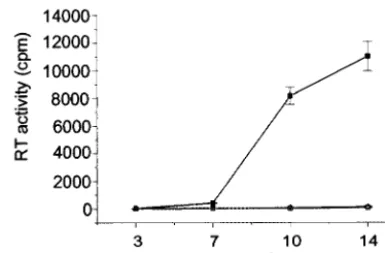
FIG. 2. Replication kinetics of wt HIV-1 and DIS stem-loop mutants in stimulated PBMCs (A) and T-cell line Sup T1 (B).
PHA-stimulated freshly isolated PBMCs or SupT1 cells were infected with either NL4.3 wt (
■
) or mutant (NL
GCGCGC[
⌬
] and NL
ACGCGT[
䊐
]) viruses.
Supernatants were collected 3, 7, 10, and 14 days after infection, and RT activity in each sample was measured. Results represent means and
standard deviations of triplicate samples and are representative of eight and six sets of experiments for PBMCs and Sup T1, respectively.
on November 8, 2019 by guest
http://jvi.asm.org/
isolated 36 h posttransfection. Briefly, supernatants were centrifuged for 30 min at 2,000⫻g, 4°C (Beckman) to remove cellular debris. The clarified supernatants were either frozen at⫺70°C or used immediately for further analysis. Cells were washed twice with 1⫻Tris-buffered saline (TBS) (50 mM Tris [pH 7.4], 150 mM NaCl) followed by protein extraction using lysis buffer containing 1⫻TBS, 10l of Nonidet P-40 per ml, 20 mM phenylmethylsulfonyl fluoride, 1M pepstatin, and 1M leupeptin.
Infectivity assay.PBMCs were isolated from buffy packs (supplied by the Red Cross Blood Bank, Melbourne) as described previously (9). PBMCs were then stimulated with phytohemagglutinin (PHA) (10g/ml; Murex Diagnostics) for 3 days and cultured in RPMI 1640 medium containing 10% fetal calf serum (RF10) and 10 units of interleukin-2 (Boehringer-Mannheim) per ml. SupT1 cells (kindly provided by Dale McPhee, The Burnet Institute, Australia) were cultured in RF10. Infectivity was tested on virus stocks obtained from either transfection or long-term infection of PBMCs (see below). Sample virus stocks with equivalent levels of RT activity, as determined by a micro-RT assay (13), were mixed with 105PBMCs or SupT1 cells in a 96-well tissue culture plate. Eight 10-fold serial dilutions of each virus were tested in triplicate. Supernatants were collected on days 3, 7, 10, and 14 postinfection and subsequently stored at ⫺70°C. Viral infectivity was measured by monitoring the production of viral RT activity by using a micro-RT assay (13). The infectivities of wt and mutant viral particles were quantified by using a 50% tissue culture infective dose (TCID50) method as previously described (9).
Measurement of virion RNA packaging and virion RNA dimer formation.
Pelleted wt and mutant virions derived from transfected 293T cells were nor-malized according to p24 levels (HIV-1 p24 antigen immunoassay; Abbott Lab-oratories). Virion RNAs were extracted as previously described (11, 12). Virion RNAs were electrophoretically separated on a denatured agarose gel to examine the impact of DIS mutations on virion RNA packaging (40). A dilution series of wt virion RNA was used to construct a standard curve to quantify the amount of genomic RNA being packaged into the mutant HIV-1 for the equivalent levels of virion-associated p24.
The stability and the conformation of virion RNA dimers isolated from wt and DIS mutant HIV-1 derived from both transfected 293T cells and infected PBMCs were assessed. PHA-stimulated PBMCs (2⫻107) were infected with wt and DIS mutant HIV-1 for 10 days as described below. Virion RNA was isolated, and a native RNA dimerization gel assay was utilized as previously described (11, 12). Briefly, dimeric RNAs were heated at 4, 25, 37, 42, 48, and 52°C for 10 min in an RNA dimerization buffer (10 mM Tris [pH 7.5], 1 mM EDTA, 25 mM NaCl), immediately followed by a quick chill on ice. The dimeric and monomeric RNAs were then separated by electrophoresis (18, 40–43). The electrophoreti-cally separated RNAs were then transferred onto nitrocellulose (Amersham) as previously described (18, 40–43). The virion RNA on the nitrocellulose was detected by use of a 150-nt in vitro-synthesized radioactive riboprobe (pGEM7zHIV-1), which is complementary to the R-U5 regions of the HIV-1 genomic RNA (NL4.3 RNA sequences nt 77 to 227) (18, 40–43).
Intracellular viral protein analysis.Cell lysates were rapidly freeze-thawed three times to weaken the cellular membrane. Cell debris was subsequently removed by centrifugation for 30 min at 4°C, 20,000⫻g. The transfection efficiency of the samples was determined by measuring the level of enhanced green fluorescent protein from the reporter plasmid by use of a Bio Imaging Analyser (Fuji Photo Film Co.). Cellular protein from each sample, normalized for equivalent levels of en-hanced green fluorescent protein, was mixed with 3l of sample buffer (100 mM Tris [pH 6.8], 3% sodium dodecyl sulfate [SDS], 33% glycerol, and 0.03% bromo-phenol blue), denatured for 10 min at 95°C, and resolved by SDS–10% polyacryl-amide gel electrophoresis (PAGE). Resolved proteins were transferred to a
nitro-cellulose membrane (Amersham). The membrane was blocked for 2 h in 3% casein dissolved in 1⫻TBS containing 0.3% Tween 20 (TBST) and probed overnight at 4°C with pooled HIV-1 seropositive patient sera. After three washes with 1⫻TBST buffer, the membrane was incubated with anti-human horseradish peroxidase-con-jugated secondary antibody (DAKO) for 2 h at room temperature. An enhanced chemiluminescence technique (Amersham) was used for visualization of HIV-1 proteins present in the cellular lysates.
Virion purification and protein analysis.Supernatants from transfected cells were purified and concentrated by ultracentrifugation through a 20% sucrose cushion by using a Beckman ultracentrifuge L-90 model (SW 41 rotor) at 100,000 ⫻gfor 1 h at 4°C. Pellets were resuspended in 50l of TBS lysis buffer. Equal amounts of virion protein (as determined by virion-associated p24) from each sample were mixed with 3l of sample buffer containing 5 mM -mercapto-ethanol, heated for 10 min at 95°C. Virion proteins were then resolved by SDS–10% PAGE as described above. The resolved virion protein samples were transferred onto nitrocellulose membranes by electrophoresis using a Bio-Rad transfer apparatus. Virion HIV-1 protein profiles of the samples were deter-mined by Western analysis as described above.
Long-term culture and sequencing analysis of the viral genome in the infected PBMCs.PHA-stimulated PBMCs were infected with wt and DIS mutant HIV-1 derived from transfected 293T cells. Virus stocks were treated with DNase to prevent contamination of plasmid DNA prior to the initial infection. PBMCs infected with wt and mutant HIV-1 were maintained in culture for 10 days. Cell-free culture fluids were then collected, and RT activities were compared. Cell pellets were collected and stored at⫺70°C for DNA sequencing. Collection of culture fluids and cell pellets at day 10 of infection marked the end of the first passage of wt and mutant HIV-1 in primary cells. A total of four passages in PBMCs were performed in parallel for wt and DIS mutant HIV-1. Equivalent amounts of viruses (as determined by the levels of RT activity) were used as input viruses in each of the four passages of PBMC infection. Similar levels of RT activities were detected in the culture fluids collected from wt and DIS mutant HIV-1-infected PBMCs within each passage. The resultant viral supernatants from each passage were tested for infectivity in SupT1 cells as described above. DNA was extracted from infected PBMCs by incubating cell pellets with 1⫻DNA extraction buffer (20 mM Tris-HCl [pH 8.0], 50 mM KCl, 0.45% NP-40, 0.45% Tween 20, and 60g of proteinase K/ml) at 37°C for 16 h. Heating at 95°C for 10 min subsequently inactivated the proteinase K in the samples. Viral DNA fragments containing nt 37 to 517, nt 947 to 1197, and nt 1387 to 1627 (numbering corresponds to the RNA sequences) were amplified via specific PCR primers pairs containing ApaI andEcoRI sequences. The three sets of primers were DIS/MA sense (5⬘CCC GAA TTC CTG AGC CTG GGA GCT CTC TGG C3⬘) and DIS/MA antisense (5⬘CCC GGG CCC ACG CGT CTA GCT CCC TGC TTG CCC3⬘) (set 1); CA sense (5⬘CCC GAA TTC GAG ACC ATC AAT GAG GAA GCT GCA GAA TGG GAT3⬘) and CA antisense (5⬘CCC GGG CCC ACG CGT TTT GGT CCT TGT CTT ATG TCC AGA ATG C3⬘) (set 2); and p2/NC sense (5⬘CCC GAA TTC AGG GAG TGG GGG GAC CCG GCC ATA AAG3⬘) and p2/NC antisense (5⬘CCC GGG CCC ACG CGT AGC CTG TCT CTC AGT ACA ATC TTT C3⬘) (set 3). The DIS stem-loop region and the selected matrix (MA), capsid (CA), p2 and NC coding sequences were monitored because compensatory mutations have been found in these regions after long-term culturing of partial DIS stem-loop deleted mutants in MT4 cells (25, 26). PCR products were digested with the restric-tion enzymesApaI and EcoRI and cloned into the pGEM7z vector for DNA sequencing. Sequencing was performed with an automating fluorescence DNA se-quencer (Applied Biosystems) at the Baker Institute, Melbourne, Australia. For each passage of a given wt or mutant virus, four to five separate clones were sequenced with each primer set.
NL
GCGCGC5,600
56
32,000
320,000
32
56
460,000
32,000
SupT1
NL4.3
32,000
3,200
3,200
1,800
180,000
320,000
NL
ACGCGT3.2
0.56
0.56
3.2
3.2
0.32
NL
GCGCGC3.2
3.2
0.56
3.2
5.6
0.56
aTCID
50was measured as described in Materials and Methods. cpm, counts per minute.
on November 8, 2019 by guest
http://jvi.asm.org/
[image:3.603.42.548.81.188.2]RESULTS AND DISCUSSION
The DIS stem-loop is not required for HIV-1 replication in
PHA-stimulated PBMCs.
Previous work has shown that the
DIS stem-loop is critical for HIV-1 replication in T-cell lines
(4, 8, 16, 23, 37), but the role of DIS in HIV-1 replication in
PBMCs
has
not
been
examined.
Two
DIS
mutants
(NL
GCGCGCand NL
ACGCGT) were used in this study.
NL
GCGCGChas the natural palindrome DIS sequence (5
⬘
GC
GCGC3
⬘
) in place of the 39-nt DIS stem-loop in NL4.3.
NL
ACGCGThas an arbitrary palindrome sequence, 5
⬘
ACGCG
T3
⬘
, in place of the 39-nt DIS stem-loop in NL4.3 (Fig. 1).
Mutant and wt HIV-1 were generated by transfecting the
in-dicated proviral DNAs into 293T cells. Parallel infections were
carried out by using the T-cell line SupT1 and PHA-stimulated
PBMCs. Mutant and wt HIV-1 collected from the supernatant
of the transfected 293T cells were normalized for RT activity
prior to infection. Equivalent amounts of wt and DIS mutant
virions were used to infect both PBMCs and SupT1 cells. The
replication kinetics and TCID
50of wt and DIS mutants were
determined through infections with PBMCs from eight
differ-ent donors and six independdiffer-ent SupT1 infections.
The HIV-1 DIS mutants were replication defective in SupT1
cells (Fig. 2B), which is consistent with reported data from
assays using T-cell lines (37). However, NL
GCGCGCand
NL
ACGCGTwere replication competent in PBMCs (Fig. 2A).
The TCID
50of each virus was measured to assess the relative
infectivity among wt (NL4.3) and the DIS mutants in both
PBMCs and Sup T1 cells (Table 1). Our data show that for
equivalent levels of RT activity the HIV-1 DIS mutants were
consistently replication competent in PBMCs from eight
do-nors (Table 1). In contrast, the parallel infection study in
SupT1 cells showed that the same NL
GCGCGCand NL
ACGCGTvirus stocks were approximately 1,000 to 10,000 times less
infectious than the wt HIV-1 (Table 1).
The DIS stem-loop is important for virion RNA packaging
and the formation of discrete RNA dimers.
Cell-free in vitro
RNA binding studies have shown that the DIS loop sequence
(Fig. 1, RNA nt 257 to 262) is critical for viral RNA
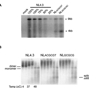
dimeriza-FIG. 3. (A) Mutations in DIS stem-loop inhibit virion packaging of genomic RNA. Virion particles were produced by transfecting the indicated
proviral DNA into 293T cells. Virion RNA samples that were normalized via a quantitative p24 assay were separated by electrophoresis on a
denatured agarose gel. A dilution series of wt virion RNA was used to construct a standard curve. The impact of mutations on virion RNA
packaging was visualized by Northern analysis and quantified by phosphorimaging. Results are representative of three sets of experiments. (B) The
conformation but not the stability of HIV-1 virion RNA dimers from transfected 293T cells was affected by mutations of the DIS stem-loop. Virion
RNA dimers were isolated from transfected 293T cells and prepared as described in Materials and Methods. Viral monomeric and dimeric RNA
species were separated on a 1% native agarose gel via electrophoresis after the samples were heated for 10 min at various temperatures (4, 25,
37, 42, 48, and 52°C, lanes 1 to 6, respectively). Viral RNAs were visualized by Northern analysis using a radioactive riboprobe that specifically
recognizes HIV-1 RNA. Results are representative of five sets of experiments.
on November 8, 2019 by guest
http://jvi.asm.org/
[image:4.603.126.465.62.396.2]tion (20, 44). Previous reports have shown that deletions
sim-ilar to those used in this study (NL
GCGCGCand NL
ACGCGT)
reduce the packaging of virion RNA and/or impair the
forma-tion of discrete RNA dimers (4, 8, 16, 23, 37). Genomic RNA
packaging and dimerization analyses were carried out with our
DIS mutants to verify the demonstrated impact of DIS
muta-tions on virion RNA genomes. The virion packaging of
genomic RNA in these DIS mutants was reduced to
approxi-mately 50% of that of the wt normalized for the same amount
of p24 proteins (Fig. 3A). While the amount of virion genomic
RNA packaged for both of the DIS mutants was reduced, there
was an increased level of 4-kb viral RNA compared to that of
the wild type (Fig. 3A). One possibility for this phenomenon is
that RNA packaging-deficient virus-like particles may
nonspe-cifically incorporate excess singly spliced viral RNA and/or
cellular RNA to compensate for the reduction of genomic
RNA packaging during retroviral assembly (28, 34). These data
also highlighted that reduction of genomic RNA packaging by
50% compared to the wt was sufficient for the replication of
HIV-1 DIS mutants in primary cells.
RNA dimerization analysis of wt and DIS mutant RNA
derived from transfected 293T cells demonstrated that while
the formation of discrete RNA dimers was impaired in each of
the DIS mutants, the stability of the virion RNA dimers, as
determined by the first appearance of the monomeric RNA
band after heating at 42°C, was not affected (Fig. 3B).
In-creased levels of spliced mRNA are visible in each of the DIS
mutants compared to the levels observed with the wt when
heated to 48 or 52°C, which is consistent with the increased
level of 4-kb viral RNA observed for the DIS mutants in the
RNA packaging analysis using a denaturing RNA gel. These
results suggest that the diffuse RNA dimer may, at least in part,
be a consequence of the spliced RNA that is packaged within
the DIS mutant virions. To determine whether the
conforma-tion of the DIS mutant RNA dimers was rescued following
successful infection of PBMCs, RNA dimerization analysis was
carried out with virion RNA isolated after wt and DIS mutants
had been used to infect PBMCs for 10 days (Fig. 4). Again, the
formation of discrete RNA dimers was impaired in each of the
DIS mutants while the stability of the virion RNA dimers was
not altered. The finding that the conformation but not the
stability of the RNA dimers was affected by DIS mutations is in
agreement with the hypothesis that other non-DIS stem-loop
RNA sequences are involved in the process of HIV-1 RNA
dimer formation (1, 4), and retroviral RNA dimerization may
rely on multiple segments of the retroviral RNA genomes (29,
36, 39).
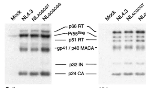
Mutations in the HIV-1 DIS stem-loop structure do not
alter the processing of viral proteins or yield compensatory
mutations in p2 and NC sequences in the viral genome of the
HIV-1-infected PBMCs.
It has been reported that the DIS
deletion is associated with defects in precursor protein
pro-cessing and this propro-cessing defect can be rescued by
compen-satory mutations in p2 and NC sequences (25). Furthermore,
compensatory mutations in the matrix and capsid can also
rescue the defects of viral infectivity in DIS deletion mutants
(26). Western blot analysis of intracellular viral protein (Fig. 5)
and pelleted virion proteins from 293T cells transfected with
the various DIS mutants showed that the virion protein profiles
were similar to those of wt virus (Fig. 5), suggesting that our
DIS mutations did not influence the processing of viral
pro-teins.
DNA sequencing was performed for wt- and DIS mutant
(NL
GCGCGCand NL
ACGCGT)-infected PBMC cultures 40 days
postinfection in three discrete experiments. Four to five clones
were sequenced for each sample, and in all cases the sequences
of the clones were identical. Previous studies have reported
that compensatory mutations are required to rescue the
infec-tivity of DIS-deleted HIV-1 mutants in T cells (25, 26); no such
mutation was found in the corresponding MA, CA, p2, and NC
sequences in the viral genome of NL
GCGCGC- and NL
ACGCGT-infected primary cells. The respective DIS deletions were
maintained in the NL
GCGCGC- and NL
ACGCGT-infected
PBMC cultures 40 days postinfection (data not shown). DNA
sequencing (as described in Materials and Methods) revealed
no changes to the DNA sequence when compared to the input
viruses. To rule out the possibility of contamination with a
replication-competent
virus
in
the
NL
GCGCGC-
and
NL
ACGCGT-infected PBMC cultures, replication kinetics were
[image:5.603.44.278.71.144.2]determined for SupT1 cells by using supernatant collected
FIG. 4. The conformation but not the stability of HIV-1 virion
RNA dimers isolated from PBMCs was affected by mutations of the
DIS stem-loop. Virion RNA dimers were isolated from infected
PBMCs and prepared as described in Materials and Methods. Viral
monomeric and dimeric RNA species were separated on a 1% native
agarose gel by electrophoresis after the samples had been heated for
10 min at various temperatures (4, 25, 37, 42, 48, and 52°C, lanes 1 to
6, respectively). Viral RNAs were visualized by Northern analysis with
a radioactive riboprobe that specifically recognizes HIV-1 RNA.
Re-sults are representative of three sets of experiments.
FIG. 5. Mutations in the HIV-1 DIS stem-loop do not alter the
intracellular viral protein and virion protein profile. Intracellular
(293T cells) and virion proteins were resolved by SDS–10% PAGE.
The viral proteins were visualized by Western blotting using pooled
HIV-1-positive patient sera and anti-human horseradish
peroxidase-conjugated secondary antibody. Results are representative of five sets
of experiments.
on November 8, 2019 by guest
http://jvi.asm.org/
[image:5.603.309.544.72.214.2]from each passage of two sets of the long-term infections (eight
in total). Although replication competent in PBMCs, viruses
present in the supernatants of NL
GCGCGC- and NL
ACGCGT-infected PBMC cultures were replication defective in SupT1
cells (Fig. 6), which indicates that there was no contamination.
These results correspond with the DNA sequencing data
show-ing that the sequences of the NL
GCGCGCand NL
ACGCGTinput
viruses were unaltered after 40 days in PBMCs. Consequently,
our data show that there is no strong selective pressure for a wt
DIS stem-loop sequence to support HIV-1 replication in
PB-MCs.
In this study, we have demonstrated that the requirement of
the HIV-1 DIS stem-loop in virus replication is cell type
de-pendent. The finding that the DIS deletion mutants cannot
replicate in SupT1 cells while the DIS is largely dispensable for
replication in PBMCs suggests the involvement of a
DIS-de-pendent cellular factor. Our data also demonstrate that wt
levels of genomic RNA packaging are not critical for HIV-1
replication in primary cells.
Paillart et al. (37) have shown that the DIS stem-loop is
important for the synthesis of cDNA during reverse
transcrip-tion in the T-cell line SupT1. The cell type-dependent effects of
DIS deletions on HIV-1 replication suggest that this
DIS-dependent cellular factor may directly or indirectly bind to the
DIS stem-loop to enhance HIV-1 replication, perhaps by
as-sisting in the synthesis of cDNA at the early stage of the HIV-1
replication cycle. However, since the DIS stem-loop is part of
the complex RNA structure in the 5
⬘
untranslated region,
which is important at multiple stages of HIV-1 replication, the
impact of DIS mutations on other aspects of HIV-1
replica-tion, such as RNA splicing and the regulation of protein
trans-lation, should also be considered.
Using an in vitro system, Berkhout et al. (3, 5) and Huthoff
and Berkhout (19) have shown that the HIV-1 5
⬘
leader RNA
sequences assume different conformations at various stages of
viral replication. It has been suggested that the conformation
of the 5
⬘
leader RNA may play a part in regulating viral
replication (2). Fu et al. have previously shown that the HIV-1
dimeric virion RNA genome assumes two distinct
conforma-tions before and after virion particle maturation (11). Distinct
conformations of RNA dimers can also be found in other
retroviruses (12, 35, 36, 45). We have recently shown that in
addition to these two conformations of RNA dimers, HIV-1
virion RNA can assume a number of different dimeric
confor-mations depending on the presence or the absence of HIV-1
PR, RT, and IN (41). These data support the notion that the
reverse-transcription and dimerization reactions may be
cou-pled through conformational changes within the leader RNA
(2). Our data also support the suggestion that in addition to the
ascribed roles of genomic RNA packaging and the formation
of discrete RNA dimers, the DIS stem-loop is also involved in
other aspects of HIV-1 replication.
ACKNOWLEDGMENTS
We thank John Mills for helpful criticism and review of the
manu-script.
J. Mak is a recipient of an NHMRC research grant and a Monash
Logan fellowship. M. K. Hill is a recipient of a Burnet Centenary
postdoctoral fellowship and an amfAR postdoctoral fellowship. M.
Shehu-Xhilaga was a recipient of the NHMRC Ph.D. training
schol-arship and is a recipient of the NHMRC postdoctoral fellowship. S. M.
Campbell is a recipient of the NHMRC Ph.D. training scholarship. P.
Poumbourios is supported by NHMRC. S. M. Crowe is supported by
a grant from the Australian Council on HIV, AIDS and Related
Diseases through the (Australian) National Centre in HIV Virology
Research and the BI Research Fund. This work is also supported in
part by grants to J. Mak from the Clive and Vera Ramaciotti
Foun-dation, the Honda FounFoun-dation, and the Cecilia Kilkeary Foundation.
REFERENCES
1. Berkhout, B.1996. Structure and function of the human immunodeficiency virus leader RNA. Prog. Nucleic Acid Res. Mol. Biol.54:1–34.
2. Berkhout, B., M. Ooms, N. Beerens, H. Huthoff, E. Southern, and K. Ver-hoef.2002. In vitro evidence that the untranslated leader of the HIV-1 genome is an RNA checkpoint that regulates multiple functions through conformational changes. J. Biol. Chem.277:19967–19975.
3. Berkhout, B., and J. L. B. Van Wamel.2000. The leader of the HIV-1 RNA genome forms a compactly folded tertiary structure. RNA6:282–295. 4. Berkhout, B., and J. L. B. van Wamel.1996. Role of the DIS hairpin in
replication of human immunodeficiency virus type 1. J. Virol.70:6723–6732. 5. Berkhout, B., N. L. Vastenhouw, B. I. Klasens, and H. Huthoff.2001. Struc-tural features in the HIV-1 repeat region facilitate strand transfer during reverse transcription. RNA7:1097–1114.
6. Berkowitz, R., J. Fisher, and S. P. Goff.1996. RNA packaging, p. 177–218.In H.-G. Kra¨usslich (ed.), Current topics in microbiology and immunology: morphogenesis and maturation of retroviruses, vol. 214. Springer, Heidel-berg, Germany.
7. Cheung, K. S., R. E. Smith, M. P. Stone, and W. K. Joklik.1972. Comparison of immature (rapid harvest) and mature Rous sarcoma virus particles. Vi-rology50:851–864.
8. Clever, J. L., and T. G. Parslow.1997. Mutant human immunodeficiency virus type 1 genomes with defects in RNA dimerization or encapsidation. J. Virol.71:3407–3414.
9. Crowe, S. M., N. J. Vardaxis, S. J. Kent, A. L. Maerz, M. J. Hewish, M. S. McGrath, and J. Mills.1994. HIV infection of monocyte-derived macro-phages in vitro reduces phagocytosis of Candida albicans. J. Leukoc. Biol.
56:318–327.
10. Dupraz, P., S. Oertle, C. Me´ric, P. Damay, and P.-F. Spahr.1990. Point mutations in the proximal Cys-His box of Rous sarcoma virus nucleocapsid protein. J. Virol.64:4978–4987.
11. Fu, W., R. J. Gorelick, and A. Rein.1994. Characterization of human im-munodeficiency virus type 1 dimeric RNA from wild-type and protease-defective virions. J. Virol.68:5013–5018.
12. Fu, W., and A. Rein.1993. Maturation of dimeric viral RNA of Moloney murine leukemia virus. J. Virol.67:5443–5449.
13. Goff, S., P. Traktman, and D. Baltimore.1981. Isolation and properties of Moloney murine leukemia virus mutants: use of a rapid assay for release of virion reverse transcriptase. J. Virol.1:239–248.
14. Gorelick, R. J., L. E. Henderson, J. P. Hanser, and A. Rein.1988. Point mutants of Moloney murine leukemia virus that fail to package viral RNA: evidence for specific RNA recognition by a “zinc finger-like” protein se-quence. Proc. Natl. Acad. Sci. USA85:8420–8424.
[image:6.603.63.256.70.196.2]15. Gorelick, R. J., J. S. M. Nigida, J. J. W. Bess, L. O. Arthur, L. E. Henderson,
FIG. 6. Replication kinetics of wt HIV-1 and DIS stem-loop
mu-tants passaged through PBMCs in Sup T1 cells. SupT1 cells were
infected with either NL4.3 wt (
■
) or mutant viruses (NL
GCGCGC[
⌬
]
and NL
ACGCGT[
䊐
]) that had been passaged through PBMCs.
Super-natants were collected 3, 7, 10, and 14 days after infection, and RT
activity in each sample was measured. Results represent means and
standard deviations of triplicate samples and are representative of
eight sets of experiments.
on November 8, 2019 by guest
http://jvi.asm.org/
protein processing, and genomic RNA dimer stability. J. Virol.76:11245– 11253.
19. Huthoff, H., and B. Berkhout.2001. Two alternating structures of the HIV-1 leader RNA. RNA7:143–157.
20. Laughrea, M., and L. Jette.1994. A 19-nucleotide sequence upstream of the 5⬘major splice donor is part of the dimerization domain of human immu-nodeficiency virus 1 genomic RNA. Biochemistry33:13464–13474. 21. Laughrea, M., and L. Jette.1997. HIV-1 genome dimerization: kissing-loop
hairpin dictates whether nucleotides downstream of the 5⬘splice junction contribute to loose and tight dimerization of human immunodeficiency virus RNA. Biochemistry36:9501–9508.
22. Laughrea, M., and L. Jette´.1996. Kissing-loop model of HIV-1 genomic dimerization: HIV-1 RNAs can assume alternative dimeric forms, and all sequences upstream or downstream of hairpin 248–271 are dispensable for dimeric formation. Biochemistry35:1589–1598.
23. Laughrea, M., L. Jette, J. Mak, L. Kleiman, C. Liang, and M. A. Wainberg.
1997. Mutations in the kissing loop hairpin of human immunodeficiency virus type 1 reduce viral infectivity as well as genomic RNA packaging and dimer-ization. J. Virol.71:3397–3406.
24. Liang, C., L. Rong, E. Cherry, L. Kleiman, M. Laughrea, and M. A. Wain-berg.1999. Deletion mutagenesis within the dimerization initiation site of human immunodeficiency virus type 1 results in delayed processing of the p2 peptide from precursor proteins. J. Virol.73:6147–6151.
25. Liang, C., L. Rong, M. Laughrea, L. Kleiman, and M. A. Wainberg.1998. Compensatory point mutations in the human immunodeficiency virus type 1 Gag region that are distal from deletion mutations in the dimerization initiation site can restore viral replication. J. Virol.72:6629–6636. 26. Liang, C., L. Rong, Y. Quan, M. Laughrea, L. Kleiman, and M. A. Wainberg.
1999. Mutations within four distinct Gag proteins are required to restore replication of human immunodeficiency virus type 1 after deletion mutagen-esis within the dimerization initiation site. J. Virol.73:7014–7020. 27. Linial, M. L., and A. D. Miller.1990. Retroviral RNA packaging: sequence
requirements and implications. Curr. Top. Microbiol. Immunol.157:125– 152.
28. Luban, J., and S. P. Goff.1994. Mutational analysis ofcis-acting packaging signals in human immunodeficiency virus type 1 RNA. J. Virol.68:3784– 3793.
29. Ly, H., and T. G. Parslow.2002. Bipartite signal for genomic RNA dimer-ization in Moloney murine leukemia virus. J. Virol.76:3135–3144. 30. Mak, J., M. Jiang, M. A. Wainberg, M.-L. Hammarskjold, D. Rekosh, and L.
Kleiman.1994. Role of Pr160gag-pol
in mediating the selective incorporation of tRNALys
into human immunodeficiency virus type 1 particles. J. Virol.
68:2065–2072.
31. Me´ric, C., and S. P. Goff.1989. Characterization of Moloney murine leuke-mia virus mutants with single-amino-acid substitutions in the Cys-His box of the nucleocapsid protein. J. Virol.63:1558–1568.
packaging and maturation of Rous sarcoma virus genomic RNA. J. Virol.
64:5757–5763.
36. Ortiz-Conde, B. A., and S. H. Hughes.1999. Studies of the genomic RNA of leukosis viruses: implications for RNA dimerization. J. Virol.73:7165–7174. 37. Paillart, J. C., L. Berthoux, M. Ottmann, J. L. Darlix, R. Marquet, B. Ehresmann, and C. Ehresmann.1996. A dual role of the putative RNA dimerization initiation site of human immunodeficiency virus type 1 in genomic RNA packaging and proviral DNA synthesis. J. Virol.70:8348– 8354.
38. Rein, A.1994. Retroviral RNA packaging: a review. Arch. Virol. Suppl.
9:513–522.
39. Sakuragi, J.-I., and A. T. Panganiban.1997. Human immunodeficiency virus type 1 RNA outside the primary encapsidation and dimer linkage region affects RNA dimer stability in vitro. J. Virol.71:3250–3254.
40. Shehu-Xhilaga, M., S. M. Crowe, and J. Mak.2001. Maintenance of the Gag/Gag-Pol ratio is important for human immunodeficiency virus type 1 RNA dimerization and viral infectivity. J. Virol.75:1834–1841.
41. Shehu-Xhilaga, M., M. K. Hill, J. Marshall, J. Kappes, S. M. Crowe, and J. Mak.2002. The conformation of the mature dimeric human immunodefi-ciency virus type 1 RNA genome requires packaging of Pol protein. J. Virol.
76:4331–4340.
42. Shehu-Xhilaga, M., H. G. Kraeusslich, S. Pettit, R. Swanstrom, J. Y. Lee, J. A. Marshall, S. M. Crowe, and J. Mak.2001. Proteolytic processing of the p2/nucleocapsid cleavage site is critical for human immunodeficiency virus type1 RNA dimer maturation. J. Virol.75:9156–9164.
43. Shehu-Xhilaga, M., J.-Y. Lee, S. M. Campbell, J. A. Marshall, S. M. Crowe, and J. Mak.2002. Overexpression and incorporation of GagPol precursor does not impede packaging of HIV-1 tRNA(Lys3) but promotes intracellular budding of virus-like particles. J. Biomed. Sci.9:697–705.
44. Skripkin, E., J.-C. Paillart, R. Marquet, B. Ehresmann, and C. Ehresmann.
1994. Identification of the primary site of the human immunodeficiency virus type 1 RNA dimerization. Proc. Natl. Acad. Sci. USA91:4945–4949. 45. Stewart, L., G. Schatz, and V. M. Vogt.1990. Properties of avian retrovirus
particles defective in viral protease. J. Virol.64:5076–5092.
46. St. Louis, D. C., D. Gotte, E. Sanders-Buell, D. W. Ritchey, M. O. Salminen, J. K. Carr, and F. E. McCutchan.1998. Infectious molecular clones with the nonhomologous dimer initiation sequences found in different subtypes of human immunodeficiency virus type 1 can recombine and initiate a spread-ing infection in vitro. J. Virol.72:3991–3998.
47. Stoltzfus, C. M., and P. N. Snyder.1975. Structure of B77 sarcoma virus RNA: stabilization of RNA after packaging. J. Virol.64:1161–1170. 48. Swanstrom, R., and J. W. Wills.1997. Retroviral gene expression. II.
Syn-thesis, processing, and assembly of viral proteins, p. 263–334.InJ. M. Coffin, S. H. Hughes, and H. E. Varmus (ed.), Retroviruses. Cold Spring Harbor Laboratory Press, Cold Spring Harbor, N.Y.
on November 8, 2019 by guest
http://jvi.asm.org/