Copyright © 1977 AmericanSocietyforMicrobiology Printed inU.S.A.
Expression
of
an
Early,
Nonstructural
Antigen of Herpes
Simplex Virus in Cells Transformed In
Vitro
by Herpes
Simplex
Virus
V. L. FLANNERY, R. J. COURTNEY, AND P. A. SCHAFFER*
DepartmentofVirology andEpidemiology, Baylor College of Medicine, Houston,Texas77030
Received for publication 26 July 1976
Hyperimmune rabbit antiserum to an early, nonstructural herpes simplex virus type 2 (HSV-2)-induced polypeptide (VP143) reacted in immunofluores-cence tests with a variety of cell lines transformed by HSV-2. Cytoplasmic fluorescence was observedin 10 to50%ofHSV-2-transformedcells, whereasno fluorescence wasobservedincells transformed by other oncogenic DNA viruses orby achemical carcinogen. VP143-specific reactivity could be adsorbed from
anti-VP143serumwith HSV-2-transformed cells butnotwith cells transformed
by other agents. When HSV-2-transformed cellsweresynchronized in mitosis
and examinedatvarioustimespostmitosisforVP143-specific fluorescence, the
expressionof VP143wasshowntobe cellcycle dependent.
Herpes simplex virus (HSV) has been sero-logically associated with human malignancy (24, 26). The demonstration of in vitro onco-genictransformation of mammalian cells with HSV types 1 (6)and2(5, 16)represents
signifi-cant supportive evidence for the in vivo
onco-genicpotential ofthese viruses.
Definitive proofthat transformed cells con-tainviral information liesinthe demonstration
of viral DNA or its products, viral RNA, and
virus-specificproteins. Viral RNA (2)and,more
recently, viral DNA (4, 8, 17, 22) have been
identified by molecular hybridization tech-niques in cells transforned in vitro by HSV
type2. Virus-specific proteinshave been
dem-onstrated inHSV-transformed cellsbya
num-ber of techniques. (i) HSV-specific antigens
have been detected by immunofluorescence testsusinghyperimmuneantiseratoHSV (1, 5, 6, 16, 18-20, 27, 30). (ii) Antibodies whichreact with HSVantigensinneutralization tests have
beendemonstratedintheseraof animals
bear-ing tumors induced by HSV-transformed cells
(1, 5, 16). (iii) The presence of HSV-specific
thymidine kinase (TK) has been demonstrated
inTK- mouseLcellsafterinvitro transforma-tion with HSV (23). (iv) Complementation of HSV type 2 temperature-sensitive mutants in
HSVtype2-transformedcells at the
nonpermis-sive temperature has been demonstrated (15, 21). Although viral genetic information has been showntobepresentinHSV-transformed cells, the identity of the specific viral genes involved in the induction and maintenance of transformation is currently not known.
Clearly, studies of viral gene expression in
transformed cells would benefit greatly from
theuseoftechniques that are capable of detect-ing individual viral gene products in these
cells. Of the four methods just described for
detectingviral proteins, only the latter two are
capable of detecting the product of a single viral gene.
Courtney and Benyesh-Melnick (3) have
re-centlydemonstratedtheusefulness of hyperim-mune antiserum prepared against an
individ-ual HSV-specificpolypeptide (VP175) for
stud-ies of the expression of this polypeptide in vi-rus-infected cells. The use of this antiserum in indirect immunofluorescence tests has facili-tated studiesof thekineticsof synthesisandthe intracellular location of an individual viral
geneproductinvirus-infected cells.The
poten-tial usefulness ofsuchpolypeptide-specific anti-serum instudiesofHSV-transformed cellswas first reported by Kimura et al. (16). In
Jhese
studies hyperimmune antiserum prepared
against anonstructural HSV-specific
polypep-tide (VP143)synthesized earlyinthe infectious
cycle was used todemonstrate the presence of VP143 in fixed preparations of HSV-trans-formed cells.
In the present report we have attempted to furthercharacterize the expression of VP143 in HSV-transformed cells with regard to the sensi-tivity and specificity of the reaction and as a function of the stage of the cell cycle during whichmaximum expression occurs.
MATERIALS AND METHODS
Cellsand cell culture. Primary hamsterembryo fibroblasts were obtained from inbred Lakeview 284
on November 10, 2019 by guest
http://jvi.asm.org/
Syrian hamster embryos (strain LSH) inthe thir-teenthday of gestation (Lakeview Hamster Colony, Newfield, N.J.). Cultures were used in low passage (passages1to5, LSHlp)andhighpassage (passages 39to 86, LSH hp). An HSV type 2 (HSV-2)-trans-formed LSH tumor cell line (line 333-8-9 Tu) was kindly provided by F. Rapp and R. Duff (Milton S. Hershey Medical Center, Hershey, Pa.). The cell line was received in high passage (>100). It was passed one additional time in hamsters and four times in cell culturebefore use in thisstudy. Addi-tional transformed LSH cell lines and tumor (Tu) cell lines derived from them werekindly supplied by S. Tevethia (Tufts University School ofMedicine, Boston, Mass.). These lines include the following: LSH-DMBA Tu (passages 8-15), LSH-SV40 Tu (pas-sages 6-27), LSH-Adl2 (passage 3), and LSH-Ad7 Tu(passage 9) transformed by dimethyl-benzanthra-cene(DMBA), simian virus type40(SV40), adenovi-rus 12,andadenovirus 7, respectively. A laboratory strainof BHK-21cellsinpassage 30wasalso used in this study. Allcellswere grown inEagle medium supplemented with 10% fetal bovine serum and 0.075% NaHCO3 for cultures in closed vessels and 0.225%NaHCO3 forcultures in openvessels.
Preparation of antiserum. Two types of antisera were used in these studies: hyperimmune rabbit antiserum to HSV-2 was prepared as describedby Esparza et al. (7) and antiserum to VP143 was pre-paredas described by Courtney and Benyesh-Mel-nick (3).Itshould be noted that thispolypeptidewas previously designated as VP134based on the molec-ular weightdetermination of HSV type 1 polypep-tides. The corresponding polypeptidein HSV-2-in-fectedcells hasa slightly higher molecularweight and will be subsequently designated as VP143. Briefly, the latter antiserum was prepared as fol-lows. The nuclear fraction of HSV-2 (strain 186)-infected human embryonic lung fibroblasts har-vested24 h postinfection was subjected to sodium dodecylsulfate preparativepolyacrylamide gel elec-trophoresis. Polypeptide VP143 was removed from peak fractions and further purified by two addi-tional runs onanalytical cylindrical gels. Rabbits wereimmunized withgel slicescontainingVP143as previouslydescribed (3).
Immunofluorescencetest. The indirect immuno-fluorescence (IF)testdescribedbyPorteretal. (25) was employed with minor modifications. Approxi-mately 5x 10⁴cells wereseededoncoverslips.After incubation for 6 h at 37°C, cover slipswerewashed threetimesinTris, pH7.4,dried for 25 minat room temperature, and fixed in coldacetone (4°C) for15 min. Beforestaining, cellswererehydratedwith 0.1 ml ofTris for 5 minat roomtemperature.Cellswere then treated for 30min at roomtemperature with either antisera orpreimmune rabbitserumdiluted 1:2 in Tris. Cover slips were then washed three times inTris and treated for30 min with fluores-cein-conjugated goat anti-rabbit gamma globulin (Hyland, Div. of TravenolLaboratories, Inc., Costa Mesa, Calif.). Coverslipswerewashed three times in Tris andonce indistilled water, airdried, and mountedinElvanol.Control, infected cultureswere prepared byinoculating primary LSH cellson cover slipswith HSV-2 (strain 333) ata multiplicityof5
PFU/cell. Aftera4-hincubationat37°C,coverslips wereprocessedandstainedasdescribedabove.
Absorption technique. LSH hp, LSH-SV40 Tu,
and 333-8-9 Tu cells were seeded in 100-mm petri dishesto containapproximately 107cells per dish. After6 h, cells in three dishes were harvested by scrapingintothemedium andpelleting. Pellets con-taining approximately 3 x 10⁷ cells were washed three times incold Tris andresuspended in 0.4 ml of undiluted rabbit anti-VP143 serum. Suspensions were then subjected to sonic oscillation in a Ray-theon sonicoscillator at 10 kc for two 30-sintervals. Suspensions were shaken gently in a water bath at 37°C for 1 h and centrifuged at 100,000 x g for 20 min. Supernatant fluids were removed, diluted 1:2 inTris to yield a total of 0.8 ml, and absorbed a second time for 1 hat 37°C and overnight at 4°C with cell pellets containing 6.0 x 10⁷ cells. Suspensions were then spun at 100,000 x g for 20 min, and absorbed sera were tested forVP143 reactivity by IF.
VP143 expression in synchronized 333-8-9 Tu cells. (i)Determination of celldoubling time. Ap-proximately2 x 105cellsfrom asynchronous 333-8-9 Tucultures were used to seed 60-mm petri dishes. At 6-h intervals postseeding, duplicate monolayers were trypsinized, and cells were counted until the number of cells had doubled.
(ii) Preparation of synchronized cell cultures. Suspensions of synchronous cells were preparedby the method ofTerashima and Tolmach (31). This method isbased upon the observation that cells in mitosis adhere lessstrongly to the culture substrate and canthereforeeasily be detached andselectively removed from the monolayer. Therefore, plastic flasks (150 cm2) were seeded with 107 cells, and the cultures were incubated at 37°C for an interval equivalent to onedoubling time. At this time mi-totic cells weredetached fromthe vessel surfaceby vigorous shaking andpelletedby low-speed centrif-ugation.Pellets were held in anice bath overnight, counted, diluted, and seeded inculture dishes the followingmorning.Examination of cell suspensions atthe time ofseeding revealed that approximately 98% of cells were viable and that the nuclei of greater than 90% of cells exhibited mitoticfigures.
(iii)Expression ofVP143after synchronization. Approximately4x 104cellssynchronizedinmitosis were seeded on cover slips. At designated times postseeding, coverslipswere processed and exam-ined for IF reactivity using antiserum to VP143.
DeterminationofS-phaseinsynchronized 333-8-9Tu cells. Sixty-millimeter petri dishes were seeded with 5x 105333-8-9Tucellscollectedin mitosis. At intervals postseeding, 10 j,Ciof [3H]thymidineper ml (60Ci/mM; Schwarz Bio Research,Inc., Orange-burg, N.Y.)wasaddedtoeachplate. After30min, cells wereharvested byscraping into the medium, pelleted,washedoncewith2mlof cold TNE (0.01 M Tris-hydrochloride, 0.1MNaCl,and0.001 MEDTA, pH 7.4), and repelleted. Cell pellets were resus-pendedin 3ml of TNE medium andwere lysed by treatment with0.08ml of10%sodiumlauryl sarco-sinatefor10min at roomtemperature.Suspensions were deproteinized by treatment with 0.06 ml of Pronase (1mg/ml)at37°C for30min.One-tenth
mil-285
on November 10, 2019 by guest
http://jvi.asm.org/
liliter of each suspension was placed on a Whatman GF/A filter disk. Disks were then dried, washed oncefor 5 min incold (4°C)5%trichloroaceticacid, washed twice in cold (4°C) water, redried, and placed inscintillation vials. Ten milliliters of scin-tillationfluid was added to each vial, and samples were counted in a Beckman LS250 liquid scintilla-tioncounter.
RESULTS
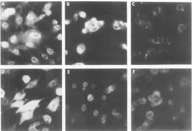
Patterns of VP143-specific fluorescence in HSV-infected and HSV-transformed cells. Typical patterns of fluorescence staining ob-served in HSV-2-infected and -transformed LSH cells treated with antisera to VP143 and to HSV-2 are shown in Fig. 1. When treated with anti-VP143 serum, HSV-2-infected cells ex-hibited predominantly nuclear fluorescence (Fig. 1A). In contrast, 10 to 50% of HSV-2-transformed cells exhibited distinct cytoplas-mic, predominantly perinuclear fluorescence (Fig. 1B). Whereas diffuse cytoplasmic and nu-clear fluorescence was observed in HSV-2 in-fected cells treated with anti-HSV-2 serum (Fig. 1D), no specific fluorescence was observed inHSV-2-transformed 333-8-9 Tu cells treated withthisantiserum (Fig. 1E). Thus, while
anti-serum to HSV-2failedtoproduce HSV-specific
fluorescence inHSV-2-transformed cells, a dis-tinct pattern of fluorescence was observed in
cells treated with antiserum to VP143.
Al-thoughinternal fluorescence was notobserved
in fixed preparations of HSV-2-transformed
cells using anti-HSV-2 serum, virus-specific
membrane fluorescence was observed in un-fixed preparations of these cells using anti-HSV-2 serum (16).
Speciflcity of the reaction. (i) Reaction of
anti-VP143 serum with LSH cells
trans-formedby otheragents.To test thespecificity
ofthe VP143 reaction, four HSV-2-transformed LSH cell lines and four LSH cell lines
trans-formedbyother agentsweretestedfor
reactiv-ity to VP143 antiserum (Table 1). A positive
cytoplasmic, predominantly perinuclear
reac-tion was observed in the four
HSV-2-trans-formed cell lines; no reaction was observed in
cellstransformedbySV40(Fig. 1C), adenovirus 7, adenovirus 12,ora knownchemical
carcino-gen (dimethylbenzanthracene). No
VP143-spe-cific reactivity was observed in either low- or high-passageLSH cells orinacommon
labora-tory line of hamstercells,BHK-21 (notshown).
FIG. 1. Patternsof IF staining observed in HSV-2-infected, virus-transformed and normal hamster cells aftertreatment with anti-VP143 andanti-HSV-2 sera. (A) HSV-2 infected cells stained with anti-VP143 serum 4hpostinfection. (B) 333-8-9Tucells stained with anti-VP143serum.(C) LSH-SV40 Tu cells stained with anti-VP143serum.(D) HSV-2-infected cells stained with anti-HSV-2serum 4hpostinfection. (E) 333-8-9Tucellsstained with anti-HSV-2serum. (F) 333-8-9 Tu cells stainedwith preimmune rabbit serum.
on November 10, 2019 by guest
http://jvi.asm.org/
[image:3.505.72.466.342.610.2](ii)Absorption experiments. Inaneffort to
further demonstrate the specificity of the VP143 reaction, antiserum to VP143 was
ab-sorbed with 333-8-9 Tu,LSH-SV40 Tu, and LSH hpcells and then tested for HSV-specific
reac-tivity intransformed cells. Typicalpatternsof fluorescent staining before and after absorption
are shown in Fig. 2. The reactivity of
anti-VP143 serum with 333-8-9 Tu cells (Fig. 2A)
was eliminated after absorption with
homolo-gous cells (Fig. 2B); however, onlyaslight de-creaseintheintensity of stainingwasobserved
afterantiserumwas adsorbed with LSH SV40
Tu (Fig. 2C)orwith LSH hp (Fig.2D).
Expression ofVP143 in synchronized 333-8-9 Tu cells. Preliminary studies had indicated that the expression of the antigen in HSV-transformed 333-8-9 Tu cells which reacted with antiseratoVP143wascell cycle dependent: the
intensity of staining in cellswasobservedtobe
greater 6 h after trypsinization and seeding than after12 to 18h.To examine theexpression of VP143 as a function of the stageof the cell
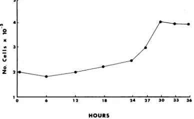
cycle, it was first necessary to determine the doubling time of 333-8-9 Tu cells. The number of 333-8-9 Tu cells doubled within 30 h after seeding (Fig. 3). Therefore, 333-8-9 Tu cells
werecollectedinmitosis, seededon coverslips,
and examinedatvarious timespostseeding for HSV-specific fluorescence. The results of this studyaresumnnarizedinFig.4.The maximum
number of cellswhich exhibited fluorescencein the30-h testperiod appearedtooccur6 h
post-seedingwhen 90% of cells werepositive. Most
cells were observed to be in pairs, indicating
recent mitosis. In cells tested at 6 h,
fluores-cence was localized to the perinuclear region
(ring-forms), and large, round, perinuclear
ac-cumulationswereprominent. By12h
postseed-ing thenumber ofcellsexhibitingfluorescence
was reduced (50 to 75%). Furthermore, the
staining reactionwas alsoslightlylessintense
and somewhat more diffuse inthe cytoplasm. The intensity ofcytoplasmic fluorescence seen
at 18 h postseedingwas greatly reduced. The
reduced intensity of the reaction made quanti-tationofIF-positivecellsmoredifficult and less
reliable. By 24 h perinuclear accumulations and ring-forms were again prominent in
ap-proximately 50% ofcells, andstaininghad
be-come more intense. By 24 to 30 h postmitosis
perinuclear accumulations predominated, and
many cells had begun to divide. Since the
in-tensity and localization of the reaction varied greatlyfrom celltocellby30h,itappearedasif mostcellswere no longer insynchrony atthis
time.
[image:4.505.250.442.89.246.2]Relationship between the expression of VP143 and DNA synthesis in synchronized
TABLE 1. Reaction of anti-VP143 serum with LSH cells transformed by HSV-2 and other agents
Cell type Reaction with VP143antiserum HSV-2-infected LSHa + (Nuclear)
333-8-9Tu (333)b + (Cytoplasmic and perinu-clear)
U-15 Tu (186) +(Cytoplasmic and perinu-clear)
U4V-8Tu (333) +(Cytoplasmic and perinu-clear)
B20V-9 Tu (333) + (Cytoplasmic and perinu-clear)
LSH-SV40 Tu
-LSH-Ad7 Tu
-LSH-Adl2 Tu
-LSH-DMBA
-LSH lp
-LSH hp
-BHK-21
-aLSHlp cells wereinfected with HSV-2 strain 333 and
harvested 4 h postinfection.
bNumbers in parentheses indicate the strain of HSV-2 used to inducetransformation. Lines U-15Tu, U4V-8Tu, andB20V-9 Tu have been described previously (16).
333-8-9 Tu cells. Todetermine therelationship
between the expression of VP143 and the time ofS-phase in the cellcycle,333-8-9 Tucells were collected in mitosis, seeded in culture dishes, pulsed with [3H]thymidine, and harvested in
parallelwith coverslips preparedfor
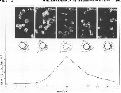
immuno-fluorescence staining. Maximum incorporation
of [3H]thymidine into DNA occurred
approxi-mately 18 h postmitosis, whereas maximum intensityof the VP143fluorescencereaction oc-curredfrom6to 12handfrom20to 30 h (Fig. 4). The results of this studythereforeindicate that the intensity ofthe VP143 staining reac-tion is cellcycledependentand that maximum intensity occurs before and after the time of
maximum DNA synthesis (i.e., during G and
earlySphaseaswellasduringlate S andG2)of
the 30-h 333-8-9 Tu cellcycle.
DISCUSSION
Thepresentstudydemonstrates the potential usefulness of antiserum prepared against an
individualviralpolypeptide forstudies of viral
geneexpressioninHSV-transformed cells.The
specificity of the reaction between anti-VP143
serumand theantigenwithwhichitreactsin
HSV-transformed cells hasbeendemonstrated,
sinceHSV-transformed hamster cells gave pos-itive reactions with VP143 antiserum whereas cells transformedby other agents did not.
Fur-thermore,anti-VP143serumreactivityin
HSV-2-transformedcells was significantly decreased afterabsorptionwithhomologouscells but not withnormal cellsorwithcells transformed by SV40, indicating that the antiserum reacted withageneproduct(s) expressed specifically in 287
on November 10, 2019 by guest
http://jvi.asm.org/
SCHAFFER
FIG. 2. Effects ofabsorptiononVP143-specific reactivity in 333-8-9Tucells. IFstaining observed in 333-8-9Tucells treated with anti-VP143 serum that was: (A) unabsorbed, (B) absorbed with 333-8-9 Tu cells, (C) absorbed withLSH-SV40 Tu cells, and (D) absorbed with LSH lp cells.
HSV-transformed cells. In addition,
prelimi-nary results of VP143 IF reactivity in HSV-2-transformed human embryoniclung cells lend
further support to the HSV-specific nature of
thereaction.
Whether the antigen in HSV-transformed cells thatreactswith anti-VP143serumis iden-tical to the antigen synthesized in
HSV-in-fectedcells isnotknown. Alternative
possibili-tiesfor the origin of VP143-reactiveantigen in HSV-2 transformed cellsinclude the following: (i)the antigen is avirus-modifiedhost protein which sharescommon antigenic determinants withthe infected cell protein; (ii) the antigen is a virus-coded precursor protein which shares antigenic determinants withthe viral protein produced ininfected cells;or(iii) the antigen is acellular geneproductwhich is absentin nor-mal cells and derepressed after infection and transformationby HSV. Of these three possibil-ities, (iii) isthe leastlikely,sincederepression
of this cellular genemust occuronlyafter
infec--n 4
0
Z2
0 6 1 2 18 24 27 30 33 36
HOURS
FIG. 3. Growth kinetics of 333-8-9 Tu cells. Ap-proximately2 x 105333-8-9Tu cellswereseeded into 60-mmpetri dishes. Culturesweretrypsinized,and cellswerecountedatthedesignatedtimes.
tionand transformationbyHSV.The definitive answer regarding the origin and nature of VP143reactiveantigencanbefoundonlywhen the antigens ininfected and transformed cells
0
on November 10, 2019 by guest
http://jvi.asm.org/
[image:5.505.101.434.67.396.2] [image:5.505.276.470.444.563.2]C
./Ik".
k--
1-27 30 36
HOURS
FIG. 4. VP143 expression and cellular DNA synthesis in synchronized 333-8-9 Tu cells. Cells were
synchronizedin mitosisandexamined for their IF reactivity with anti-VP143 rabbitserumfrom6 to30 h postseeding (photographs). Diagrammatic representations of the cytoplasmic location of VP143 reactivityin typical cellsat6-h intervalsareshown beneath photographs. Theincorporationof[3H]thymidine into cellular DNA insynchronized cellsas afunction of hours postseedingisshownatthe bottom of the figure.
can be isolated, characterized, and directly
compared.
Inthe present study antiserumtoHSV-2was
capable of detecting viral antigens in HSV-infected cells butnot inHSV-transformedcells, whereas antiserumtoVP143detected viral
an-tigens in both kinds of cell. The superiority of polypeptide-specific antisera compared with hy-perimmune antiviralserummayreflect the fol-lowing: (i) animals maymount a stronger
re-sponsewhenimmunizedwithasingle
polypep-tide than withavariety of polypeptides present
in whole viruspreparations, and (ii) thesodium dodecyl sulfate-treated polypeptide in the inoc-ulum may unfold in such a way that more
unique antigenic sitesareexposed, thus
ampli-fying the antigenicresponse. Inthisregard, it
should be noted that the IFreaction, which is detected within the transformedcell,maybea
reactiontothe unfoldedpolypeptide andnot to antigenic siteson the nativeprotein.
Further-more,werecognize that the interpretation of IF
data is a rathersubjective matter and that a more definitive method for quantitation of
VP143reactivity is desirable. Infact, confirma-tion of IF resultsby radioimmunoassayis
cur-rently inprogress.
Other characteristicsof anti-VP143serum in-clude thefollowing. (i) Anti-VP143serum does not react with the membrane of HSV-2-trans-formedcellsnordoesitneutralize HSV-2 infec-tivity. (To date, only antiserum prepared against HSV-2 VP119, the major envelope gly-coprotein, has been found to neutralize viral infectivity.) (ii) Anti-VP143 serum does react withanantigen(s) in HSV-1-transformed cells,
although the IF reactivity is slightly less in-tense. When anti-VP143 is reacted with both HSV-1-andHSV-2-infectedcells, however,the intensity of nuclear staining is the same. It should be further noted that by radioimmune precipitation this antigen (i.e., VP143)appears
tobetypecommon (T. Anzai, personal
commu-nication).
Although theuseofpolypeptide-specific anti-serumappearstobea moresensitivemeansof detecting the presence of viral antigens in transformed cells than is antiserum to whole
C
ICC
-u C.
3
.z
..
\1,I.',
.1
on November 10, 2019 by guest
http://jvi.asm.org/
[image:6.505.43.443.58.366.2]virusorvirus-infected cells, the reactionis
the-oretically capable of detecting a single gene
product. Consequently, the absence ofa
posi-tive reaction in HSV-transformed cells cannot be interpretedtoindicated the absence of viral
gene expression in these cells. Antisera
pre-pared againstaseriesofHSV-specific
polypep-tides would be essentialtoscreenforHSV-gene
expression in transformed cells. Of greater value will be the use of anti-VP143 serum in
determining the specific function of the VP143
geneproduct inHSV-transformed cells.
Of the six oncogenic HSV-transformed cell lines isolated by Kimuraetal. (16), onlythree exhibited reactivity with anti-VP143 serum.
Since cellsinall six lineswereshowntoexpress
HSV-specific surface antigens, we conclude
that detectable expression ofthe gene coding
forVP143isnotessential for the maintenance of transformation. However, preliminary data suggestthat theexpressionof thisgeneinsome
wayreflects thedegree of oncogenicityof
HSV-transformedhamster cells: in studies which at-tempttorelate theexpressionof HSVantigens with oncogenicity, we have observed that if
HSV-transformed cell lines exhibit VP143-spe-cificfluorescence, theyareoncogenic. Further-more,thegreaterthe number of VP143-positive
cells and the greatertheintensity ofstaining, the more oncogenic the cell lines have proven
to be (unpublished observation). HSV-trans-formed cell lines that do not express VP143
have been observedtobenon-oncogenicoronly
weakly oncogenic.
Using hyperimmune hamster antisera to
HSVin IF tests, DuffandRapp reportedthata
small proportion ofthe cells in several HSV-transformed cell lines contained HSV-specific antigens (5, 6). Other investigators have
re-ported both internal and surface fluorescencein HSV-transformed cells using hyperimmune anti-HSV serum (18-20, 27). However, in our
hands the reproducibility and sensitivity ofIF tests fordetecting internal antigens using hy-perimmune hamster and rabbit antisera to
HSV have beenpoor(16;unpublished
observa-tions).
Studies of the expression of an individual
viral gene in HSV-transformed cells have to
date beenlimitedtostudies of viral thymidine kinase synthesis. Thepresent report is there-fore the first to describe studies of a
virus-specificgeneproductinHSV-transformed cells employingIFtechniques. TheHSV-specific poly-peptide, VP143, is synthesized during the early stages (2 to 6 h postinfection) of virus replication. Basedonthe HonessandRoizman
(13)classificationofHSV-1polypeptides, HSV-2VP143 would be classifiedas abeta
polypep-tide (3). Preliminarystudies of the characteris-tics of this polypeptide suggest that it is not found in purified virions (K. L. Powell, per-sonal communication), it is found predomi-nantly in the nuclearfraction of HSV-2-infected cells, and its production is not affected by inhib-itors of viral DNA synthesis (unpublished ob-servations). The finding that VP143 reactivity was restricted to the cytoplasm of HSV-trans-formed cells but that reactivity in HSV-infected cells was both cytoplasmic and nuclear (the nuclear being more intense) suggests differ-ences inthe control of VP143 transport in the two typesof cell.
Thecellcycle-dependentexpression of VP143
in HSV-transformed cells suggests that viral geneexpression may be undercellular control.
Whethersynthesis andturnoverof VP143
dur-ing the cell cycle arerapid and/or whether the location of the antigen issimplyaltered during cell cycle traverse is currently being
investi-gated. With regardto the cellcycle-dependent
expressionof viral gene functions in cells
trans-formed by otherviruses, Kaplan et al. (14)
pre-viously demonstrated cell cycle dependence of SV40 induction from transformed hamster cells, and Stenman et al. (29) reported that the expression of thegene(s) forTantigenin SV40-transformed cells was cell cycle dependent.
Similarly, Greenberger and Aaronson (9) and
Schwartz et al. (28) presented evidence to sug-gestthe cell cycle dependence of drug-induced activationof type-C RNA viruses. Precedentfor the cell cycle-dependent expression of antigens in cells transformed by herpesviruses comes
from studies by Hampar et al., who described
the cell cycle-dependent activationof Epstein-Barr virus antigens (10-12).
Further studies are currentlyinprogress to elucidate the function of VP143 in both HSV-infected and -transformed cells and to examine human cervical carcinoma cells for the presence
of VP143-specific reactivity. Preliminary
re-sults of studies with cervical carcinoma cells have demonstrated that 2 of 13 lines so far examined express VP143 in a pattern similar to that described forhamster cellstransformedin vitro by HSV-2 (unpublished data with R. Lewis, E. Adam, and J. L. Melnick). The use of
polypeptide-specificantiseruminstudies of the
synthesisofother viral gene productsin
trans-formed cellsisalso inprogress.
ACKNOWLEDGMENT
This investigation was supported by research contract no.N01 CP 53526withinthe Virus CancerProgramof the National Cancer Institute.
LITERATURE CITED
1. Boyd, A. L., and T. W. Orme. 1975. Transformation of
on November 10, 2019 by guest
http://jvi.asm.org/
mouse cells afterinfection with ultraviolet
irradia-tion-inactivated herpes simplexvirus type 2.Int. J.
Cancer16:526-538.
2. Collard, W., H. Thorton, and M. Green. 1973. Cells
transformed by human herpesvirustype 2 transcribe virus-specific RNA sequences shared by herpesvirus types 1and 2. Nature (London) New Biol. 243:264-266.
3. Courtney,R.J., and M. Benyesh-Melnick. 1974. Isola-tionand characterizationof a large molecular-weight
polypeptideofherpes simplexvirus type 1.Virology
62:539-551.
4. Davis, D. B.,and D. T. Kingsbury. 1976. Quantitation
ofthe viralDNA present incellstransformedby UV-irradiatedherpes simplex virus. J. Virol. 17:788-793. 5. Duff, R., and F. Rapp. 1971. Properties of hamster
embryofibroblasts transformedin vitroafter expo-sure to ultraviolet-irradiated herpes simplex virus type 2. J.Virol. 8:469477.
6. Duff, R., and F. Rapp. 1973. Oncogenic transformation
ofhamster embryocells after exposure toinactivated herpessimplex virus type 1. J. Virol. 12:209-217. 7. Esparza, J., D. J. M.Purifoy, P. A. Schaffer, and M.
Benyesh-Melnick. 1974. Isolation, complementation
and preliminaryphenotypic characterization of
tem-perature-sensitive mutants ofherpes simplexvirus type 2. Virology 57:554-565.
8. Frenkel,N., H. Locker, B. Cox, B. Roizman, and F.
Rapp. 1976. Herpes simplex virus DNA in
trans-formed cells:sequencecomplexityinfive hamstercell lines and one derived hamster tumor. J. Virol.
18:885-893.
9. Greenberger, J. S.,andS.A.Aaronson. 1975.
Cyclohex-imide induction of xenotropic type C virus from
syn-chronized mouse cells: metabolic requirements for virus activation. J. Virol. 15:64-70.
10. Hampar,B., J. G. Derge, L. M. Martos, M. A. Taga-mets,S.-Y. Chang, and M. Chakrabarty. 1973. Iden-tification of acriticalperiod duringtheSphasefor activationof theEpstein-Barr virus by
5-iododeoxy-uridine. Nature (London) New Biol.244:214-217. 11. Hampar, B., J. G. Derge, and S. D. Showalter. 1974.
Enhanced activation of the repressed Epstein-Barr
viralgenomeby inhibitors of DNA synthesis. Virol-ogy 58:298-301.
12. Hampar, B., G. Lenoir, M. Nonoyama, J. G. Derge,
and S.-Y. Chang. 1976. Cell cycle dependence for activationof Epstein-Barrvirusby inhibitorsof pro-teinsynthesisormediumdeficient in arginine. Virol-ogy69:660-668.
13. Honess, R. W., and B. Roizman. 1974. Regulation of
herpesvirus macromolecular synthesis. I. Cascade
regulation of the synthesisof three groups of viral
proteins.J.Virol. 14:8-19.
14. Kaplan, J. C., L. F. Kleinman, and P. H. Black. 1975.
Cell cycle dependence ofsimian virus 40 induction from transformed hamster cells by ultraviolet
irra-diation. Virology68:215-220.
15. Kimura, S., J. Esparza, M. Benyesh-Melnick, and P. A.Schaffer.1974. Enhanced replicationof tempera-ture-sensitivemutantsof herpes simplexvirus type 2
(HSV-2) atthe nonpermissive temperature in cells transformed by HSV-2. Intervirology 3:162-169. 16. Kimura, S.,V. L. Flannery,B.Levy,and P. A.
Schaf-fer.1975.Oncogenic transformation of primary ham-stercells by herpes simplex virus type 2 (HSV-2) and anHSV-2temperature-sensitivemutant.Int. J. Can-cer15:786-798.
17. Kraiselburd, E., L. P.Gage,and A. Weissbach. 1975. Presence of a herpessimplex virus DNA fragment in an Lcell cloneobtainedafter infection with
irradi-atedherpes simplexvirus. J.Mol. Biol.97:533-542. 18. Kucera,L.S., andJ.P.Gusdon.1976.Transformation
of humanembryonicfibroblastsby photodynamically inactivated herpes simplex virus type 2 at
supra-optimaltemperature. J. Gen. Virol. 30:257-261. 19. Kutinova, L., V. Vonka, and J. Broucek. 1973.
In-creasedoncogenicity andsynthesisofherpesvirus an-tigens in hamster cells exposed to herpes simplex
type 2 virus. J. Natl.Cancer Inst. 50:759-766. 20. Macnab, J. C.M. 1974.Transformation ofratembryo
cellsbytemperaturesensitive mutantsofherpes
sim-plexvirus. J.Gen. Virol. 24:143-153.
21. Macnab, J. C. M.,and M. C. Timbury.1976. Comple-mentationofts mutantsofaherpes simplexvirus
ts-transformed cell line. Nature(London)262:233-235. 22. Minson, A. C.,M. E.Thouless, R. P. Eglin, and G. Darby. 1976.Thedetection ofvirus DNA sequences in aherpestype 2 transformed hamster cell line
(333-8-9). Int. J. Cancer17:493-500.
23. Munyon,W., E. Kraiselburd, D. Davis, and J. Mann.
1971.Transfer ofthymidine kinasetothymidine
ki-naselessLcellsby infectionwith
ultraviolet-irradi-ated herpes simplexvirus.J.Virol.7:813-820. 24. Nahmias,A.J., W.E.Josey,Z. M.Naib, C.F.Luce,
and B. A. Guest. 1970. Antibodiesto herpes virus
hominis type 1 and2 inhumans. II. Women with
cervicalcancer.Am. J.Epidemiol.91:547-552. 25. Porter, D.D.,I.Wimberly, andM.Benyesh-Melnick.
1969.Prevalence of antibodiestoEB virus and other herpes-viruses. J. Am.Med. Assoc.208:1675-1679. 26. Rawls,W.E., W. A.F.Tompkins, and J.L. Melnick.
1969.The association of herpesvirus type 2and carci-nomaof theuterinecervix. Am. J.Epidemiol. 89:547-554.
27. Reed, C. L.,G.H.Cohen,andF.Rapp.1975.Detection
ofavirus-specific antigen onthe surface ofherpes simplexvirus-transformed cells.J.Virol. 15:668-670.
28. Schwartz, S. A.,S. Panem, andW. H.Kirsten.1975. Distributionand virogenic effects of 5-bromodeoxy-uridineinsynchronizedratembryocells.Proc. Natl.
Acad. Sci. U.S.A.72:1829-1833.
29. Stenman, S., J. Zeuthan, and N. R. Ringertz. 1975.
ExpressionofSV40 T antigenduring the cellcycleof
SV40-transformed cells. Int.J.Cancer15:547-553. 30. Takahashi, M., and K. Yamanishi.1974.
Transforma-tionof hamsterembryoand humanembryocellsby
temperaturesensitivemutantsofherpessimplex vi-rustype 2.Virology61:306-311.
31. Terashima, T., and L. J. Tolmach. 1963. Growth and nucleic acidsynthesisinsynchronously dividing
pop-ulations ofHeLacells.Exp. CellRes. 30:344-362.
on November 10, 2019 by guest
http://jvi.asm.org/