JOURNAL OFVIROLOGY, Mar. 1995, p. 1591–1599 Vol. 69, No. 3 0022-538X/95/$04.0010
Copyrightq1995, American Society for Microbiology
Use of a Human Immunodeficiency Virus Type 1 Rev Mutant
without Nucleolar Dysfunction as a Candidate
for Potential AIDS Therapy
RIKA A. FURUTA,
1SATOSHI KUBOTA,
2MASATOSHI MAKI,
1YUKIO MIYAZAKI,
1TOSHIO HATTORI,
3ANDMASAKAZU HATANAKA
1*
Department of Molecular Virology
1and Research Center for Immunodeficiency Virus,
3Institute for Virus Research,
Kyoto University, Kyoto 606, Japan, and Laboratory of Molecular Virology and Carcinogenesis,
ABL-Basic Research Program, NCI-Frederick Cancer Research and
Development Center, Frederick, Maryland 21702-1201
2Received 14 March 1994/Accepted 6 December 1994
Applications of transdominant mutants of human immunodeficiency virus type 1 (HIV-1) regulatory
pro-teins, especially Rev mutant, have been attempted for gene therapy against AIDS, because the Rev protein is
essential for viral replication. We have previously reported that a mutant Rev protein (dRev) lacking its
nucleolar targeting signal remained out of nuclei in expressed cells and strongly inhibited the function of Rev.
To investigate the effects of dRev on HIV-1 replication, we established several dRev-expressing human cell lines
with two different vector systems and examined virus production in these cells. An HIV-1-derived vector
containing
drev
cDNA was constructed and introduced into CD4-positive HeLa cells and cells of the human
T-cell line CCRF-CEM (CEM). In dRev-expressing HeLa cells, virus replication, syncytium formation, and cell
death caused by HIV-1 infection were remarkably suppressed, and the same vector also conferred a resistant
phenotype on CEM cells. The production was also suppressed in CEM cells containing the
drev
gene driven by
a cytomegalovirus promoter. In addition, we found that dRev did not cause nucleolar dysfunction in a transient
assay, in contrast to other transdominant mutants and wild-type Rev. Since dRev cannot migrate into the
nuclei, it is expected not to interfere with nuclear/nucleolar functions of the host cell. We conclude that dRev
is one promising candidate as an antiviral molecule for gene therapy against AIDS.
The genome of the pathogenic retrovirus human
immuno-deficiency virus type 1 (HIV-1) encodes some accessory
pro-teins in addition to viral structural propro-teins (reviewed in
ref-erence 10). Among them, the nucleolar protein Rev has been
characterized most extensively (5, 9, 11, 15). Rev binds to a
region of RNA secondary structure known as Rev-responsive
element (RRE) that is a part of the env region of unspliced or
incompletely spliced HIV-1 mRNAs. These RNAs are
accu-mulated in the cytoplasm, and viral structural proteins are
synthesized (33, 40). Rev is essential for the production of
infectious virions; hence, a strategy inhibiting Rev function has
been mapped out to suppress HIV-1 production (3, 15, 26, 28,
29, 32). Rev is a 116-amino-acid protein composed of separate
functional domains. The N-terminal domain at amino acid
residues approximately 14 to 60 provides three different
func-tions: (i) a specific binding domain for RRE (7, 34), (ii) a
nucleolar targeting signal (NOS) identified as an arginine-rich
stretch of amino acids extending from positions 35 to 50 (18,
24, 37, 47), and (iii) an overlapped oligomerization domain
(40, 49). The C-terminal domain at residues approximately 78
to 90 is thought to be an activation domain of Rev (31, 33).
Mutations of the activation domain confer a dominant
nega-tive phenotype, which inhibits Rev function. Several reports
have demonstrated that the introduced trans-dominant rev
mu-tant can suppress viral replication in human T-cell lines (3, 34).
The other nonfunctional mutants with mutated N-terminal
domains do not significantly inhibit wild-type Rev function in
trans and are termed recessive negative mutants (31).
How-ever, we found that a deletion mutant of the N-terminal
do-main, named dRev, localized in the cytoplasm of transfected
cells and effectively inhibited the function of wild-type Rev
(23). In this report, we demonstrate the inhibitory effects of
this Rev mutant against HIV-1 infection in stably transduced
human cell lines and discuss the possible application of this
mutant for gene therapy against AIDS, comparing it with other
transdominant mutants.
MATERIALS AND METHODS
Plasmids.Plasmid pSE (14), which was produced by removing a 4.2-kb
SphI-EcoRI fragment from HIV-1 genomic clone pNL4-3 (1), was a gift from A.
Adachi. After disruption of the unique EcoRI site of pSE, a 1.5-kb SalI-NheI fragment was removed and an EcoRI linker was inserted. In the resultant plas-mid, pSEEcoRI, the position of the EcoRI site was transferred behind a splicing acceptor, and the 59 half of the env gene was removed. A selection marker plasmid, pSVbsr, purchased from Kaken (Kyoto, Japan) (19) was digested with
BamHI and PvuII. Then the 1.8-kb fragment containing a simian virus 40 (SV40)
early promoter, bsr gene, and poly(A) site was isolated and inserted into pSEEcoRI at the BamHI site after removal of a 0.4-kb BamHI-XhoI fragment. The resultant plasmid, pLbsr, contains (i) the HIV-1 long terminal repeat (LTR) at both the 59and 39ends, (ii) a packaging signal of HIV-1 (2, 27), (iii) a part of the env gene corresponding to RRE, and (iv) a selection marker gene in the opposite direction (Fig. 1B). pLbsr is an HIV-1 retroviral vector with an EcoRI cloning site requiring Rev expression for the packaging of the pseudogenome, and the expression of a introduced gene was upregulated by Tat protein. To construct the dRev protein expressor, pLdrevb, a 0.6-kb EcoRI fragment of pH2drev (23) was inserted at the EcoRI site of pLbsr. This plasmid is believed to express a Rev deletion mutant lacking 7 amino acid residues within its nucle-olar targeting signal (Fig. 1A) (18, 23, 46). We constructed another dRev-expressing vector, pCdrev (Fig. 1C), to allow high and constitutive dRev expres-sion. The parental vector pCMV-NEO-BAM (4), kindly given by B. Vogelstein, underwent digestion by BamHI and blunting followed by XhoI linker insertion, into which a 0.6-kb drev fragment was ligated with XhoI linker. In pCdrev, the
drev gene is expressed under the control of the cytomegalovirus (CMV)
pro-moter. The following plasmids were used to observe the cytopathic effects of Rev. Plasmids pH2rev, pH2drev, and pH2revM10 express Rev, dRev, and RevM10, respectively, under the control of the SV40 early region promoter. One of them, * Corresponding author. Phone: 4000. Fax:
81-75-751-3998.
1591
on November 9, 2019 by guest
http://jvi.asm.org/
pH2revM10, which was constructed by substituting the cDNA portion of pH2rev for that of pcREVM10 (31) (provided by B. R. Cullen), expresses a trans-dominant mutant, RevM10. A tat expression plasmid, pH2Ftat, produces a full-length Tat protein of HIV-1 (13). Plasmid pKCRH2 (36) is a parental vector and was used as a control.
Cells and virus.CD4-positive HeLa cells (HeLa cl.1022 cells [6] provided by
B. Chesebro) and COS7 cells were grown at 378C under 5% CO2in air in Dulbecco’s modified Eagle medium supplemented with 10% fetal calf serum. To establish drev-transduced HeLa cells, 30mg of linearized pLdrevb or pLbsr (as a control) were transfected into 106HeLa cl.1022 cells by a standard calcium phosphate method, selected with 2.5mg of blasticidin S per ml, and cloned by a cylinder cloning method. Cells of the human acute lymphoblastic leukemia de-FIG. 1. (A) Structures of cDNAs encoding wild-type Rev and the mutant, dRev. The deduced amino acid sequence of the deletion is shown by single-letter abbreviations. Numbers below the hatched box of rev and above the deleted sequence represent residue numbers counted from the first methionine of Rev. (B) HIV-1 vector plasmids pLbsr and pLdrevb constructed from full-length proviral clone pNL4-3 (see Materials and Methods). The heavy lines represent RRE in the HIV-1 env sequence. (C) Structure of dRev expression vector under the control of the CMV promoter. The parental vector pCMV-NEO-BAM contains a eukaryotic expression unit with the CMV promoter, BamHI cloning site, and selection maker gene. To make pCdrev, an EcoRI fragment of pLdrevb was inserted into the cloning site of pCMV-NEO-BAM with an XhoI linker. Abbreviations: bsr, blasticidin S resistance gene; NOS, nucleolar targeting signal; pA, SV40 late polyadenylation signal; SVp, SV40 early promoter; CMVp, CMV promoter; TKp, thymidine kinase promoter; neo, G418 resistance gene.
on November 9, 2019 by guest
rived T-cell line CCRF-CEM (CEM) were maintained in RPMI 1640 medium supplemented with 10% fetal calf serum at 378C under 5% CO2in air. To obtain stable transfectants of CEM cells, 30mg of linearized pLdrevb or pLbsr was transfected into 107
CEM cells by an electroporation method with a Gene Pulser (Bio-Rad, Richmond, Calif.) followed by selection with 2.5mg of blasticidin S per ml and cloning by a limiting-dilution method. Integration of pLdrevb or pLbsr was confirmed by genomic PCR (data not shown). We also transfected 30mg of pCdrev or pCMV-NEO-BAM into CEM cells by an electroporation method and selected cells with G418 at a concentration of 700mg/ml (active dose) for 3 weeks. The expression of the drev gene of these cells was examined by Northern (RNA) blot analysis (data not shown). CD4 expression of cloned HeLa and CEM cells was determined by FACScan instrument analysis using fluorescein isothio-cyanate (FITC)-conjugated Leu3a monoclonal antibody (Beckton Dickinson, Mountain View, Calif.). An HIV-1 HTLV-IIIB strain (provided by R. C. Gallo) was obtained from chronically infected H9 cells (human T-cell lymphoma cell line) grown in RPMI 1640 medium supplemented with 10% fetal calf serum.
Viral infection.For efficient infection, we cultivated a HeLa cell series with H9 cells chronically infected with the HTLV-IIIB strain of HIV-1 (H9/IIIB). In order to produce a population of nondividing cells, H9/IIIB cells were exposed to 9,000 rads from a137Cs source. One day before infection, cloned HeLa cells were seeded in a 35-mm-diameter dish. After the culture medium was changed, 43103to 53104radiated H9/IIIB cells were added and cocultivated for 48 h. Then the medium including H9/IIIB cells and virions was removed, and HeLa cells were washed with phosphate-buffered saline (PBS) four times. Afterwards, the cells were grown in fresh growth medium for an additional 6 days. In the case of CEM cells, cell-free infection was applied. Filtered culture supernatant of H9/IIIB cells was added to each CEM transfectant at a multiplicity of infection (MOI) of 0.001 to 0.05. The infected cells were maintained at 23105to 103 105/ml in 25-cm2culture bottles, and virus production was monitored every other day.
Immunofluorescence.To confirm expression and localization of dRev, COS7 cells (53104) were plated on glass coverslips in a 35-mm-diameter dish 1 day before transfection. DNAs were introduced into the cells by the DEAE-dextran method (8, 43). Forty-eight hours later, cells were fixed with 3.5% formaldehyde– PBS for 10 min at room temperature and permeabilized with 0.1% Nonidet P-40–PBS for 10 min at room temperature. Then the cells were incubated for 1 h at 378C with both anti-Rev serum (provided by B. R. Cullen or purchased from ICN, Cleveland, Ohio) and the serum of a progressive systemic scleorisis patient as an antinucleolar antibody and then stained for 1 h at room temperature with FITC-conjugated anti-rabbit immunoglobulin G (IgG) and tetramethylrhodam-ine isothiocyanate (TRITC)-conjugated anti-human IgG antibody. To investigate nucleolar deforming effects, we incubated the cells with both anti-rRNA mono-clonal antibody Y-10B (provided by J. A. Steiz) and anti-Rev serum. Then the cells were stained for 1 h at 378C with both FITC-conjugated anti-rabbit IgG (for anti-Rev) and TRITC-conjugated anti-mouse IgG (for anti-rRNA). To confirm the Tat-dependent expression of the dRev protein in pLdrevb-transduced HeLa cells, pH2Ftat was transfected into the cells by a standard calcium phosphate method. After 48 h, the cells were fixed, permeabilized as described above, and incubated with both rabbit anti-Rev antiserum and mouse anti-Tat monoclonal antibody (purchased from American BioTechnologies Inc., Cambridge, Mass.) for 1 h at room temperature. Then the cells were stained for 1 h at 378C with both FITC-conjugated anti-rabbit IgG and TRITC-conjugated anti-mouse IgG. To show subcellular localization of viral proteins of infected HeLa cells, drev-intro-duced or control CD4-positive HeLa cells were infected with filtered culture supernatant of H9/IIIB. Forty-eight hours postinfection, the cells were fixed and immunostained with both rabbit anti-Rev antiserum and mouse anti-p24Gag monoclonal antibody VAK4 (17) as described above.
Viral production assay.The multinuclear activation of a galactosidase indica-tor (MAGI) assay (20) was employed for titration of HIV-1 in culture superna-tants of infected cells. The indicator cells were established by introducing the bacterial lacZ gene under the control of the HIV-1 LTR into HeLa cells ex-pressing CD4. After infection with virus and X-Gal (5-bromo-4-chloro-3-indolyl-b-D-galactopyranoside) staining, blue-stained cells were counted to estimate infectious units. In our assay, one stained cell represents one infectious unit in a chamber of a 24-well plate. Cell-free supernatants were also assayed for virus reverse transcriptase (RT) activity (25) and virus antigen p24Gag
by a solid-phase enzyme immunoassay (Abbot Laboratories, North Chicago, Ill.). We also exam-ined syncytium formation of the HeLa cell series in which a syncytium containing more than five nuclei was regarded as an HIV-induced syncytium. Since the ratio of small syncytia in parental cells was below 1% without HIV infection, we had confirmed that such large syncytia were HIV positive by immunofluorescence with VAK4 monoclonal antibody (data not shown).
DNA amplification.To analyze proviral DNA of infected cells, we extracted DNAs 48 h postinfection from drev-introduced or control CEM cells as follows: 23106cells were suspended in STE (100 mM NaCl, 10 mM Tris-HCl [pH 7.4], 1 mM EDTA, 0.5% sodium dodecyl sulfate), digested with proteinase K, phenol extracted, and ethanol precipitated. After RNase A digestion, DNAs were re-covered by phenol extraction followed by ethanol precipitation. The DNAs were suspended with 1 ml of TE (10 mM Tris-Cl [pH 7.6], 1 mM EDTA). To amplify HIV-1 specific DNA, 1, 5, or 25ml of the DNA solution was used for semiquan-titative PCR with primer pair SRRE-3nef8978, which corresponds to part of the
env gene (nucleotides 7759 to 8978 of pNL4-3). Primer pair 5bglo-3bglo was also used to amplify part of the humanb-globin gene as an internal control. PCR was carried out using DNA thermal cycler PJ1000 (Perkin-Elmer Cetus Instrument) in 50ml of the following reaction mixture: 10 mM Tris-HCl (pH 8.3), 50 mM KCl, 1.5 mM MgCl2, 100mM each deoxynucleoside triphosphate, and 0.25mM each primer. The reaction mixture was incubated at 958C for 5 min and chilled on ice for 5 min, and then 2 U of Taq polymerase (Perkin-Elmer Cetus Instrument) was added. PCR was repeated for 30 cycles of denaturation at 948C for 30 s, anneal-ing at 528C for 30 s, and extension at 728C for 90 s. DNA fragments were analyzed by 1.2% agarose gel electrophoresis with ethidium bromide staining. Synthetic oligonucleotide sequences of the primers were as follows: SRRE, 59-ATAGGAGCTCAGGAGCTTTGTTCCTTGGGT-39; 3nef8978, 59-CTCCT CTTGTGCTTCTAGCCA-39; 5bglo, 59-GGTTGGGCCAATCTACTCCCAG G-39; and 3bglo, 59-GCTCACTCACTGTGGCAAAG-3.
RESULTS
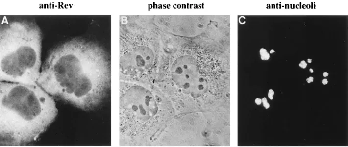
Efficient expression of dRev in COS7 cells by pLdrevb.
We
[image:3.612.128.483.71.221.2]subcloned the cDNA of a Rev mutant with a 7-amino-acid
deletion in its nucleolar targeting signal, named dRev (Fig. 1A)
(23), into HIV-1 vector pLbsr (Fig. 1B). Expression and
sub-cellular localization of dRev from pLdrevb were confirmed in
COS7 cells in a transient transfection assay by an
immunoflu-orescence technique (Fig. 2). In contrast to wild-type Rev and
other transdominant mutants (31), the dRev protein efficiently
expressed by pLdrevb remained in the cytoplasm of transfected
cells like pH2drev with the SV40 promoter (23). We also
examined expression and localization of the dRev protein
un-der the control of a CMV promoter (Fig. 1C). pCdrev gave a
FIG. 2. Intracellular localization of dRev expressed by pLdrevb. COS7 cells were transfected with 6mg of pLdrevb, fixed 48 h after transfection, and stained with both FITC for anti-Rev antiserum (A) and TRITC for serum from a patient with progressive systemic sclerosis (C). (B) Phase-contrast microscopic view of the same cells.
VOL. 69, 1995 NOVEL Rev MUTANT FOR AIDS THERAPY 1593
on November 9, 2019 by guest
http://jvi.asm.org/
high level of expression of the dRev protein in the cytoplasm of
transfected COS7 and HeLa cells (data not shown).
Establishment of dRev-introduced HeLa and CEM cell
lines.
First, we established a CD4-positive HeLa cell line stably
transduced with the drev gene in order to show subcellular
localization of introduced gene products and to examine the
effects of dRev on virus infection. We chose HeLa cl.1022
constitutively expressing CD4 as a parental cell line because it
was originally established for a focal immunoassay of HIV
infectants with high sensitivity (6). After introduction of dRev
expression vector pLdrevb or control plasmid pLbsr by
trans-fection followed by selection with blasticidin S and cell cloning,
integration of each vector was confirmed by PCR (data not
shown). The dRev proteins were detected in established cells
only when Tat was expressed by transfection with plasmid
pH2Ftat (Fig. 3A and B), whereas control plasmid pKCRH2
did not give such an effect (data not shown). We also analyzed
the level of CD4 expression in the cloned cell lines and
paren-tal HeLa cl.1022 cells. FACScan analysis using an anti-CD4
monoclonal antibody, Leu3a, revealed that there is no
signifi-cant difference in the expression levels of CD4 molecules
among the three cell clones (data not shown), assuming that
the susceptibility to HIV of these cells could be the same.
Doubling times of three cell lines were as follows: 19.6 h in
HeLa cl.1022 cells, 21.9 h in the cell lines containing pLdrevb
(HeLa/dRev), and 23.2 h in the cell line containing pLbsr
(HeLa/bsr).
In order to examine the inhibitory effects of dRev on virus
production in T cells in long-term infection, we next
intro-duced pLbsr or pLdrevb into CEM cells and obtained stable
transfectants after selection by blasticidin S and cell cloning.
FACScan analysis also revealed that the mean fluorescence
intensities given by an anti-CD4 monoclonal antibody were the
same among the parental CEM cells, CEM bsr cells (CEM/
bsr), CEM dRev mixed population (CEM/dRev mix.), and
CEM dRev cloned cells (CEM/dRev cl.) (data not shown). The
doubling times of these cell lines were also almost the same. In
addition, we established CEM cell lines expressing
constitu-tively the dRev protein under the control of a CMV promoter.
CEM cells were transfected with the dRev-expressing plasmid,
pCdrev, or parental vector pCMV-NEO-BAM (Fig. 1C) and
selected in G418-containing medium for three weeks. The
es-tablished cells (CEM/CdRev and CEM/neo) were subjected to
virus infection assays.
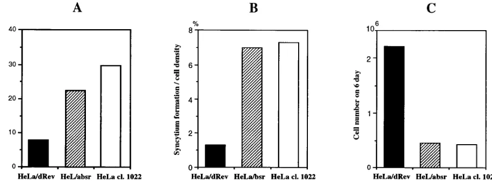
HIV-resistant phenotype of a HeLa/dRev cell clone.
We
ex-amined whether the stably transduced drev gene can inhibit
HIV-1 replication in established cell lines. In the HeLa cell
series, three clones, HeLa/dRev, HeLa/bsr, and HeLa cl.1022,
were cocultivated with H9/IIIB, and then HIV-1 replication
was monitored by MAGI assay. The representative data of four
different experiments with similar results are shown (Fig. 4A).
The replication of virus was suppressed in HeLa/dRev cells
about 27% compared with that in HeLa cl.1022 cells and 35%
compared with that in HeLa/bsr cells at 4 days postinfection
without cell passage. Next, we compared the rate of syncytium
formation among these cell lines (Fig. 4B). While we observed
many syncytia 4 days after infection in control cells, the rate of
syncytium formation was very low in HeLa/dRev cells. Since
HIV-mediated syncytium formation depends upon the
inter-action of CD4 and gp120-gp41 complex (22, 30), it reflects the
expression of the env gene induced by the Rev protein,
sug-gesting that dRev efficiently blocked the Rev function of
HIV-1 in stably transduced cells. In order to clarify that dRev
conferred such an HIV-1-resistant phenotype on HeLa cells
FIG. 3. (A and B) Tat-dependent expression of the dRev proteins in a dRev-introduced cell clone, HeLa/dRev; (C to F) subcellular localization of viral proteins in infected HeLa cells. HeLa/dRev cells were transfected with 4mg of pH2Ftat, fixed 48 h after transfection, and stained with both TRITC for anti-Tat monoclonal antibody (A) and FITC for anti-Rev antiserum (B). HeLa/dRev and HeLa/bsr cells were infected with HTLV-IIIB, fixed 48 h postinfection, and stained with both TRITC for anti-p24Gagmonoclonal antibody (C and E) and FITC for anti-Rev antiserum (D and F).
on November 9, 2019 by guest
through inhibition of the function of Rev, we examined
sub-cellular localization of viral and antiviral proteins in the
fected HeLa cell. Rev was localized in nuclei/nucleoli in
in-fected HeLa/bsr, and viral structural protein p24
Gagwas
strongly expressed in all of these cells (Fig. 3C and D). These
results confirmed that Rev effectively functions in nuclei and
induced the synthesis of viral structural proteins. On the other
hand, in HeLa/dRev cells infected with HIV-1, Rev was
de-tected only in the cytoplasm, and p24
Gagwas not detected in
the same cells (Fig. 3E and F). We believe that the cells
positively stained in the cytoplasm by anti-Rev antibody were
infected, because the antibody used here recognizes both
wild-type and mutant Rev and also because the gene expression of
both rev and drev depended on Tat, as shown in Fig. 3A and B.
These data indicate that dRev interfered with the localization
of Rev and that the viral replication was suppressed in these
cells.
In addition, many HeLa/dRev cells were alive, unlike other
control cells on day 6 postinfection (Fig. 4C). Since the
dou-bling times and the levels of CD4 expression of these three cell
lines have been almost equal, the viable cell numbers represent
actual populations that escaped cell death by HIV-1 infection.
Syncytium formation and cell viability were almost the same in
HeLa/bsr and HeLa cl.1022 cells, suggesting that the titration
effects of Tat and Rev by the trans-activation-responsive region
(TAR) and RRE of pLbsr vector were not observed in this
assay system. Considering the above findings together, we
con-cluded that the dRev-harboring HeLa cells escaped cell death
by HIV-1 infection through inhibition of the function of Rev.
The inhibitory effects of dRev on virus production in CEM
cells.
We established drev-introduced CEM cells to examine
whether dRev does work as an antiviral molecule in human
T-cell lines as well as in CD4-positive HeLa cells. First, we
examined the effects of dRev under the control of the HIV-1
LTR. Parental CEM cells and stably transduced CEM series
(CEM/bsr, CEM/dRev mix., and CEM/dRev cl.) cells were
infected with HTLV-IIIB via cell-free infection at a MOI of
0.001 to 0.05. Virus production of each cell type was quantified
by MAGI assay. Figure 5A shows one of the typical results
among five experiments independently performed with various
MOIs. The production of virus in control CEM cells increased
from day 18 postinfection. An increase in the number of
in-fectious units of CEM/bsr cells was observed on day 20. Virus
production in both the dRev-introduced mixed population and
cloned cell line remained very low, though a little increase was
observed on days 20 and 24. This increase could be explained
by the insufficient amount of dRev, because dRev was
ex-pressed under the control of the HIV-1 LTR in these cells.
Therefore, we next performed another infection assay using
cells expressing dRev by the CMV promoter. The RT activities
of control CEM/neo cells peaked on day 12 (Fig. 5B). In
CEM/CdRev cells, however, no significant virus production
was seen through the experimental period.
In order to confirm that these cells were equally susceptible
to HIV-1, we analyzed the viral DNA in the infected cells by a
PCR method (Fig. 5C). Forty-eight hours postinfection,
genomic DNAs were prepared from the CMV/CdRev and
CMV/neo cells and subjected to semiquantitative PCR.
Al-most the same levels of HIV-1 specific fragments were
ampli-fied from the DNA of both CEM/neo and CEM/CdRev cells.
PCR products of the human
b
-globin gene of CEM/neo and
CEM/CdRev cells 48 h postinfection were observed as the
same, assuming that the DNA of these cells had been amplified
equally under these PCR conditions; therefore, there was no
great difference in the amounts of the viral DNA between
these cells. These data clearly demonstrated that the HIV-1
infection in these cells occurred equally. We therefore
con-cluded that dRev inhibited the virus production in T cells as
well as in HeLa cells.
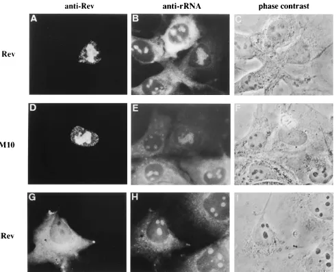
dRev is free from nucleolar side effects observed in another
transdominant Rev mutant.
We have previously found that
overexpressed Rev proteins have cytotoxic effects in some cell
lines, and these effects were also observed in the cells
intro-duced with Rev mutants, including a transdominant mutant
(38). It has been reported that transdominant mutant RevM10
was able to block HIV-1 replication in stably transduced cells,
and its potential use for gene therapy has been suggested (3,
32). Cytotoxic effects by dRev, Rev, and RevM10 were
exam-ined. We transfected wild-type or mutant Rev expression
vec-tors into COS7 cells and doubly immunostained the cells with
anti-Rev and anti-rRNA antibodies. The nucleoli of COS7
cells were deformed and ballooned 48 h after transfection with
wild-type Rev or RevM10 expressor, and rRNAs mostly
dis-appeared in those cells (Fig. 6A to F). These findings are not
dependent on cell cycle (36a). In contrast, dRev has shown
neither nucleolar destructive effects nor an aberrant
distribu-FIG. 4. Effects of the drev gene stably transduced in CD4-positive HeLa cells against infection by HIV-1. Cloned HeLa/dRev, HeLa/bsr, and parental HeLa cl.1022 cells were cocultivated with chronically infected H9 cells for 48 h. Four days after infection, HeLa cells were assayed for the virus replication that was monitored by MAGI assay (A) (see Materials and Methods). The rate of syncytium formation (B) and viable cell numbers counted by trypan blue exclusion (C) are also displayed.
VOL. 69, 1995 NOVEL Rev MUTANT FOR AIDS THERAPY 1595
on November 9, 2019 by guest
http://jvi.asm.org/
[image:5.612.67.548.71.249.2]tion of rRNA in transduced cells, although the excess amounts
of the dRev proteins were expressed (Fig. 6G, H, and I) under
the same conditions in experiments using Rev and RevM10.
DISCUSSION
In this study, we demonstrated that dRev suppressed HIV-1
production in the several stable transformants with the drev
gene and clearly show that this suppression was due to
inter-ference with Rev localization by dRev in infected HeLa cells,
which we have previously reported in a cotransfection assay
(23).
We introduced the drev gene into two human cell lines,
CD4-positive HeLa cells and CEM cells, by two different
vec-tors and found that virus production by the dRev-harboring
cells was apparently lower than that by control cells in the all
cells tested here. The vector, pLbsr, used in this study was
designed as a retroviral vector so that the introduced gene is
expressed under the control of the HIV-1 LTR and that the
genomic RNA of the vector is expected to be packaged into a
virion in the presence of the Rev and viral structural proteins.
Furthermore, mRNA of pLbsr contains two important
second-ary structural regions, TAR and RRE, which are expected to
titrate Tat and Rev of HIV. We, however, transduced it into
the cells by transfection in this study because packaging devices
for HIV vectors are still under development (42, 44). We
measured virus production in culture supernatants from cells
expressing drev by this vector by MAGI assay, because mRNA
transcribed from pLbsr or pLdrevb containing the packaging
signal of HIV-1 might be packaged into virions and so that the
amount of p24 and RT activity in culture supernatants of these
cells may reflect production of such pseudovirions in addition
to wild-type ones.
The dRev protein was efficiently expressed in the cytoplasm
of the transfected COS7 cells from this vector (Fig. 2), and the
expression of dRev was regulated in stably transduced HeLa
cells in a Tat-dependent manner (Fig. 3A and B). Moreover,
dRev was highly expressed and worked as an antiviral molecule
when the drev-introduced HeLa cells were infected with HIV-1
(Fig. 3C to F), in which case we found different localization of
the functional or nonfunctional Rev by immunofluorescence.
In the infected HeLa/dRev cells, syncytium formation, which is
one of the typical cytopathic effects observed in HIV-infected
cultured cells, was suppressed (Fig. 4B) and virus production
was also suppressed compared with that in parental HeLa
cl.1022 cells (Fig. 4A). A slight inhibition was also seen in
HeLa/bsr cells, probably reflecting the titration effects of TAR
and RRE against Tat and Rev as discussed above.
Further-more, the number of viable HeLa/dRev cells on day 6
postin-fection far exceeded than that of the control cells (Fig. 4C).
Therefore, we concluded that dRev conferred an
HIV-resis-tant phenotype on the introduced HeLa cells.
[image:6.612.322.556.71.246.2]We also performed a cell-free infection study using CEM
cells and found that virus production was also suppressed
in CEM/dRev cells. The delay in virus production seen in
CEM/bsr cells was probably due to the same effects observed
in HeLa/bsr cells. Virus production was not completely
suppressed by dRev in the HIV-1 vector system, probably
FIG. 5. The inhibitory effect of dRev on virus production in CEM cells. (A) pLbsr or pLdRevb was stably transduced into CEM cells, and HTLV-IIIB was added to 23105parental CEM cells and each transfectants (CEM/bsr, CEM/ dRev mix., and CEM/dRev cl. cells) at a MOI of 0.001. The titer of culture supernatant was quantitated every other day by MAGI assay. (B) RT activities of culture supernatant of infected CEM cells containing pCdRev (CEM/CdRev) or pCMV-NEO-BAM (CEM/neo) at a MOI of 0.05. (C) Viral DNAs of these cells on day 4 postinfection were analyzed by semiquantitative PCR with primer pairs to amplify HIV-1 specific DNA (see Materials and Methods). (Upper panel) Ten microliters of PCR mixture was applied to each lane. Lane 1, size maker (lphage DNA digested with HindIII). DNAs used as a template were as follows: lane 2, 25ml of DNA of uninfected CEM/neo cells (negative control); lanes 3 to 5, 1, 5, and 25ml of infected CEM/neo DNA, respectively; lanes 6 to 8, 1, 5, and 25ml of infected CEM/Cdrev DNA, respectively; lanes 9 to 12, 1, 5, 25, and 125 pg of pNL4-3 DNA. (Lower panel) An internal control PCR was also carried out with a primer pair for the humanb-globin gene. Five microliters of PCR mixture was applied to each lane; lanes correspond to template DNAs of the upper lanes.
on November 9, 2019 by guest
because the expression level of dRev was not sufficient for
complete inhibition in this experimental condition. In
con-trast, almost complete inhibition was observed in CEM/
CdRev cells in which drev was driven by the CMV promoter.
Bahner et al. (3) also reported that the retroviral vectors
con-taining a transdominant mutant Rev gene driven by the CMV
promoter worked more effectively than that driven by the
HIV-1 LTR. We believe that Tat-dependent expression of
anti-HIV molecules may be safe in gene therapy against
AIDS because transduced gene products are not expected
to be expressed at a high level without HIV infection and the
HIV vector could transfer genes into the HIV target cells
in vivo, yet the suppressive effect in this system was not
com-plete. Further investigation for a more efficacious transmission
and expression system of anti-HIV molecules would be
neces-sary.
Although the precise mechanism of the inhibitory effect of
dRev has not been proved yet, it is evident that Rev cannot
migrate into the cell nucleus/nucleolus and does not function
in the presence of dRev. Very recently, Duan et al. reported an
intracellularly expressed anti-Rev single-chain antibody. They
claimed that the antibody changed the localization of Rev and
decreased HIV-1 replication in human cells, results very
sim-ilar to ours (12). The mechanism of how dRev can retain Rev
in the cytoplasm has not been clarified. It is possible that dRev
competes with Rev in the cytoplasm for some cellular factors
which carry Rev into the nucleus/nucleolus. We presume,
how-ever, that dRev may not interact with such molecules, since
dRev lacks its nucleolar targeting signal. Alternatively, dRev
may form a hetero-oligomer with Rev which cannot migrate
into the nucleus/nucleolus as hypothesized previously (23). It
was reported that an excess amount of Rev makes an oligomer
without RRE and some Rev mutants with substitutions in the
nuclear targeting signal form a hetero-oligomer with wild-type
Rev (40). Multimer formation of Rev with or without RRE,
however, remains unsettled.
It is important to mention that dRev works only in the
cytoplasm of the introduced cell. Until now, a variety of
ret-FIG. 6. Distribution of rRNA in COS7 cells transfected with Rev or mutant Rev expression vector. Cells transfected with 5mg of pH2rev (A to C), pH2revM10 (D to F), or pH2drev (G to I) were doubly stained 48 h after transfection. Panels A, D, and H were stained with rabbit anti-Rev serum and FITC-conjugated anti-rabbit IgG antibody. Panels B, E, and I were stained with mouse anti-rRNA monoclonal antibody Y-10B and TRITC-conjugated anti-mouse IgG antibody. Panels C, F, and G were viewed by phase-contrast microscopy.
VOL. 69, 1995 NOVEL Rev MUTANT FOR AIDS THERAPY 1597
on November 9, 2019 by guest
http://jvi.asm.org/
[image:7.612.64.555.69.467.2]fected cells and in T cells acutely infected with HIV-1 but not
in chronically infected cells (38). These observations indicated
that the high expression of Rev may be lethal to cells. Here, we
also demonstrated that highly expressed transdominant mutant
RevM10 induced nucleolar ballooning and deformity with
ab-errant accumulation of rRNAs like wild-type Rev in
transfec-tion experiments (Fig. 6). Although these phenomena could be
explained by the excess amount of expression, dRev did not
show such effects at the same level of expression. To sum up,
dRev remains in the cytoplasm, inhibits viral replication
effec-tively, and does not affect nuclear events directly. Malim et al.
showed that the transduced RevM10 gene produced an
effec-tive blockade against HIV-1 in CEM cells without remarkable
side effects on some T-cell functions using selected clones of
transduced cells (32). Also similar results have been shown by
Bahner et al., and they noted that it is important to check the
toxicity of these genes on normal cellular function (3). We
think that much should be done to examine the toxicity of
transduced gene products in various conditions.
Our data reported previously (23, 38) and here suggest that
it is possible that the dRev protein blocks cell death caused by
HIV-1 through two pathways. One is an inhibitory effect on
viral protein production, including the env gene product, which
induced cell death (reviewed in references 16 and 21). The
other is the fact that dRev is able to retain Rev in the
cyto-plasm to prevent Rev from nucleolar accumulation which may
cause nucleolar dysfunction, occasionally leading to cell death.
Considering the above aspects together, we may reasonably
conclude that dRev is a good candidate for gene therapy
against AIDS.
ACKNOWLEDGMENTS
We thank B. R. Cullen for anti-Rev antiserum and various rev mutants, A. Adachi for pSE, J. A. Steiz for anti-rRNA monoclonal antibody, B. Chesebro for HeLa cl.1022 cells, R. C. Gallo for HTLV-IIIB, and S. Oroszlan for devices of the MAGI assay. We thank K. Sano and the Diagnostic Research and Development Department, Asahi Chemical Industry Co., Ltd., for help in measurement of the RT activity and also thank A. Kondo, K. Imada, and H. Sakaida for help in FACSan analysis.
This work was supported in part by grants from the Life Insurance Association of Japan and from the Ministry of Education, Science and Culture of Japan.
REFERENCES
1. Adachi, A., H. E. Gendelman, S. Koenig, T. Folks, R. Willey, A. Rabson, and M. A. Martin.1986. Production of acquired immunodeficiency syndrome-associated retrovirus in human and nonhuman cells transfected with an infectious molecular clone. J. Virol. 59:284–291.
2. Aldovini, A., and R. A. Young. 1990. Mutations of RNA and protein se-quences involved in human immunodeficiency virus type 1 packaging result in production of noninfectious virus. J. Virol. 64:1920–1926.
3. Bahner, I., Z. Chen, X. J. Yu, Q. L. Hao, J. C. Guatelli, and D. B. Kohn. 1993.
10. Cullen, B. R. 1991. Regulation of HIV-1 gene expression. FASEB J. 5:2361– 2368.
11. Cullen, B. R., and W. G. Green. 1989. Regulatory pathways governing HIV-1 replication. Cell 58:423–426.
12. Duan, L., O. Bagasara, M. A. Laughlin, J. W. Oakes, and R. J. Pomerantz. 1994. Potent inhibition of human immunodeficiency virus type 1 replication by an intracellular anti-Rev single-chain antibody. Proc. Natl. Acad. Sci. USA 91:5075–5079.
13. Endo, S., S. Kubota, H. Siomi, A. Adachi, S. Oroszlan, M. Maki, and M. Hatanaka.1989. A region of basic amino-acid cluster in HIV-1 Tat protein is essential for trans-acting activity and nucleolar localization. Virus Genes 3:99–110.
14. Gendelman, H. E., W. Phelps, L. Feigenbaum, J. M. Ostrove, A. Adachi, P. M. Howley, G. Khoury, H. S. Ginsberg, and M. A. Martin.1986. Trans-activation of the human immunodeficiency virus long terminal repeat se-quence by DNA viruses. Proc. Natl. Acad. Sci. USA 83:9759–9763. 15. Green, M. R. 1993. Molecular mechanism of Tat and Rev. AIDS Res.
3:41–55.
16. Haseltine, W. A. 1991. Molecular biology of the human immunodeficiency virus type 1. FASEB J. 5:2349–2360.
17. Hattori, T., K. Sagawa, S. Matsushita, A. Koito, H. Suto, M. Matsuoka, M. Yokoyama, and K. Takatsuki.1987. Characterization of three monoclonal antibodies (VAK3–5) that identify p24, core protein of human immunode-ficiency virus, and its precursors. Jpn. J. Cancer Res. 78:235.
18. Hope, T. J., D. McDonald, J. Low, and T. G. Parslow. 1990. Mutational analysis of the human immunodeficiency virus type 1 Rev transactivator: essential residues near the amino terminus. J. Virol. 64:5360–5366. 19. Izumi, M., H. Miyazawa, T. Kamakura, I. Yamaguchi, T. Endo, and F.
Hanaoka.1991. Blasticidin S-resistance gene (bsr): a novel selectable marker for mammalian cells. Exp. Cell Res. 197:229–233.
20. Kimpton, J., and M. Emerman. 1992. Detection of replication-competent and pseudotyped human immunodeficiency virus with a sensitive cell line on the basis of activation of an integratedb-galactosidase gene. J. Virol. 66: 2232–2239.
21. Koga, Y., M. Sasaai, H. Yoshida, H. Wigzell, G. Kimura, and K. Nomoto. 1990. Cytopathic effect determined by the amount of CD4 molecules in human cell lines expressing envelope glycoprotein of HIV. J. Immunol. 144:94–102.
22. Kowalski, M., J. Potz, L. Basiripour, T. Dorfman, W. C. Goh, E. Terwilliger, A. Dayton, C. Rosen, W. A. Haseltine, and J. Sodroski.1987. Functional regions of the human immunodeficiency virus envelope glycoprotein. Sci-ence 237:1351–1355.
23. Kubota, S., R. Furuta, M. Maki, and M. Hatanaka. 1992. Inhibition of human immunodeficiency virus type 1 Rev function by a mutant which interferes with nuclear/nucleolar localization of Rev. J. Virol. 66:2510–2513. 24. Kubota, S., H. Siomi, T. Satoh, S. Endo, M. Maki, and M. Hatanaka. 1989. Functional similarity of HIV-1 rev and HTLV-I rex proteins: identification of a new nucleolar-targeting signal in rev protein. Biochem. Biophys. Res. Commun. 162:963–970.
25. Lee, M. H., K. Sano, F. E. Morales, and D. T. Imagawa. 1987. Sensitive reverse transcriptase assay to detect and quantitate human immunodefi-ciency virus. J. Clin. Microbiol. 25:1717–1721.
26. Lee, T. C., B. A. Sullenger, H. F. Gallardo, G. E. Ungers, and E. Gilboa. 1992. Overexpression of RRE-derived sequences inhibits HIV-1 replica-tion in CEM cells. New Biol. 4:66–74.
27. Lever, A., H. Gottlinger, W. Haseltine, and J. Sodroski. 1989. Identification of a sequence required for efficient packaging of human immunodeficiency virus type 1 RNA into virions. J. Virol. 63:4085–4087.
28. Li, G., J. Lisziewicz, D. Sun, G. Zon, S. Daefler, F. Wong-Staal, R. C. Gallo, and M. E. Klotman.1993. Inhibition of Rev activity and human immunode-ficiency virus type 1 replication by antisense oligodeoxynucleotide phospho-rothioate analogs directed against the Rev-responsive element. J. Virol. 67:6882–6888.
on November 9, 2019 by guest
29. Liem, S. E., A. Ramezani, X. Li, and S. Joshi. 1993. The development and testing of retroviral vectors expressing trans-dominant mutants of HIV-1 resistance. Hum. Gene Ther. 4:625–634.
30. Lifson, J. D., M. B. Finberg, G. R. Reyes, L. Rabin, B. Banapour, S. Chakrabarti, B. Moss, F. Wong-Staal, K. S. Steiner, and E. G. Engleman. 1986. Induction of CD4-dependent cell fusion by the HTLV-III/LAV enve-lope glycoprotein. Nature (London) 323:725–728.
31. Malim, M. H., S. Bohnlein, J. Hauber, and B. R. Cullen. 1989. Functional dissection of the HIV-1 Rev trans-activator-derivation of a trans-dominant repressor of Rev function. Cell 58:205–214.
32. Malim, M. H., W. W. Freimuth, J. Liu, T. J. Boyle, H. K. Lyerly, B. R. Cullen, and G. J. Nabel.1992. Stable expression of transdominant Rev protein in human T cells inhibits human immunodeficiency virus replication. J. Exp. Med. 176:1197–1201.
33. Malim, M. H., D. F. McCarn, L. S. Tiley, and B. R. Cullen. 1991. Mutational definition of the human immunodeficiency virus type 1 Rev activation do-main. J. Virol. 65:4248–4254.
34. Malim, M. H., L. S. Tiley, D. F. McCarn, J. R. Rusche, J. Hauber, and B. R. Cullen.1990. HIV-1 structural gene expression requires binding of the Rev trans-activator to its RNA target sequence. Cell 60:675–683.
35. Matsukura, M., G. Zon, K. Shinozuka, M. Robert-Guroff, T. Shimada, C. A. Stein, H. Mitsuya, F. Wong-Staal, J. S. Cohen, and S. Broder.1989. Regu-lation of viral expression of human immunodeficiency virus in vivo by an antisense phosphorothioate oligonucleotide against rev (art/trs) in chronically infected cells. Proc. Natl. Acad. Sci. USA 86:4244–4248.
36. Mishina, M., T. Kurosaki, T. Tobimatsu, Y. Morimoto, M. Noda, T. Yamamoto, M. Terao, J. Lindstrom, T. Takahashi, M. Kuno, and S. Numa. 1984. Expression of functional acetylcholine receptor from cloned cDNAs. Nature (London) 307:604–608.
36a.Nosaka, T. Personal communication.
37. Nosaka, T., H. Siomi, Y. Adachi, M. Ishibashi, S. Kubota, M. Maki, and M. Hatanaka.1989. Nucleolar targeting signal of human T-cell leukemia virus type I rex-encoded protein is essential for cytoplasmic accumulation of un-spliced viral mRNA. Proc. Natl. Acad. Sci. USA 86:9798–9802.
38. Nosaka, T., T. Takamatsu, Y. Miyazaki, K. Sano, A. Sato, S. Kubota, M. Sakurai, Y. Ariumi, M. Nakai, S. Fujita, and M. Hatanaka.1993. Cytotoxic activity of rev protein of human immunodeficiency virus type 1 by nucleolar
dysfunction. Exp. Cell Res. 209:89–102.
39. Ohkawa, J., N. Yuyama, Y. Takebe, S. Nishikawa, and K. Taira. 1993. Importance of independence in ribozime reactions: kinetic behavior of trimmed and of simply connected multiple ribozymes with potential activity against human immunodeficiency virus. Proc. Natl. Acad. Sci. USA 90: 11302–11306.
40. Olsen, H. S., A. W. Cochrane, P. J. Dillon, C. M. Nalin, and C. A. Rosen. 1990. Interaction of the human immunodeficiency virus type 1 Rev protein with a structured region in env mRNA is dependent on multimer formation mediated through a basic stretch of amino acids. Genes Dev. 4:1357–1364. 41. Perkins, A., A. Cochrane, S. Ruben, and C. Rosen. 1989. Structural and functional characterization of the human immunodeficiency virus rev pro-tein. J. Acquired Immune Defic. Syndr. 2:256–263.
42. Poznansky, M., A. Lever, L. Bergeron, W. Hasltine, and J. Sodroski. 1991. Gene transfer into human lymphocytes by a defective human immunodefi-ciency virus type 1 vector. J. Virol. 65:532–536.
43. Queen, C., and D. Baltimore. 1983. Immunoglobulin gene transcription is activated by downstream sequence elements. Cell 33:741–748.
44. Richardson, J. H., L. A. Child, and A. M. L. Lever. 1993. Packaging of human immunodeficiency virus type 1 RNA requires cis-acting sequences outside the 59leader region. J. Virol. 67:3997–4005.
45. Sarver, N., E. M. Cantin, P. S. Chang, J. A. Zaia, P. A. Lande, D. A. Stephens, and J. J. Rossi.1990. Ribozymes as potential anti-HIV-1 thera-peutic agents. Science 247:1222–1225.
46. Siomi, H., H. S. Shida, S. H. Nam, T. Nosaka, M. Maki, and M. Hatanaka. 1988. Sequence requirements for nucleolar localization of human T cell leukemia virus type I pX protein, which regulates viral mRNA processing. Cell 55:197–209.
47. Sullenger, B. A., H. F. Gallardo, G. E. Ungers, and E. Gilboa. 1990. Over-expression of TAR sequences renders cells resistant to human immunode-ficiency virus replication. Cell 63:601–608.
48. Trono, D., and D. Baltimore. 1990. A human cell factor is essential for HIV-1 Rev action. EMBO J. 9:4155–4160.
49. Zapp, M. L., T. J. Hope, T. G. Parslow, and M. R. Green. 1991. Oligomer-ization and RNA binding of the HIV-1 Rev protein: a dual function for an arginine-rich binding motif. Proc. Natl. Acad. Sci. USA 88:7734–7738.
VOL. 69, 1995 NOVEL Rev MUTANT FOR AIDS THERAPY 1599