R E S E A R C H
Open Access
Improving the population genetics toolbox for
the study of the African malaria vector
Anopheles
nili: microsatellite mapping to chromosomes
Ashley Peery
1, Maria V Sharakhova
1, Christophe Antonio-Nkondjio
2, Cyrille Ndo
2, Mylene Weill
3, Frederic Simard
4,5and Igor V Sharakhov
1*Abstract
Background:Anopheles niliis a major vector of malaria in the humid savannas and forested areas of sub-Saharan Africa. Understanding the population genetic structure and evolutionary dynamics of this species is important for the development of an adequate and targeted malaria control strategy in Africa. Chromosomal inversions and microsatellite markers are commonly used for studying the population structure of malaria mosquitoes. Physical mapping of these markers onto the chromosomes further improves the toolbox, and allows inference on the demographic and evolutionary history of the target species.
Results:Availability of polytene chromosomes allowed us to develop a map of microsatellite markers and to study polymorphism of chromosomal inversions. Nine microsatellite markers were mapped to unique locations on all five chromosomal arms ofAn. niliusing fluorescent in situhybridization (FISH). Probes were obtained from 300-483 bp-long inserts of plasmid clones and from 506-559 bp-bp-long fragments amplified with primers designed using theAn. niligenome assembly generated on an Illumina platform. Two additional loci were assigned to specific
chromosome arms ofAn. nilibased onin silicosequence similarity and chromosome synteny with Anopheles gambiae. Three microsatellites were mapped inside or in the vicinity of the polymorphic chromosomal inversions 2Rband2Rc. A statistically significant departure from Hardy-Weinberg equilibrium, due to a deficit in heterozygotes at the2Rbinversion, and highly significant linkage disequilibrium between the two inversions, were detected in naturalAn. nilipopulations collected from Burkina Faso.
Conclusions:Our study demonstrated that next-generation sequencing can be used to improve FISH for
microsatellite mapping in species with no reference genome sequence. Physical mapping of microsatellite markers inAn. nilishowed that their cytological locations spanned the entire five-arm complement, allowing genome-wide inferences. The knowledge about polymorphic inversions and chromosomal locations of microsatellite markers has been useful for explaining differences in genetic variability across loci and significant differentiation observed among natural populations ofAn. nili.
Keywords:Chromosome inversions, genome sequence, malaria vector, microsatellite markers, population structure
Background
Anopheles gambiae, An. arabiensis, An. funestus, and
An. nili are the major malaria vectors in sub-Saharan Africa because they are anthropophilic and susceptible toPlasmodium falciparum [1-3]. These species belong
to species complexes or groups, and members within these complexes/groups vary significantly in their vec-torial capacity. Moreover, species can be further sub-divided into populations adapted to different environ-ments. Some malaria control initiatives have failed because they targeted the wrong species or population [4,5]. Understanding and targeting the heterogeneity and complexity of all major vector species and populations
* Correspondence: [email protected]
1
Department of Entomology, Virginia Polytechnic and State University, West Campus Drive, Blacksburg, VA 24061, USA
Full list of author information is available at the end of the article
is necessary for effective vector control and malaria era-dication [6].
Most studies of African malaria vectors have involved only An. gambiae, An. arabiensis, and An. funestus, while research on other important malaria vectors has critically lagged behind. For An. nili, this is partly because molecular and cytogenetic tools for characteriz-ing population structure, ecological adaptation, and
taxonomic status have been lacking. Anopheles nili is
widely distributed and contributes substantially to malaria transmission in the African savannah and forested areas, where it breeds in lotic streams and riv-ers [7,8]. Sporozoite rates in this species can reach 3%, and the annual entomological inoculation rates can be over 100 [9]. For example, An. niliis highly anthropo-phagous and responsible for 10.2% of malaria transmis-sion in the densely populated area surrounding Yaounde, the capital of Cameroon [10]. Gaps in our knowledge of this vector represent a critical barrier to progress in the field of vector biology. Recent findings
of circulation ofP. falciparum and otherPlasmodium
species in great apes and other primates [11-13] raise concerns about pathogen transfer between humans and primates, and highlight the need to improve our knowl-edge of malaria vectors that inhabit forested areas in Central Africa.
Multi-allelic microsatellites are informative markers for inferring the population and taxonomic status of dis-ease vectors and parasites [1,14-26]. Microsatellites are hyper-variable markers that tend to evolve neutrally. Eleven polymorphic microsatellite markers have been developed forAn. nili[27]. Recently, the level of genetic variability and differentiation has been explored among nine populations ofAn. nilifrom Senegal, Ivory Coast, Burkina Faso, Nigeria, Cameroon, and The Democratic Republic of Congo (DRC) [1]. Genetic variability was determined by assessing polymorphisms at these 11 microsatellite markers, together with sequence variations in four genes within the ITS2, 28S rDNA subunit D3,
and mitochondrial DNA. High FSTestimates based on
microsatellites (FST> 0.118,P< 0.001) were observed in all comparisons between Kenge in the DRC, and all other populations sampled from Senegal to Cameroon. Sequence variation in mtDNA genes matched these results; however, low polymorphism in rDNA genes pre-vented detection of any population substructure at this geographical scale. Both local adaptation and geographic isolation could cause this differentiation. Geographic isolation should affect all markers, even if they are unlinked (i.e. located in different chromosomes). How-ever, chromosomal locations of the microsatellite mar-kers and, therefore, the degree of their physical independence in the genome were unknown. Further-more, because reduced recombination and increased
selection within or near polymorphic inversions can result in estimates of gene flow that may differ signifi-cantly from those based on loci elsewhere in the gen-ome [28,29], it would also be important to know the location of microsatellite markers with respect to
poly-morphic inversions inAn. niliwhen performing
popula-tion genetic analyses.
Polymorphic chromosomal inversions are usually under selection and, thus, are useful markers for study-ing ecological adaptations of malaria mosquitoes [30-32]. The polymorphic inversions of chromosome 2 ofAn. gambiaehave been associated with the arid Sahel Savanna [33-37] and with tolerance to desiccation and heat [38,39]. Moreover, frequencies of these inversions are higher indoors where the nocturnal saturation deficit is higher than outdoors [35]. Such ecological heteroge-neity has important consequences for vector control. For example, indoor residual spraying of insecticides
affected only indoor populations ofAn. gambiae in the
Garki malaria control project in Nigeria [40]. Our pre-vious cytogenetic analysis demonstrated that two
poly-morphic inversions, 2Rb and 2Rc, are present
simultaneously in an An. nilimosquito. However, they
display very different patterns of polymorphism.
Fre-quencies of inverted and standard 2Rbvariants were
almost equal (with a deficiency of heterozygotes) in Bur-kina Faso, whereas only the standard arrangement was
found in Cameroon. In contrast, inversion2Rcoccurred
at higher frequency (without a deficiency of heterozy-gotes) in the dry savannah of Burkina Faso (83%) and at lower frequency in the humid rainforest of Cameroon
(0.6%) [32]. Moreover, inversion 2Rcwas found in the
mountainous area (Magba), but not in the forested area (Mbebe) of Cameroon. These observations suggest the involvement of inversions in local adaptation (2Rb) or in an ecogeographic adaptive cline from dry to more humid environments (2Rc). BecauseAn. nili is a forest-savannah transition species, polymorphic inversions could provide genetic plasticity that allows this species to expand its range from dry savannah to deforested areas of Central Africa, where most of the human popu-lation is present. The repopu-lationship between these two inversions has not been studied. For example, it would be useful to know if inversions 2Rband2Rcare in
link-age disequilibrium (LD) in natural populations of An.
nili.
In this study, we mapped nine microsatellite markers
to polytene chromosomes ofAn. nili using fluorescent
in situhybridization (FISH). Plasmid clones of theAn. nilimicrosatellites and/orad hocDNA fragments
ampli-fied from a low coverage assembly of the An. nili
gen-ome were used as probes. The microsatellites hybridized to unique locations on all chromosomes both inside and
demonstrated highly significant linkage disequilibrium
between inversions2Rband2Rc. This knowledge about
polymorphic inversions and chromosomal locations of microsatellite loci helped us to better understand genetic variations and differentiation in natural popula-tions ofAn. nili.
Results
Experimental approaches to microsatellite mapping
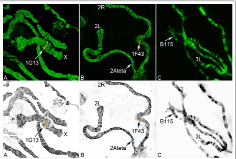
In the current study, we used three experimental approaches to map microsatellite markers to the poly-tene chromosomes from ovarian nurse cells of wild femaleAn. nili specimens collected in Burkina Faso. In the first approach, microsatellites were amplified from genomic DNA using specific primers, which were pre-viously developed [27]. All microsatellites were success-fully amplified from the genomic DNA. However, because of the small size of the products (approximately 90-230 bp), a majority of the probes failed to hybridize to chromosomes. Only one microsatellite, 1F43, was mapped by this method. In the second approach, inserts containing microsatellites previously cloned in the pUC18 plasmid [27] were amplified using M13 forward and reverse primers. The insert sizes in this case ranged from 300 to 483 bp. Most of the microsatellites, except F41, B115, 2C157, and A154 were successfully labeled and hybridized to polytene chromosomes. Marker 1F43 was also mapped by the second approach to the same chromosomal region as in the first approach. In the third approach, we used a recently obtained genomic sequence assembly ofAn. nilito identify the microsatel-lite loci via BLASTN search and to design primers for PCR. These primers allowed the amplification of 506-559 bp-long PCR products containing the microsatellites
that could not be hybridized previously. The An. nili
genome was sequenced by Illumina 72 bp paired-end method using genomic DNA isolated from two indivi-dual larvae collected in Dinderesso, Burkina Faso. The assembly consisted of 51,048 contigs with a total length of 98,320,874 bp. The average contig length was 1,926 bp and the maximum contig length was 30,512 bp. Pri-mers were designed for microsatellites B115, 2C157, and A154 (accession numbers: JF742787, JF742788, JF742789) based on sequences identified by BLASTN (Table 1). We successfully mapped microsatellites B115 and 2C157 to polytene chromosomes using this approach. However, microsatellite A154 failed to
hybridize to chromosomes despite several attempts. The BLASTN search yielded multiple hits for microsatellite
locus F41 in theAn. niligenome because of widespread
occurrence of the (CT)11TT(CT)8 repeats. The BLASTN
search of the flanking regions did not yield any signifi-cant hits in theAn. niligenome.
Locations of microsatellite markers on the chromosomal map ofAnopheles nili
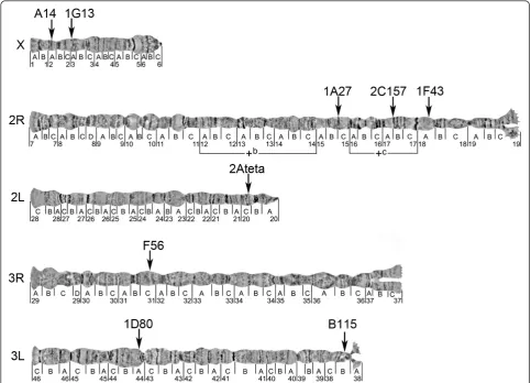
The An. nili chromosomal complement in ovarian nurse cells consists of five chromosomal arms: X, 2R, 2L, 3R and 3L. All nine microsatellites were mapped to unique locations on all autosomes and the X chromo-some using FISH (Figure 1). We assigned these micro-satellites to the precise positions on the recently
developed polytene chromosome map of An. nili
(Fig-ure 2, Table 2). Two microsatellites hybridized to the X chromosome in subdivisions 2A and 3A; three microsatellites localized to the 2R arm in subdivisions 15C, 17AB and 18A; two microsatellites were mapped to the 3L arm in regions 38B and 44A; and arms 2L and 3R each hybridized with only one microsatellite marker in sections 20C and 31C, respectively. Only one microsatellite, 2C157, was mapped inside the
pre-viously described polymorphic inversion2Rc.
Microsa-tellite 1A27 localized to subdivision 15C located
between inversions 2Rb and 2Rc. Microsatellite 1F43
was mapped to subdivision 18A located next to the proximal breakpoint of inversion 2Rc.
Microsatellite mapping through comparative genomics withAnopheles gambiae
In this study, we identified sequences in the An.
gam-biae genome that are homologous to six microsatellite
loci of An. nili (Table 2). The remaining five loci did not have significantly similar sequences in the An.
gam-biaegenome. Markers A14, 2C157, 2Ateta, and B115,
which we mapped in An. niliby FISH, were placed to
specific regions of homologous chromosome arms in
An. gambiae by BLASTN. The BLASTN results con-firmed arm homologies between the two species that we determined in our previous study [32]. In addition, we
mapped in silicomicrosatellites A154 and F41, which
were not previously mapped by FISH. We used the
Illu-mina-based genome sequence assembly of An. nilifor
A154 and the clone sequence for F41 to perform
[image:3.595.55.538.677.731.2]BLASTN against theAn. gambiaegenome. According to
Table 1 Primers designed for the microsatellite loci using theAn. niligenome sequence.
Locus Forward primer Reverse primer Size of PCR product
B115 CACGCTGGCTAAGAGGAAAC CTGGTCTCTAGCACCCGAAG 559 bp
2C157 GAGAGCGGCTGTTCGTAATC CGGACTGGCGAAATAAACAT 506 bp
the established arm homology, we assigned microsatel-lites A154 and F41 to 3R and 2R chromosome arms of
An. nili, respectively (Table 2).
Inversion polymorphism inAnopheles nili
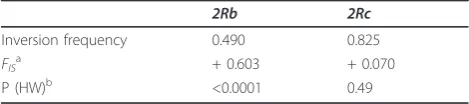
To test if inversions2Rband2Rcare in LD, we karyotyped 44An. nilifemales collected in Dinderesso, Burkina Faso. Inversion frequencies were calculated jointly for these individuals and for 56 previously karyotyped females from the same village [32]. We found a highly significant LD between the two inversions (P = 0.00054), i.e., these inver-sions occur together much more often than expected.
Fre-quencies of inverted and standard2Rb variants were
almost equal (0.51 and 0.49 for the standard and inverted arrangement, respectively). However, a highly significant departure from Hardy-Weinberg proportions due to a def-icit in heterozygotes (e.g., positiveFISvalue) was observed at this locus (FIS= +0.603, P < 0.0001 single test level).
Inversion2Rc occurred at high frequency in the sample
(0.825), with no significant deviation from Hardy-Wein-berg equilibrium (HWE) (P = 0.49) (Table 3).
Discussion
Availability of readable polytene chromosomes in An.
niliallowed us to develop a map of microsatellite mar-kers and to study polymorphism of chromosomal inver-sions. Among the three experimental approaches used to map microsatellite markers to chromosomes, using
cloned inserts and genome sequence assembly of An.
nili to amplify and hybridize microsatellites was more
successful than using microsatellite fragments amplified with primers for population genetics studies [1,27] (Fig-ure 1). Larger DNA fragments were more suitable for effective labeling by the random primer method than smaller fragments obtained with primers for population genetic studies [27]. In addition to these experimental
approaches, we conducted BLASTN searches of theAn.
nili genome fragments with microsatellites (both
Illu-mina generated and cloned) against the An. gambiae
genome to assign microsatellite loci to chromosome
arms according to the synteny betweenAn. niliandAn.
gambiae [32] (Table 2). Although, X, 2R, and 3R are
[image:4.595.58.539.88.413.2]homologous between the two species, the 2L arm ofAn.
gambiaecorresponds to the 3L arm ofAn. nili,and the
3L arm of An. gambiae corresponds to the 2L arm of
An. nili,indicating the presence of a whole-arm translo-cation. Because of the high number of inversions fixed
between the two species, the genome of An. gambiae
[image:5.595.56.540.89.437.2]cannot be used as a reference for precise positioning of microsatellites on theAn. nili chromosomes. In our pre-vious study, we calculated the minimum number of
Figure 2Physical chromosome map of theAn. nilimicrosatellites. Chromosomal locations of nine microsatellite markers on polytene chromosomes are shown by arrows. Two polymorphic chromosomal inversions are indicated by brackets.
Table 2 Location of theAn. nilimicrosatellite markers on polytene chromosomes.
Locus Accession numbera(with length) Location on theAn. nili
map
Location on theAn. gambiae
map
Best e-value for BLASTN
1. A14 AJ536274 (467 bp) X: 2A X: 3D 3e-08
2. 1G13 AJ536271 (441 bp) X: 3A -
-3. 1A27 AJ536268 (300 bp) 2R: 15C -
-4. 2C157 AJ536273 (404 bp) JF742788 (2,967 bp) 2R: 17AB 2R: 10A 7e-04
5. 1F43 AJ536270 (462 bp) 2R: 18A -
-6. F41 AJ536277 (423 bp) 2R:unknownb 2R: 16D 2e-27
7. 2Ateta AJ536272 (479 bp) 2L: 20C 3L: 45A 3e-17
8. F56 AJ536278 (474 bp) 3R: 31C -
-9. A154 JF742789 (1,522 bp) 3R:unknownb 3R: 30E 6e-43
10. B115 AJ536276 (350 bp) JF742787 (10,057 bp)
3L: 38B 2L: 20D 3e-24
11. 1D80 AJ536269 (483 bp) 3L: 44A -
-a
AJ refers to clone sequences; JF refers to assembled sequences.
b
Arms determined based on the chromosome homology established earlier [19].
[image:5.595.55.540.552.719.2]fixed inversions amongAn. nili, An. gambiae, andAn. stephensi and concluded that An. niliis, at least, as
diverged from An. gambiae as An. stephensi[32]. In
addition to the fixed inversion differences, An. nilihas a distinct pattern of polymorphic inversions. Therefore, the chromosomal positions of homologous loci with respect to polymorphic inversions will be different in the two species.
The developed microsatellite map (Figure 2) improved our understanding of the population genetic structure of
An. nili. A recent study using 11 microsatellite markers demonstrated significant genetic differentiation of the
An. nili population of Kenge in the DRC as compared
to theAn. nili populations in Central and West Africa
[1]. Both local adaptation and geographic isolation could cause this differentiation. Extensive allele sharing between populations and homogeneity across loci sug-gested that enhanced genetic drift rather than selection was responsible for the observed pattern. Although it is unlikely that all loci would be within or close to the same inversion, chromosomal mapping of the markers was needed to determine the degree of their indepen-dence. Our study demonstrated that the microsatellite locations are not limited to one or a few specific regions in the genome but spanned the entire five-arm comple-ment (Figure 2). Because most of these markers are phy-sically unlinked, we conclude that enhanced genetic drift, rather than selection was responsible for reduced variability and increased differentiation of the Kenge, DRC population (see also Additional file 1). These data strongly suggest the role of the equatorial forest block as a barrier to gene flow between the south-African and north-African populations ofAn. nili.
Among the mapped microsatellite loci, 1A27 and A14 were found to be in particularly strong and significant departure from HWE due to a deficiency of heterozy-gotes in West Africa (Burkina Faso and Senegal) but not in Central Africa (Cameroon) [1]. We also detected a highly statistically significant departure from HWE due to a deficit in heterozygotes (FIS = +0.603, P < 0.0001 single test level, Table 3) at inversion2Rbin the village of Dinderesso in Burkina Faso among 100 karyotyped females. It is possible that the2Rb inversion plays a role
in local adaptation and subdivides An. nili into
populations with limited gene flow. This process or the presence of null alleles could cause heterozygote defi-ciency at microsatellite loci. In contrast, inversion2Rc
demonstrated no significant deviation from HWE (Table 3). However, we found a highly significant LD between the two inversions (P = 0.00054). Microsatellite 1A27 is
located between 2Rb and2Rc and it could be affected
by the LD and reduced recombination in the vicinity of chromosomal breakpoints (Figure 2). Future studies should determine whether this LD is caused by physical linkage or selection. 2C157 is the only microsatellite located inside an inversion; and it does not demonstrate deficiency of heterozygotes. This locus is in the middle
of inversion2Rcwhere recombination could be close to
normal. Moreover, significant departure from HWE due to a deficiency of heterozygotes was demonstrated for inversion2Rbbut not for2Rc.Marker A14 is located on the X chromosome, which lacks polymorphic inversions, suggesting that genetic differentiation is not limited to the inversions (see Additional file 1 for locus-specific
FSTestimates). Microsatellites in Hardy-Weinberg dise-quilibrium could also be associated with genes
responsi-ble for epidemiologically important ecological
adaptations. Indeed, the microsatellite motif of A14 is located 259 bp upstream from the start codon of an
open reading frame in the An. niligenome, and the
sequence homologous to the A14 clone is found in the
5’UTR and the first exon of the An. gambiae gene
AGAP000275. According to gene ontology annotation, the protein encoded by this gene has oxidoreductase activity. The transcript of AGAP000275 has demon-strated significant differential expression in a variety of mosquito tissues and life stages. Significant differences have been shown between: different stages of embryonic development, between embryonic serosa and embryo [41], between blood-fed and non-blood-fed females, between fat body and ovaries, between males and females, between adults and larva [42], between hemo-lymph and carcass [43], between West and East African strains of S form gravid females [44], between larval anterior midgut and hindgut [45], between larval salivary gland and whole organism [46]. Significant 1.2-fold increase in the transcription level of AGAP000275 has also been found between females 6 hours and 24 hours after mating [47]. Altogether, these data suggest strong
selection acting on AGAP000275 in An. gambiae that
might translate into non-neutral polymorphism distribu-tion at locus A14 in An. nili. Sequences homologous to
other An. nili microsatellite loci with significant
BLASTN hits in the An. gambiaegenome were found
outside genes, except microsatellite B115, which was located within the second intron of gene AGAP004824.
Our recent mapping of 12 microsatellites to An.
[image:6.595.56.291.111.163.2]ste-phensi chromosomes has demonstrated that the
Table 3 Chromosome inversion polymorphism in theAn. nilipopulation from Dinderesso, Burkina Faso.
2Rb 2Rc
Inversion frequency 0.490 0.825 FIS
a
+ 0.603 + 0.070
P (HW)b <0.0001 0.49
a
FISwas computed as in [59].
b
chromosomal position of microsatellites may affect esti-mates of population genetic parameters [48]. In a similar study ofAn. funestus, 16 microsatellites were physically mapped to polytene chromosomes, and the location of microsatellites based on the inversions were determined [49]. Interestingly, microsatellites located between
inver-sions 3Raand 3Rb in An. funestus were found in LD
with these inversions in Burkina Faso [50] but not in Cameroon [30], reflecting different evolutionary out-comes in different eco-geographic regions. Altogether, these studies point to the importance of physical map-ping of molecular markers exposed to contrasted evolu-tionary dynamics for unravelling the demographic and evolutionary history of malaria vectors. This paper pro-vides the necessary toolbox for such endeavour to be pursued inAn. nili.
Conclusions
Our study demonstrates that the chromosomal position of microsatellites is informative for interpretation of population genetics data and highlights the importance of developing physical maps for nonmodel organisms. Next-generation sequencing can be used for designing microsatellite primers to obtain longer microsatellite-containing probes and improve FISH mapping. An Illu-mina-based genome sequence assembly can also be used for identifying homologous loci in the reference gen-omes and assigning microsatellite markers to chromoso-mal arms in a species of interest based on synteny. The integrated chromosomal map of microsatellites and inversions will allow for more complete characterization of An. niliin future population genetics studies. It will be possible to test for a LD among and between inver-sions and microsatellites, genetic differentiation at microsatellite loci located inside and outside inversions, and genetic differentiation according to the distance from inversion breakpoints. In addition, the new genetic map could be used for designing quantitative trait loci mapping studies for this species.
Methods
Wild mosquito collection, preservation, and species identification
Anopheles nili adult females were collected by pyre-thrum spraying and bednet traps in the village of Din-deresso (11°14’N; 4°23’W) in Burkina Faso.Anopheles nililarvae were collected in a river in Dinderesso, Bur-kina Faso. Specimens were identified in the field as
members of the An. niligroup by using morphological
identification keys [51-53] and were further character-ized by molecular assays as An. nili s.s. [54]. Females were dissected under a microscope, and their ovaries at the appropriate stage were preserved in Carnoy’s fixative solution (3 parts of ethanol: 1 part of glacial acetic acid
by volume). Ovaries were kept at room temperature overnight before being stored at -20°C. Larvae were pre-served in Carnoy’s fixative solution and stored at -20°C.
Genome sequencing and BLASTN
The genome assembly for An. nili was obtained by
sequencing of genomic DNA isolated from two larvae collected in Dinderesso, Burkina Faso. Genomic DNA was isolated using the Qiagen DNeasy Blood and Tissue Kit (Qiagen Science, Germantown, MD, USA). The library preparation and sequencing was performed on the Illumina Genome Analyzer IIx, using 72 bp paired-end processing at Ambry Genetics Corp. (Aliso Viejo, CA, USA). Samples were prepared using the Illumina protocol outlined in“Preparing Samples for Sequencing
Genomic DNA” (Part # 11251892 Rev. A 2007). Briefly,
DNA fragment ends were repaired and phosphorylated using Klenow, T4 DNA Polymerase and T4 Polynucleo-tide Kinase. Next, an‘A’base was added to the 3’end of the blunted fragments, followed by ligation of Illumina paired-end adaptor via T-A mediated ligation. The ligated products were size selected by gel purification and then PCR amplified using Illumina Paired-End pri-mers. The library size and concentration were deter-mined using an Agilent Bioanalyzer. The library was seeded onto the flowcell at 8 pM, yielding approximately 275 K clusters per tile, and it was sequenced using 73 cycles of chemistry and imaging (73 cycles) for read 1 and read 2. Initial data processing, including extraction of cluster intensities and base calling, was done using RTA 1.6.47 (SCS version 2.6.26). Sequence quality filter-ing scripts were executed in the Illumina CASAVA soft-ware (ver 1.6.0, Illumina, Hayward, CA). Quality metric data included the approximate proportion of sequences with 1, 2, 3 or 4 errors, IVC plots, and visualizations of cluster intensity over the duration of the sequencing run. The BLASTN algorithm was used to identify
homo-logous sequences in the An. gambiaegenome, which is
available at VectorBase [55]. The BLASTN algorithm was also used to find larger genomic fragments with microsatellite loci in theAn. niligenome using a server and the Geneious 5.1.5 software http://www.geneious. com, a bioinformatics desktop software package pro-duced by Biomatters Ltd http://www.biomatters.com.
Probe preparation
Three approaches were utilized for the microsatellite probe preparation. First, microsatellites were directly
amplified from the An. nili genomic DNA using
using standard M13 forward and reverse primers (Fer-mentas, Inc., Glen Burnie, MD, USA). Third, primers were designed for three microsatellites using the Pri-mer3 program [56] based on sequences identified by
BLASTN in the genome assembly of An. nili(accession
numbers: JF742787, JF742788, JF742789). The size of these fragments was about 506-559 bp. PCR conditions were as follows: 94°C for 5 min; 45 cycles of 94°C for 45 s, 50°C for 45 s and 72°C for 30 s; and 72°C for 5 min. DNA was purified using the GE healthcare illustra GFX PCR DNA and Gel Band Purification Kit (GE Health-care UK Ltd, Buckinghamshire, UK). Probes were labeled using Cy3-AP3-dUTP or Cy5-AP3-dUTP (GE Healthcare UK Ltd., Buckinghmashire, UK) fluorophores by a Random Primer DNA Labelling System (Invitrogen Corporation, Carlsbad, CA, USA).
Chromosome preparation and FISH
To obtain chromosomal slides, follicles of ovaries were separated in 50% propionic acid. Then a cover slip was used to squash the follicles. The quality of slides and the banding pattern of polytene chromosomes were ana-lyzed using an Olympus CX-41 phase contrast micro-scope (Olympus America Inc., Melville, NY, USA). Slides then were dipped into liquid nitrogen, cover slips were removed, and slides were dehydrated in 50%, 70%, 90% and 100% ethanol. Slides were air dried and used for further experiments. Labelled probes were hybridized
at 42°C to An. nili polytene chromosome slides
over-night. Then, slides were washed in 0.2 X SSC (Saline Sodium citrate, 0.03 M sodium chloride and 0.03 M sodium citrate) at 42°C and room temperature. Chromo-somes were stained using YOYO-1 (Invitrogen Corpora-tion, Carlsbad, CA, USA), and slides were mounted in 1,4-diazabicyclo[2.2.2]octane (DABCO) antifade solution. A Zeiss LSM 510 Laser Scanning Microscope (Carl Zeiss MicroImaging, Inc., Thornwood, NY, USA) was used to detect fluorescent signals. Microscopic images were taken from the signal, and the locations of signals were determined using a standard cytogenetic photo map ofAn. nili[32].
Image processing
Confocal images were processed using ImageJ and Adobe Photoshop software as described elsewhere [57]. Briefly, color channels were split from the initial RGB image into separate images. Each channel image was converted into the monochrome image by using a
‘Channel mixer’ and then inverted. The inverted
monochrome image was adjusted by using a ‘Curves’
tool until the background is removed and each chro-mosome of the spread becomes fuzzy-edged. The reduction of noise was achieved by blurring of each pixel with the Gaussian blur filter tool. The quality of
the image was improved by additional application of the ‘Curves’ and/or subtraction of the‘Relative white’. Finally, green channel image with chromosomes was merged with monochrome image FISH signals. Pro-cessing yielded contrasted, inverted, grayscale images with color labels, which are more suitable for mapping.
Population genetics analyses
Homozygous and heterozygous inversions were scored using the chromosomal map published earlier [32]. Alternative chromosomal arrangements were considered as different alleles of the same locus, and conformance to Hardy-Weinberg equilibrium was tested with Fisher’s exact tests available in GENEPOP V4.0 [58]. AFISvalue was computed as in [59]. LD between the inversions
2Rband2Rcwas assessed using the log likelihood ratio statistic (G-test) available in GENEPOP V4.0 [58].
Additional material
Additional file 1: Re-analysing genetic differentiation between Anopheles nilipopulations from West and Central Africa. The file contains the genotypic data re-analyzed according to microsatellite loci cytological location. Locus-specificFSTvalues are shown in Table S1, together withFSTestimates across each chromosomal arm and overall. Locus-specific jackknifed meanFSTestimates (+/- standard deviation) betweenAn. nilipopulations from West and Central Africa are shown in Figure S1.
List of abbreviations
BLASTN: Basic Local Alignment Search Tool for Nucleotide sequences; DABCO: 1,4-diazabicyclo [2.2.2] octane; FISH: fluorescentin situhybridization; HWE: Hardy-Weinberg equilibrium; LD: linkage disequilibrium.
Acknowledgements
We are grateful to Z. Tu for hosting the genome sequence ofAn. nilion his server and conducting BLASTN. We thank S. Demin for explaining the image processing procedure. This work was supported by the National Institute of Allergy and Infectious Diseases, National Institutes of Health (grant R21AI079350 to IVS).
Author details 1
Department of Entomology, Virginia Polytechnic and State University, West Campus Drive, Blacksburg, VA 24061, USA.2Malaria Research Laboratory,
Organisation de Coordination pour la lutte contre les Endémies en Afrique Centrale (OCEAC), Yaounde, BP 288, Cameroon.3Institut des Sciences de
l’Evolution, Université Montpellier 2, Centre National de la Recherche Scientifique, Place Eugène Bataillon, C.C. 065, 34095 Montpellier, France.
4Unité Mixte de Recherche 224‘Maladies Infectieuses et Vecteurs: Ecologie,
Genetique, Evolution et Controle (MIVEGEC)’, Team‘Biology, Ecology and Evolution of vector Systems (BEES)’, Institut de Recherche pour le Developpement (IRD), BP 64501, Montpellier 34394, France.5Institut de Recherche en Sciences de la Santé (IRSS), 399 Avenue de la Liberte, Bobo Dioulasso, 01BP171, Burkina Faso.
Authors’contributions
which was critically revised by CAN, FS, and MVS. All authors read and approved the final manuscript.
Competing interests
The authors declare that they have no competing interests.
Received: 5 August 2011 Accepted: 19 October 2011 Published: 19 October 2011
References
1. Ndo C, Antonio-Nkondjio C, Cohuet A, Ayala D, Kengne P, Morlais I, Awono-Ambene PH, Couret D, Ngassam P, Fontenille D, Simard F:
Population genetic structure of the malaria vectorAnopheles niliin sub-Saharan Africa.Malar J2010,9:161.
2. Antonio-Nkondjio C, Kerah CH, Simard F, Awono-Ambene P, Chouaibou M, Tchuinkam T, Fontenille D:Complexity of the malaria vectorial system in Cameroon: contribution of secondary vectors to malaria transmission.J Med Entomol2006,43:1215-1221.
3. Okara RM, Sinka ME, Minakawa N, Mbogo CM, Hay SI, Snow RW:
Distribution of the main malaria vectors in Kenya.Malar J2010,9:69. 4. Van Bortel W, Harbach RE, Trung HD, Roelants P, Backeljau T, Coosemans M:
Confirmation ofAnopheles varunain Vietnam, previously misidentified and mistargeted as the malaria vectorAnopheles minimus.Am J Trop Med Hyg2001,65:729-732.
5. Coluzzi M:Malaria vector analysis and control.Parasitol Today1992,
8:113-118.
6. Enayati A, Hemingway J:Malaria Management: Past, Present, and Future.
Annual Review of Entomology2010,55:569-591.
7. Sinka ME, Bangs MJ, Manguin S, Coetzee M, Mbogo CM, Hemingway J, Patil AP, Temperley WH, Gething PW, Kabaria CW,et al:The dominant
Anophelesvectors of human malaria in Africa, Europe and the Middle East: occurrence data, distribution maps and bionomic precis.Parasit Vectors2010,3:117.
8. Djenontin A, Bio-Bangana S, Moiroux N, Henry MC, Bousari O, Chabi J, Osse R, Koudenoukpo S, Corbel V, Akogbeto M, Chandre F:Culicidae diversity, malaria transmission and insecticide resistance alleles in malaria vectors in Ouidah-Kpomasse-Tori district from Benin (West Africa): A pre-intervention study.Parasit Vectors2010,3:83.
9. Carnevale P, Le Goff G, Toto J, VR:Anopheles nilias the main vector of human malaria in villages of southern Cameroon.Med Vet Entomol1992,
6:135-138.
10. Antonio-Nkondjio C, Awono-Ambene P, Toto JC, Meunier JY, Zebaze-Kemleu S, Nyambam R, Wondji CS, Tchuinkam T, Fontenill D:High malaria transmission intensity in a village close to Yaounde, the capital city of Cameroon.J Med Entomol2002,39:350-355.
11. Prugnolle F, Durand P, Neel C, Ollomo B, Ayala FJ, Arnathau C, Etienne L, Mpoudi-Ngole E, Nkoghe D, Leroy E,et al:African great apes are natural hosts of multiple related malaria species, includingPlasmodium falciparum.Proc Natl Acad Sci USA2010,107:1458-1463.
12. Duval L, Fourment M, Nerrienet E, Rousset D, Sadeuh SA, Goodman SM, Andriaholinirina NV, Randrianarivelojosia M, Paul RE, Robert V,et al:African apes as reservoirs ofPlasmodium falciparumand the origin and diversification of theLaveraniasubgenus.Proceedings of the National Academy of Sciences of the United States of America2010,107:10561-10566. 13. Prugnolle F, Ollomo B, Durand P, Yalcindag E, Arnathau C, Elguero E,
Berry A, Pourrut X, Gonzalez JP, Nkoghe D,et al:African monkeys are infected byPlasmodium falciparumnonhuman primate-specific strains.
Proc Natl Acad Sci USA2011,108:11948-11953.
14. Ramirez CA, Requena JM, Puerta CJ:Identification of the HSP70-II gene in
Leishmania braziliensisHSP70 locus: genomic organization and UTRs characterization.Parasit Vectors2011,4:166.
15. Hiwat H, Bretas G:Ecology ofAnopheles darlingiRoot with respect to vector importance: a review.Parasit Vectors2011,4:177.
16. Cohuet A, Dia I, Simard F, Raymond M, Rousset F, Antonio-Nkondjio C, Awono-Ambene PH, Wondji CS, Fontenille D:Gene flow between chromosomal forms of the malaria vectorAnopheles funestusin Cameroon, Central Africa, and its relevance in malaria fighting.Genetics 2005,169:301-311.
17. Lehmann T, Licht M, Elissa N, Maega BT, Chimumbwa JM, Watsenga FT, Wondji CS, Simard F, Hawley WA:Population structure ofAnopheles gambiaein Africa.J Hered2003,94:133-147.
18. Gakuya F, Rossi L, Ombui J, Maingi N, Muchemi G, Ogara W, Soriguer RC, Alasaad S:The curse of the prey: Sarcoptes mite molecular analysis reveals potential prey-to-predator parasitic infestation in wild animals from Masai Mara, Kenya.Parasit Vectors2011,4:193.
19. Hamarsheh O, Amro A:Characterization of simple sequence repeats (SSRs) fromPhlebotomus papatasi(Diptera: Psychodidae) expressed sequence tags (ESTs).Parasit Vectors2011,4:189.
20. Alasaad S, Oleaga A, Casais R, Rossi L, Min AM, Soriguer RC, Gortazar C:
Temporal stability in the genetic structure ofSarcoptes scabieiunder the host-taxon law: empirical evidences from wildlife-derived Sarcoptes mite in Asturias, Spain.Parasit Vectors2011,4:151.
21. Melachio TT, Simo G, Ravel S, De Meeus T, Causse S, Solano P, Lutumba P, Asonganyi T, Njiokou F:Population genetics ofGlossina palpalis palpalis
from central African sleeping sickness foci.Parasit Vectors2011,4:140. 22. Ouma JO, Beadell JS, Hyseni C, Okedi LM, Krafsur ES, Aksoy S, Caccone A:
Genetic diversity and population structure ofGlossina pallidipesin Uganda and western Kenya.Parasit Vectors2011,4:122.
23. Kovalenko DA, Razakov SA, Ponirovsky EN, Warburg A, Nasyrova RM, Ponomareva VI, Fatullaeva AA, Nasereddin A, Klement E, Alam MZ,et al:
Canine leishmaniosis and its relationship to human visceral leishmaniasis in Eastern Uzbekistan.Parasit Vectors2011,4:58.
24. Echodu R, Beadell JS, Okedi LM, Hyseni C, Aksoy S, Caccone A:Temporal stability ofGlossina fuscipes fuscipespopulations in Uganda.Parasit Vectors2011,4:19. 25. Hamarsheh O:Distribution ofLeishmania major zymodemesin relation to
populations ofPhlebotomus papatasisand flies.Parasit Vectors2011,4:9. 26. Peacock L, Ferris V, Bailey M, Gibson W:Intraclonal mating occurs during
tsetse transmission ofTrypanosoma brucei.Parasit Vectors2009,2:43. 27. Berthomieu A, Kengne P, Awono-Ambene P, Raymond M, Fontenille D,
Weill M:Isolation and characterization of microsatellite DNA markers in the malaria vectorAnopheles nili.Molecular Ecology Notes2003,3:394-396. 28. Tripet F, Dolo G, Lanzaro GC:Multilevel analyses of genetic differentiation
inAnopheles gambiae s.s.reveal patterns of gene flow important for malaria-fighting mosquito projects.Genetics2005,169:313-324. 29. Lanzaro GC, Toure YT, Carnahan J, Zheng L, Dolo G, Traore S, Petrarca V,
Vernick KD, Taylor CE:Complexities in the genetic structure ofAnopheles gambiaepopulations in west Africa as revealed by microsatellite DNA analysis.Proc Natl Acad Sci USA1998,95:14260-14265.
30. Ayala D, Fontaine MC, Cohuet A, Fontenille D, Vitalis R, Simard F:
Chromosomal inversions, natural selection and adaptation in the malaria vectorAnopheles funestus.Mol Biol Evol2011,28:745-758.
31. Costantini C, Ayala D, Guelbeogo WM, Pombi M, Some CY, Bassole IH, Ose K, Fotsing JM, Sagnon N, Fontenille D,et al:Living at the edge: biogeographic patterns of habitat segregation conform to speciation by niche expansion inAnopheles gambiae.BMC Ecol2009,9:16.
32. Sharakhova MV, Antonio-Nkondjio C, Xia A, Ndo C, Awono-Ambene P, Simard F, Sharakhov IV:Cytogenetic map forAnopheles nili: Application for population genetics and comparative physical mapping.Infect Genet Evol2011,11:746-754.
33. Coluzzi M, Sabatini A, della Torre A, Di Deco MA, Petrarca V:A polytene chromosome analysis of theAnopheles gambiaespecies complex.Science 2002,298:1415-1418.
34. Coluzzi M, Sabatini A, Petrarca V, Di Deco MA:Chromosomal
differentiation and adaptation to human environments in theAnopheles gambiaecomplex.Trans R Soc Trop Med Hyg1979,73:483-497.
35. Powell JR, Petrarca V, della Torre A, Caccone A, Coluzzi M:Population structure, speciation, and introgression in theAnopheles gambiae
complex.Parassitologia1999,41:101-113.
36. Toure YT, Petrarca V, Traore SF, Coulibaly A, Maiga HM, Sankare O, Sow M, DiDeco MA, Coluzzi M:The distribution and inversion polymorphism of chromosomally recognized taxa of theAnopheles gambiaecomplex in Mali, West Africa.Parassitologia1998,40:477-511.
37. Simard F, Ayala D, Kamdem GC, Pombi M, Etouna J, Ose K, Fotsing JM, Fontenille D, Besansky NJ, Costantini C:Ecological niche partitioning betweenAnopheles gambiaemolecular forms in Cameroon: the ecological side of speciation.BMC Ecol2009,9:17.
38. Gray EM, Rocca KA, Costantini C, Besansky NJ:Inversion 2La is associated with enhanced desiccation resistance inAnopheles gambiae.Malar J 2009,8:215.
39. Rocca KA, Gray EM, Costantini C, Besansky NJ:2La chromosomal inversion enhances thermal tolerance ofAnopheles gambiaelarvae.Malar J2009,
40. Coluzzi M, Sabatini A, Petrarca V, Di Deco MA:Behavioural divergences between mosquitoes with different inversion karyotypes in polymorphic populations of theAnopheles gambiaecomplex.Nature1977,
266:832-833.
41. Goltsev Y, Rezende GL, Vranizan K, Lanzaro G, Valle D, Levine M:
Developmental and evolutionary basis for drought tolerance of the
Anopheles gambiaeembryo.Dev Biol2009,330:462-470.
42. Marinotti O, Nguyen QK, Calvo E, James AA, Ribeiro JMC:Microarray analysis of genes showing variable expression following a blood meal in
Anopheles gambiae.Insect Molecular Biology2005,14:365-373. 43. Pinto SB, Lombardo F, Koutsos AC, Waterhouse RM, McKay K, An C,
Ramakrishnan C, Kafatos FC, Michel K:Discovery ofPlasmodium
modulators by genome-wide analysis of circulating hemocytes in
Anopheles gambiae.Proc Natl Acad Sci USA2009,106:21270-21275. 44. Cassone BJ, Mouline K, Hahn MW, White BJ, Pombi M, Simard F,
Costantini C, Besansky NJ:Differential gene expression in incipient species ofAnopheles gambiae.Mol Ecol2008,17:2491-2504.
45. Neira Oviedo M, Vanekeris L, Corena-McLeod MD, Linser PJ:A microarray-based analysis of transcriptional compartmentalization in the alimentary canal ofAnopheles gambiae(Diptera: Culicidae) larvae.Insect Mol Biol 2008,17:61-72.
46. Neira Oviedo M, Ribeiro JM, Heyland A, VanEkeris L, Moroz T, Linser PJ:The salivary transcriptome ofAnopheles gambiae(Diptera: Culicidae) larvae: A microarray-based analysis.Insect Biochem Mol Biol2009,39:382-394. 47. Rogers DW, Baldini F, Battaglia F, Panico M, Dell A, Morris HR, Catteruccia F:
Transglutaminase-mediated semen coagulation controls sperm storage in the malaria mosquito.PLoS Biol2009,7:e1000272.
48. Kamali M, Sharakhova M, Baricheva E, Karagodin D, Tu Z, Sharakhov IV:An integrated chromosome map of microsatellite markers and inversion breakpoints for an Asian malaria mosquito,Anopheles stephensi.Journal of Heredity2011.
49. Sharakhov I, Braginets O, Grushko O, Cohuet A, Guelbeogo WM, Boccolini D, Weill M, Costantini C, Sagnon N, Fontenille D,et al:A microsatellite map of the African human malaria vectorAnopheles funestus.J Hered2004,95:29-34.
50. Michel AP, Guelbeogo WM, Grushko O, Schemerhorn BJ, Kern M, Willard MB, Sagnon N, Costantini C, Besansky NJ:Molecular differentiation between chromosomally defined incipient species ofAnopheles funestus.
Insect Mol Biol2005,14:375-387.
51. Gillies MT, Coetzee M:A supplement to the Anophelinae of Africa south of the Sahara.Publ S Afr Inst Med Res1987,55:1-143.
52. Awono-Ambene HP, Kengne P, Simard F, Antonio-Nkondjio C, Fontenille D:
Description and bionomics ofAnopheles(Cellia)ovengensis(Diptera: Culicidae), a new malaria vector species of theAnopheles niligroup from south Cameroon.J Med Entomol2004,41:561-568.
53. Brunhes J, Cuisance D, Cuny G, Manguin S, de La Rocque S, Geoffrey B:
Medical entomology: the technological explosion.Med Trop (Mars)1998,
58:15-20.
54. Kengne P, Awono-Ambene P, Antonio-Nkondjio C, Simard F, Fontenille D:
Molecular identification of theAnopheles niligroup of African malaria vectors.Med Vet Entomol2003,17:67-74.
55. Lawson D, Arensburger P, Atkinson P, Besansky NJ, Bruggner RV, Butler R, Campbell KS, Christophides GK, Christley S, Dialynas E,et al:VectorBase: a data resource for invertebrate vector genomics.Nucleic Acids Res2009,
37:D583-587.
56. Rozen S, Skaletsky H:Primer3 on the www for general users and for biologist programmers.Methods Mol Biol2000,132:365-386.
57. Demin S, Pleskach N, Svetlova M, Solovjeva L:High-resolution mapping of interstitial telomeric repeats in Syrian hamster metaphase
chromosomes.Cytogenet Genome Res2011,132:151-155. 58. Rousset F:GENEPOP‘007: a complete re-implementation of the
GENEPOP software for Windows and Linux.Molecular Ecology Resources 2008,8:103-106.
59. Weir BS, Cockerham CC:Estimating F-Statistics for the Analysis of Population-Structure.Evolution1984,38:1358-1370.
doi:10.1186/1756-3305-4-202
Cite this article as:Peeryet al.:Improving the population genetics toolbox for the study of the African malaria vectorAnopheles nili: microsatellite mapping to chromosomes.Parasites & Vectors20114:202.
Submit your next manuscript to BioMed Central and take full advantage of:
• Convenient online submission
• Thorough peer review
• No space constraints or color figure charges
• Immediate publication on acceptance
• Inclusion in PubMed, CAS, Scopus and Google Scholar
• Research which is freely available for redistribution