Neuron specific reduction in CuZnSOD is not sufficient to initiate a full sarcopenia phenotype
Kavithalakshmi Sataranatarajan1*, Rizwan Qaisar1*, Carol Davis4, Giorgos K Sakellariou3, Aphrodite Vasilaki3, Yiqiang Zhang6, Yuhong Liu7, Shylesh Bhaskaran1, Anne McArdle3, Malcolm Jackson3, Susan V.
Brooks4, Arlan Richardson2, 5 and Holly Van Remmen1, 2
* denotes equal contribution by these authors
1 Free Radical Biology and Aging Program, Oklahoma Medical Research Foundation, Oklahoma City, OK 73104.
2 Oklahoma VA Medical Center, Oklahoma City, OK 73104.
3Liverpool -MRC Arthritis Research UK Centre for Integrated research into Musculoskeletal Ageing (CIMA), Department of Musculoskeletal Biology, Institute of Ageing and Chronic Disease, University of Liverpool, UK.
4Department of Molecular and Integrative Physiology, University of Michigan
5 Reynolds Oklahoma Center on Aging, University of Oklahoma Health Sciences Center and Oklahoma City VA Medical Center, Oklahoma, OK, USA
6 Department of Biochemistry, UT Southwestern Medical Center, Dallas, TX,USA
7 The Sam and Ann Barshop Institute for Longevity and Aging Studies, The University of Texas Health Science Center at San Antonio, San Antonio, TX, USA
Keywords: CuZnSOD, oxidative stress, neuromuscular junction, muscle atrophy, sarcopenia
Corresponding Author Holly Van Remmen, PhD
Free Radical Biology and Aging Research Program, Oklahoma Medical Research Foundation,
825 N.E. 13th Street, Oklahoma City, OK 73104.
Phone: (405) 271-2520 Fax: (405) 271-3470
Highlights
The sarcopenia phenotype of mice lacking CuZnSOD in all tissues is not recapitulated in mice with a neuron specific deletion of CuZnSOD (nSod1KO mice)
Neuronal loss of CuZnSOD reduces force generation to a small extent, but mass is not reduced in the gastrocnemius muscle in 20 month old mice, a muscle that is highly sensitive to age related loss of muscle mass and strength
Loss of CuZnSOD in neuronal tissue alters NMJ morphology and induces moderately markers of muscle denervation; however, it does not initiate neuromuscular junction degeneration, loss of innervation or increases in oxidative stress or mitochondrial generation of ROS.
Abstract.
Introduction
Our previous studies have provided significant insight into potential mechanisms of sarcopenia using mice that lack CuZn-superoxide dismutase (CuZnSOD), i.e., Sod1KO mice, as a model of age-related muscle atrophy. Although indistinguishable from wild type mice at birth, by 5-8 months of age, gastrocnemius muscles of Sod1KO mice display significant reductions in mass and function that progress through adulthood, such that by 20 months of age Sod1KO mice resemble 30-month-old wild type mice (Muller et al., 2006, Jang et al., 2010, Larkin et al., 2011). In addition, both Sod1KOmice and old wild type mice exhibit profound alterations in neuromuscular innervation in conjunction with the initiation of skeletal muscle atrophy (Jang et al., 2010). Post-synaptic endplates are severely disrupted, the number of acetylcholine receptors (AChRs) is reduced and the receptors are significantly fragmented. Finally, mitochondrial function is reduced and mitochondrial ROS generation is elevated in both adult Sod1KO mice and old wild type mice. Overall, our findings in the Sod1KO mice point to the significance of neuromuscular interaction in maintenance of mitochondrial function and muscle mass and suggest that disruption of the neuromuscular junction (NMJ) might be a trigger for declines in muscle mass and function during aging.
Materials and Methods
Generation of neuron-specific Sod1-knockout (nSod1KO) mice. Details of the generation of the Sod1flox/flox mice was reported in our earlier publication (Zhang et al., 2013). To generate nSod1KO mice, Sod1flox/flox mice were bred with a mouse strain that expresses Cre recombinase under the control of the nestin promotor [(B6.Cg(SJL)-Tg(Nes-cre)1Kln/J)] that we obtained from Jackson laboratory (Bar Harbor, ME,USA). Mice were maintained on a 12-hr dark/light cycle and provided with food and water ad libitum. At sacrifice, mice were euthanized by CO2 inhalation and tissues were immediately excised and weighed. All the tissues, except those used for immunostaining, were snap frozen and stored at −80°C. All animal protocols were consistent with The Guide for the Care and Use of Laboratory Animals and approved by the Institutional Animal Care and Use Committee at Oklahoma Medical Research Foundation (OKC, OK, USA).
Measurement of CuZnSOD and MnSOD activity. CuZnSOD and MnSOD activity in brain, spinal cord and muscle were determined using native gels as described (Van Remmen et al., 1999).
CuZnSOD immunoblot analysis. Equal amounts of protein from brain, spinal cord, gastrocnemius and quadriceps muscle were resolved by SDS-PAGE and transferred to PVDF membrane. The membranes were blocked and probed with CuZnSOD (Enzo Life Sciences, Inc. Farmingdale, NY, USA) and GAPDH (Sigma, St. Louis, MO, USA) antibodies overnight at 4 C. Membranes were washed extensively and incubated with secondary antibodies linked to horseradish peroxidase (Santa Cruz Biotechnology, Dallas, TX, USA). Proteins were visualized using enhanced chemiluminescence reagent and signal intensities were quantified using Image J 1.45b software (developed by Wayne Rasband, National Institute of Health, Bethesda, MD)
IgG, (Cell Signalling, Hitchin, UK) was used as secondary antibody. Peroxidase activity was detected using an ECL Plus substrate (Amersham International Cardiff, UK), and band intensities were analysed using Quantity One Software (Biorad Laboratories Ltd). The specificity of the bands was identified in comparison with a sample that had not been exposed to the primary antibody and the molecular weight was determined by using molecular weight markers. All protein contents were normalized to the GAPDH content of the same sample.
Analyses of the 3-nitrotyrosine (3-NT) content of muscle proteins. Total cellular protein was isolated and 20µg was separated by SDS-PAGE followed by western blotting as describe above. The separated proteins were transferred onto polyvinylidene difluoride (PVDF) membranes. The content of 3-NT was analysed by using a rabbit monoclonal antibody (Cell Biolabs, San Diego, USA), as per the manufacturer’s instructions. Bands were visualized and densitometric quantification was undertaken using Quantity One Software (Biorad Laboratories Ltd).
Analysis of F2-isoprostanes. Levels of F2-isoprostanes in quadriceps muscle was measured as decribed
(Ward et al, 2005, Zhang et al., 2013). Briefly, 200mg of tissue was homogenized in 10 ml of ice-cold Folch solution (CHCl3: MeOH, 2:1) containing butylated hydroxytoluene (BHT). The mixture was incubated at room temperature for 30min. 2 ml of 0.9% NaCl was added and mixed well. The homogenate was centrifuged at 3000 × g for 5 minutes at 4°C. The aqueous layer was discarded while the organic layer was secured and evaporated to dryness under N2 at 37°C. Esterified F2-isoprostanes were measured using gas chromatography-mass spectrometry. The level of F2-isoprostanes in muscle tissues was expressed as nanograms of 8-Iso-PGF2α, per gram of muscle tissue.
muscle was isolated from surrounding tissues and the distal tendon was securely tied to the lever arm of a servomotor (model 6650LR, Cambridge Technology). The knee and foot were clamped to the platform. A continual drip of saline warmed to 37°C was applied to the gastrocnemius muscle to maintain its temperature. The muscle was activated by stimulation of the tibial nerve. With the muscle held at optimal length (Lo), 300-ms trains of stimulus pulses were applied at increasing stimulation frequencies until the maximum isometric tetanic force (Po) was achieved. Contractile properties were measured for extensor digitorum longus and soleus muscles in vitro as described by Brooks and Faulkner (1988). Muscles were placed in a horizontal bath containing buffered mammalian Ringer solution maintained at 25°C and tied to a force transducer (model BG-50, Kulite Semiconductor Products, Leonia, NJ) and a servomotor (model 305B, Aurora Scientific, Aurora, ON). Muscles were stimulated by platinum plate electrodes connected to a high-power biphasic current stimulator (model 701B, Aurora Scientific). Muscles were held at Lo and subjected to trains of pulses of 300 ms duration for EDL muscles and 900 ms for soleus muscles. Stimulus frequency was increased until the Po was achieved.
After muscles were removed, deeply anesthetized mice were euthanized by pneumothorax and removal of a vital organ. Muscles were trimmed of their tendons, blotted and weighed. Fiber lengths (Lf) were estimated by multiplying Lo by previously determined Lf-to-Lo ratios (Larkin et al., 2011, Brooks and Faulkner, 1988), and physiological cross-sectional areas (CSA) of the muscles were determined by dividing the mass of the muscle by the product of Lf and 1.06 g/cm3, the density of mammalian skeletal muscle. The specific Po was determined by dividing Po by the physiological CSA.
Neuromuscular Junction (NMJ) imaging. For imaging acetylcholine receptors (AchRs), longitudinal muscle sections were permeabilized with 1% triton X-100 for 1 hour at room temperature and then incubated with Alexa Fluor 647-conjugated bungarotoxin (Molecular Probes, Eugene, OR, USA) diluted in PBS and 1% triton X-100 for one hour at room temperature. Sections were washed with PBS and mounted in Vectashield (Vector Laboratories, Peterborough, UK). Immune fluorescence imaging was obtained with a Nikon TE2000U microscope.
After a brief permeabilization with 0.1% Triton X-100, fibers were stained with rhodamine-phalloidin (1:200, Molecular Probe Inc., Eugene, OR, USA) for 45 minutes and DAPI (Invitrogen, Molecular Probes, OR, USA) for 5 minutes followed by final washes. Linear fiber segments of ≈350-450µm were chosen for imaging. All images were captured with a Nikon TE2000U inverted fluorescence microscope equipped with a 20 x phase-contrast objective (NA 0.45) and imported into Image J software (developed by Wayne Rasband, National Institute of Health, Bethesda, MD) for analysis.
MyHC isoforms composition by gel electrophoresis. In short, the myosin heavy chain (MyHC) isoform composition of the muscle was determined by SDS-PAGE as previously described (Larsson et al., 1993). The total acrylamide and bis concentration were 4% (w/v) in the stacking gel and 8% in the running gel, and the gel matrix included 30% glycerol. Sample loads were kept small to improve the resolution of the MyHC bands, and the electrophoresis was performed at 120 V for 24 h with a Tris-glycine electrode buffer (pH = 8.3) at 4°C.
Mitochondrial H2O2 generation. Intact isolated mitochondria from the gastrocnemius muscles were used to measure reactive oxygen species (ROS) generation indirectly as H2O2 release using the fluorescent probe Amplex Red (Molecular Probes, Eugene, OR, USA) as described previously (Muller et al., 2007, Zhang et al., 2013). The assay was performed in 100ul of reaction buffer that contained 125 mM KCl, 10 mM HEPES, 5 mM MgCl2, and 2 mM K2HPO4 (pH 7.44) along 20-50 μg of mitochondrial protein with and without respiratory substrates GM (2.5 mM glutamate and 2.5 mM malate) at 37°C. Amplex Red oxidation by H2O2 every 2 s for ∼10 min at an excitation of 545 nm and an emission of 590 nm was measured using a Fluoroskan-FL Ascent Type 374 multiwell plate reader (Labsystems, Helsinki, Finland).
Results.
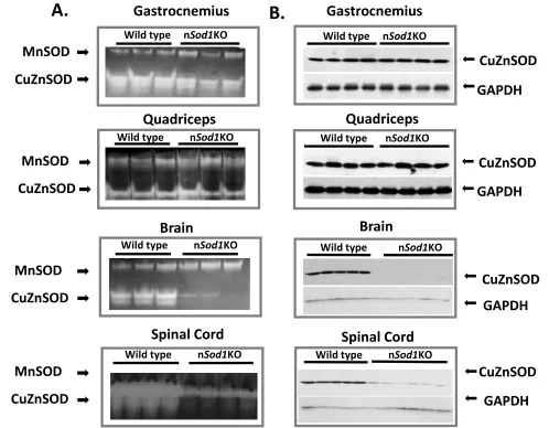
Generation and characterization of a conditional mouse model with reduced CuZnSOD targeted to neuronal tissue. NesCre x Sod1flox/flox mice (nSod1KO mice) are expected to show a reduction in CuZnSOD expression in neuronal tissue with no change in CuZnSOD expression in other tissues (i.e., in muscle). As shown in Figure 1, nSod1KO mice show a dramatic reduction in the activity (Figure 1A) and protein levels (Figure 1B) of CuZnSOD in brain and spinal cord. In contrast, CuZnSOD activity and level in gastrocnemius and quadriceps muscle from nSod1KO mice are equivalent to the values measured in wild type mice. Thus, the nSod1KO mice we have generated shows a neuron specific loss of CuZnSOD and normal levels of muscle CuZnSOD, giving us a model to test directly the role that reduced CuZnSOD in neurons plays in the loss of muscle mass and and NMJ disaggregation that is observed in Sod1KO mice.
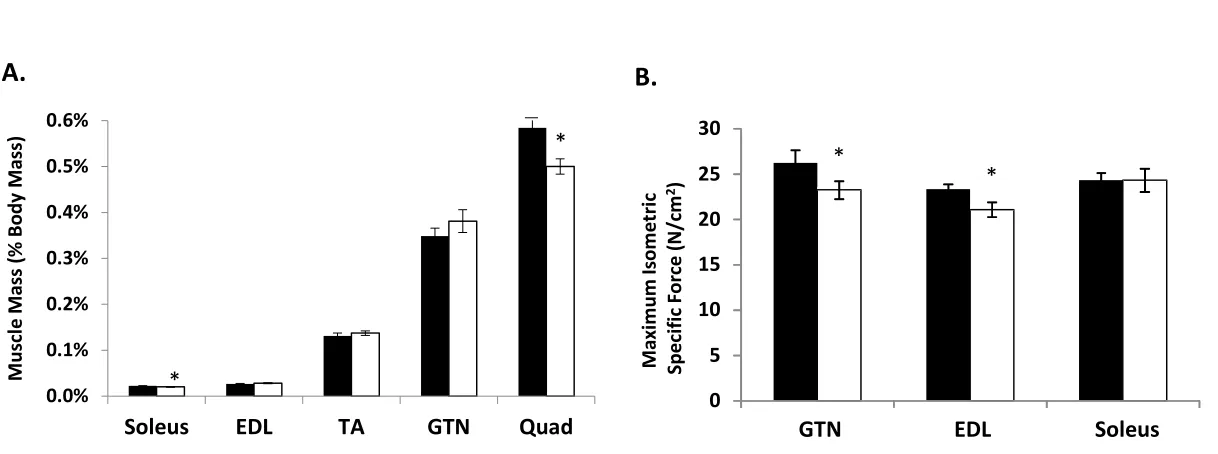
Effect of neuronal depletion of CuZnSOD on muscle mass and strength. Our previous studies showed that Sod1KO mice have significant muscle atrophy in a number of muscles. For example, the mass of lower hind limb muscles, in particular the gastrocnemius muscle, a muscle that undergoes a significant loss of mass during aging (Muller et al., 2006, Larkin et al., 2011), is reduced by 30-45% by 20 months of age (Muller et al., 2006, Larkin et al., 2011, Jang et al., 2012). We have also reported that muscle strength, as measured by specific force, is reduced in Sod1KO mice approximately 50% at 8 month of age (Larkin et al., 2011). We measured muscle mass in a number of individual hind limb muscles in 17- to 20-month-old wild type and nSod1KO mice. As shown in Figure 2A, mass was not altered in the gastrocnemius, tibialis anterior or extensor digitorum longus muscles, while quadriceps (~14%) and soleus muscles (less than 10%) showed small but significant reduction in mass. Muscle-specific effects were also evident in measures of contractile force (Figure 2B). Despite no change in mass, both the extensor digitorum longus and gastrocnemius showed small but significant reductions (~10%) in specific force. In contrast, the soleus muscle, which showed a reduction in mass, showed no loss of force generation.
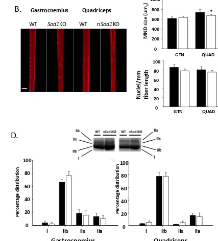
(N=5, n=225) and nSod1KO (N=3, n=136) mice. In contrast to muscles of Sod1KO mice, the data in Figure 3A show there was no significant difference in fiber diameter between the two experimental groups in either muscle. We also measured the number of nuclei per mm of fiber length and calculated the size of the MNDs along the length of single muscle fiber linear segments. We found no difference in the number of nuclei per unit length between the wild type and nSod1KO mice in either muscle (Figure 3B). The MND was not altered in the gastrocnemius muscle, but was slightly reduced in fibers from the quadriceps muscle from nSod1KO mice, reflecting a trend for a reduction in fiber size but unchanged nuclei count when compared to wild type muscle (Figure 3B). Both gastrocnemius and quadriceps muscle from nSod1KO mice showed increased numbers of fibers with central nuclei (Figure 3C). This is in agreement with our previous finding of an increased numbers of central nuclei in gastrocnemius muscle from Sod1KO mice (Jang et al., 2012). The increase in central nuclei may suggest an increase in degeneration and regeneration of quadriceps muscle fibers in the nSod1KO mice compared to wild type mice. Single muscle fiber size, force generation and the nuclei count vary between glycolytic and oxidative fibers and the changes described above can be influenced by the change in muscle fiber type composition. However, as shown in Figure 3D, we do not report a fiber type transition between gastrocnemius and quadriceps muscles from wild type and nSod1KO mice.
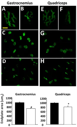
(right panel in Figure 4B). However, the expression of two additional genes associated with the NMJ (MuSK and rapsyn) whose expression is increased with denervation, was not increased. These data suggest a morphological change in the AChR consistent with changes seen in the initiation of denervation; however, the NMJ fragmentation and denervation that occurs in muscle from the Sod1KO mice is not seen in the nSod1KO mice.
Changes in mitochondrial function and oxidative damage. One of the most robust alterations we have measured in response to denervation is mitochondrial dysfunction, especially an increase in generation of H2O2 by isolated muscle mitochondria and increased oxidative damage. For example, we have reported dramatic increases in H2O2 in isolated muscle mitochondria from muscle after sciatic nerve transection and nerve crush (Muller et al., 2007, Walsh et al., 2015). Using the fluorescent probe Amplex Red, we measured H2O2 generation in mitochondria isolated from gastrocnemius of wild type and 17-22 month old nSod1KO mice (Figure 5A). H2O2 generation was not significantly increased during State 1 respiration (no exogenous substrate provided) or in response to glutamate and malate as substrates in the gastrocnemius muscle of the nSod1KO mice.
Discussion.
Aging is accompanied by a 30% to 40% reduction in skeletal muscle mass and function that is the main cause of frailty and loss of independence in the elderly (Roubenoff and Hughes, 2000). This phenomenon, called sarcopenia, is universal to all mammalian species, and is observed even in the absence of disease. The loss of muscle mass involves both loss of muscle fibers and, to a lesser extent, atrophy of the remaining muscle fibers. Aging in skeletal muscle is associated with an increased number of fibers per motor unit and fiber type grouping consistent with a cycle of continual denervation and reinnervation (Essen-Gustavsson and Borges, 1986, Larsson and Edstrom, 1986). In addition to the changes that occur in muscle tissue, parallel changes have also been reported in motor neurons during aging that have been proposed to play an important role muscle atrophy, including loss of motor neurons and alterations in axonal sprouting (Fagg et al., 1981). While the etiology of sarcopenia is poorly understood, several potential contributing factors have been identified, including loss of innervation, muscle mitochondrial dysfunction, and oxidative stress (Fulle et al., 2004, Kadhiresan et al., 1996).
function are prevented in both adult Sod1KO and old wild type mice subjected to dietary restriction ( Jang et al., 2012, Zhang et al.,2013b).
change in the force generating capacity of the tissue.
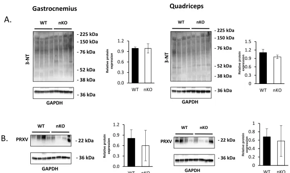
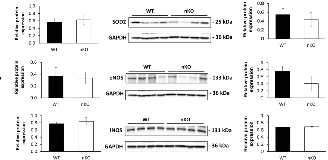
In contrast to the mSod1KO mice, we found that the nSod1KO mice at 17-22 months of age showed changes in NMJ morphology (reduced endplate area) and increased expression of AChRa, Runx1 and gadd45a, genes associated with denervation (Jang et al., 2010, Wang et al., 2005, Bongers et al., 2013). However, we did not observe any evidence of NMJ fragmentation in the nSod1KO mice. In contrast, Sod1KO mice show evidence of NMJ fragmentation as early as 5 months of age (Jang et al., 2012). These results suggest that deleting Sod1 in neuronal tissue has an effect on NMJ structure/function; however, this effect is much less severe than that which occurs in the Sod1KO mouse. We have found the induction of production of reactive oxygen species by isolated muscle mitochondrial (mtROS, as measured by H2O2 generation) and increased oxidative damage and stress in muscle to be consistently associated with NMJ fragmentation and denervation. For example, mtROS is induced over 30-fold following sciatic nerve transection (Muller et al, 2007). In addition, the level of mtROS production by muscle is correlated with the severity of muscle atrophy [e.g, mtROS increased approximately 30% in 30-month-old wild type mice, more than 50% in 20-month-old Sod1KO mice, and approximately 75% in muscle from symptomatic amyotrophic lateral sclerosis (ALS) mice (Muller et al, 2007) and dietary restriction dramatically reduces muscle atrophy and mtROS generation in muscle of the Sod1KO mice (Jang et al., 2012). We did not observe any significant change in either mtROS generation, oxidative damage (measured by F2-isoprostane or protein nitration levels), or oxidative stress (measured by RONS regulatory enzymes) in the muscle of the nSod1KO mice. Thus, the changes we observed in the NMJs of the nSod1KO mice do not lead to changes in the muscle that we observe in the Sod1KO mice.
Acknowledgments.
Figure legends
Figure 1: Generation of neuron-specific Sod1-knockout (nSod1KO) mice. A) Native gels stained for CuZnSOD and MnSOD enzyme activities in gastrocnemius, quadriceps, brain and spinal cord of wild type and nSod1KO mice. B) Western blot analysis of CuZnSOD expression in gastrocnemius, quadriceps, brain and spinal cord of wild type and nSod1KO. (n=3-6 mice in each group between 7 and 18 months of age). Both males and females were used to generate these data.
Figure 2: Muscle mass and function of the nSOD1KO mice. A) Muscle mass expressed as percentage of body mass. B) Maximum isometric specific force (N/cm2) of gastrocnemius, extensor digitorum longus and soleus muscle. (*p<0.05 vs wild type by Students t-test, n=8-10 mice per group, 20 month old mice, wild type -black bars, nSod1KO- white bars)
Figure 3: Fiber diameter, type, myonuclear domain (MND) and number of nuclei in gastrocnemius and quadriceps muscle of the nSOD1KO mice. A) Percentage distribution of fiber diameter of gastrocnemius and quadriceps muscle. B) MND size from single muscle fiber segments of gastrocnemius and quadriceps. Rhodamine-phalloidin labeled actin is shown in red and nuclei visualized by DAPI in blue. Scale bar denotes 50µm. C) Fibers with central nuclei in the gastrocnemius and quadriceps. D) Percentage of fiber type distribution in the gastrocnemius and quadriceps (*p=0.05 vs wild type by t-test, n= 3-7 mice in each group). 5 months and 20 months old mice were used.
Figure 4: Acetylcholine receptor morphology and denervation associated markers in nSod1KO mice A). Acetylcholine receptors from gastrocnemius (GTN) (A, B, C, D) and quadriceps (Quad) muscles (E, F, G, H). (scale bar = 10mm for receptors and 40mm for clusters). B). mRNA levels of the denervation markers. In gastrocnemius,*p<0.001 nSod1KO vs wild type, *p<0.01 Sod1KO vs wild type for AChRα mRNA; *p<0.01 nSod1KO vs wild type, *p<0.0001 Sod1KO vs wild type for Runx1 mRNA;*p<0.05 nSod1KO vs wild type, *p<0.001 Sod1KO vs wild type for GADD45α mRNA by analysis of variance (ANOVA) with comparisons between groups performed using Newman-Keuls post –hoc test using Prism6 software. In quadriceps,*p<0.01 nSod1KO vs WT for AChRα mRNA; *p<0.0001 nSod1KO vs WT for Runx1 mRNA,*p<0.05 nSod1KO vs wild type for GADD45a mRNA by t-test. Mice were 16-18months of age and n= 4-13 mice per group. (WT -black bars, nSod1KO- white bars, Sod1KO- grey bars).
Figure 5: Mitochondrial ROS measurement and F2-isoprostane levels in nSod1KO mice A) Hydrogen
per group) (wild type -black bars, Sod1KO – grey bars, nSod1KO- white bars).
Figure 6: Markers of oxidative damage to proteins in nSOD1KO mice. A) Protein nitration in gastrocnemius and quadriceps. B) Expression of peroxynitrite reductase, PRXV in the gastrocnemius and quadriceps. (n=4 mice per group, 17-22 month old mice with quantification on the right) (wild type- black bars, nSOD1KO- white bars).
References
Bongers, K.S., Fox, D.K., Ebert, S.M., Kunkel, S.D., Dyle, M.C., Bullard, S.A., Dierdorff, J.M., and Adams, C.M. (2013). Skeletal muscle denervation causes skeletal muscle atrophy through a pathway that
involves both Gadd45a and HDAC4. American journal of physiology. Endocrinology and Metabolism 305, E907-915.
Brooks, S.V., and Faulkner, J.A. (1988). Contractile properties of skeletal muscles from young, adult and aged mice. J Physiol 404, 71-82.
Elchuri, S., Oberley, T.D., Qi, W., Eisenstein, R.S., Jackson Roberts, L., Van Remmen, H., Epstein, C.J., and Huang, T.T. (2005). CuZnSOD deficiency leads to persistent and widespread oxidative damage and hepatocarcinogenesis later in life. Oncogene 24, 367-380.
Essen-Gustavsson, B., and Borges, O. (1986). Histochemical and metabolic characteristics of human skeletal muscle in relation to age. Acta Physiol Scand 126, 107-114.
Fagg, G.E., Scheff, S.W., and Cotman, C.W. (1981). Axonal sprouting at the neuromuscular junction of adult and aged rats. Exp Neurol 74, 847-854.
Fulle, S., Protasi, F., Di Tano, G., Pietrangelo, T., Beltramin, A., Boncompagni, S., Vecchiet, L., and Fano, G. (2004). The contribution of reactive oxygen species to sarcopenia and muscle ageing. Exp Gerontol 39, 17-24.
Hamilton, R.T., Bhattacharya, A., Walsh, M.E., Shi, Y., Wei, R., Zhang, Y., Rodriguez, K.A., Buffenstein, R., Chaudhuri, A.R., and Van Remmen, H. (2013). Elevated protein carbonylation, and misfolding in sciatic nerve from db/db and Sod1(-/-) mice: plausible link between oxidative stress and demyelination. PLoS One 8, e65725.
Jang, Y.C., Lustgarten, M.S., Liu, Y.H., Muller, F.L., Bhattacharya, A., Liang, H.Y., Salmon, A.B., Brooks, S.V., Larkin, L., Hayworth, C.R., et al. (2010). Increased superoxide in vivo accelerates age-associated muscle atrophy through mitochondrial dysfunction and neuromuscular junction degeneration. FASEB J 24, 1376-1390.
Jang, Y.C., and Van Remmen, H. (2011). Age-associated alterations of the neuromuscular junction. Exp Gerontol 46, 193-198.
Jang, Y.C., Liu, Y., Hayworth, C.R., Bhattacharya, A., Lustgarten, M.S., Muller, F.L., Chaudhuri, A., Qi, W., Li, Y., Huang, J.Y., et al. (2012). Dietary restriction attenuates age-associated muscle atrophy by lowering oxidative stress in mice even in complete absence of CuZnSOD. Aging Cell 11, 770-782.
Kadhiresan, V.A., Hassett, C.A., and Faulkner, J.A. (1996). Properties of single motor units in medial gastrocnemius muscles of adult and old rats. J Physiol 493 ( Pt 2), 543-552.
Larkin, L.M., Davis, C.S., Sims-Robinson, C., Kostrominova, T.Y., Van Remmen, H., Richardson, A., Feldman, E.L., and Brooks, S.V. (2011). Skeletal muscle weakness due to deficiency of CuZn-superoxide dismutase is associated with loss of functional innervation. Am J Physiol Regul Integr Comp Physiol 301, R1400-1407.
R. L. (1993). Maximum velocity of shortening in relation to myosin isoform composition in single fibers from human skeletal muscles. J Physiol 472, 595-614
McFadden, S.L., Ding, D., Burkard, R.F., Jiang, H., Reaume, A.G., Flood, D.G., and Salvi, R.J. (1999). Cu/Zn SOD deficiency potentiates hearing loss and cochlear pathology in aged 129,mCD-1 mice. The Journal of comparative neurology 413, 101-112.
Muller, F.L., Song, W., Liu, Y., Chaudhuri, A., Pieke-Dahl, S., Strong, R., Huang, T.T., Epstein, C.J., Roberts, L.J., 2nd, Csete, M., et al. (2006). Absence of CuZn superoxide dismutase leads to elevated oxidative stress and acceleration of age-dependent skeletal muscle atrophy. Free Radic Biol Med 40, 1993-2004. Muller, F.L., Song, W., Jang, Y.C., Liu, Y., Sabia, M., Richardson, A., and Van Remmen, H. (2007).
Denervation-induced skeletal muscle atrophy is associated with increased mitochondrial ROS production. Am J Physiol-Reg I 293, R1159-R1168.
Roubenoff, R., and Hughes, V.A. (2000). Sarcopenia: current concepts. J Gerontol A Biol Sci Med Sci 55, M716-724.
Sakellariou, G.K., Pye, D., Vasilaki, A., Zibrik, L., Palomero, J., Kabayo, T., McArdle, F., Van Remmen, H., Richardson, A., Tidball, J.G., et al. (2011). Role of superoxide-nitric oxide interactions in the accelerated age-related loss of muscle mass in mice lacking Cu,Zn superoxide dismutase. Aging Cell 10, 749-760. Sakellariou, G.K., Davis, C.S., Shi, Y., Ivannikov, M.V., Zhang, Y., Vasilaki, A., Macleod, G.T., Richardson, A., Van Remmen, H., Jackson, M.J., et al. (2014). Neuron-specific expression of CuZnSOD prevents the loss of muscle mass and function that occurs in homozygous CuZnSOD-knockout mice. FASEB J.
Shi, Y., Ivannikov, M.V., Walsh, M.E., Liu, Y., Zhang, Y., Jaramillo, C.A., Macleod, G.T., and Van Remmen, H. (2014). The lack of CuZnSOD leads to impaired neurotransmitter release, neuromuscular junction destabilization and reduced muscle strength in mice. PLoS One 9, e100834.
Van Remmen, H., Salvador, C., Yang, H., Huang, T.T., Epstein, C.J., and Richardson, A. (1999). Characterization of the antioxidant status of the heterozygous manganese superoxide dismutase knockout mouse. Arch Biochem Biophys 363, 91-97.
Vasilaki, A., van der Meulen, J.H., Larkin, L., Harrison, D.C., Pearson, T., Van Remmen, H., Richardson, A., Brooks, S.V., Jackson, M.J., and McArdle, A. (2010). The age-related failure of adaptive responses to contractile activity in skeletal muscle is mimicked in young mice by deletion of Cu,Zn superoxide dismutase. Aging Cell 9, 979-990.
Walsh, M.E., Bhattacharya, A., Liu, Y., and Van Remmen, H. (2015). Butyrate prevents muscle atrophy after sciatic nerve crush. Muscle & nerve. In Press
Wang, X., Blagden, C., Fan, J., Nowak, S.J., Taniuchi, I., Littman, D.R., and Burden, S.J. (2005). Runx1 prevents wasting, myofibrillar disorganization, and autophagy of skeletal muscle. Genes & development 19, 1715-1722.
Zhang, Y., Davis, C., Sakellariou, G.K., Shi, Y., Kayani, A.C., Pulliam, D., Bhattacharya, A., Richardson, A., Jackson, M.J., McArdle, A., et al. (2013a). CuZnSOD gene deletion targeted to skeletal muscle leads to loss of contractile force but does not cause muscle atrophy in adult mice. FASEB J 27, 3536-3548. Zhang, Y., Ikeno, Y., Bokov, A., Gelfond, J., Jaramillo, C., Zhang, H.M., Liu, Y., Qi, W., Hubbard, G.,
A.
B.
Figure 1
Gastrocnemius
Quadriceps
CuZnSOD
MnSOD
Gastrocnemius
Quadriceps
CuZnSOD
CuZnSOD
GAPDH
GAPDH
CuZnSOD
MnSOD
Brain
CuZnSOD
MnSOD
Spinal Cord
CuZnSOD
Brain
Spinal Cord
CuZnSOD
GAPDH
GAPDH
CuZnSOD
MnSOD
Wild type nSod1KO Wild type nSod1KO
Wild type nSod1KO
Wild type nSod1KO
Wild type nSod1KO Wild type nSod1KO
Figure 2
A.
B.
0.0% 0.1% 0.2% 0.3% 0.4% 0.5% 0.6%
Soleus
EDL
TA
GTN
Quad
M
usc
le
M
ass (
%
B
od
y M
ass)
*
*
0 5 10 15 20 25 30
GTN
EDL
Soleus
M
ax
im
um
Is
om
et
ric
Sp
eci
fic
Fo
rce
(N
/cm
2)
*
Figure 3
C.
WT
Sod1
KO WT n
Sod1
KO
Gastrocnemius
Quadriceps
GTN QUAD
N
uc
lei
/mm
fibe
r l
eng
th
0 20 40 60 80 100*
M ND siz e (u m3 ) 0 200 400 600 800 1000GTN QUAD
B.
Fiber diameter (um)
20 30 40 50 60 70 80 90
Pe
rc
en
ta
ge
di
str
ibuti
on o
f f
ibe
rs
0 10 20 30 40 M ea n f iber di ame te r ( um) 0 10 20 30 40 50 60Fiber diameter (um)
20 40 60 80 100
Pe
rc
en
ta
ge
di
str
ibuti
on o
f f
ibe
rs
0 10 20 30 40 M ea n f iber di ame te r ( um) 0 10 20 30 40 50 60 70Gastrocnemius
Quadriceps
A.
D.
I IIb IIx IIa
Per cen ta ge d ist rib ut io n 0 20 40 60 80 100
I IIb IIx IIa
Per cen ta ge d ist rib ut io n 0 20 40 60 80 100
Gastrocnemius
Quadriceps
Figure 4
A.
Gastrocnemius
Quadriceps
E
F
G
H
*
0 200 400 600 800 1000 1200*
Endpl
at
e
ar
ea
(um
2)
0 200 400 600 800 10001200 Gastrocnemius Quadriceps
B
.
AChRa Musk Rapsyn Runx1 GADD45a AChRa Musk Rapsyn Runx1 GADD45a
Figure 5.
pmo
lH
2O
2/m
g
m
itoch
on
dr
ia
l p
rot
ei
n
A.
WT n
Sod1
KO
0.0 10.0 20.0 30.0 40.0 50.0 60.0 70.0
Glutamate/Malate
State 1
n
Sod1
KO
WT
0.00 0.50 1.00 1.50 2.00 2.50 3.00 3.50WT
Sod1
KO n
Sod1
KO
Figure 6
3-NT
WT
GAPDH
- 36 kDa
nKO
- 76 kDa
- 52 kDa - 38 kDa - 150 kDa - 225 kDa
0.0 0.3 0.6 0.9 1.2 WT nKO Rel at iv e pr ot ei n ex pr es sio n WT
PRXV - 22 kDa
GAPDH
- 36 kDa
nKO 0.0 0.3 0.6 0.9 1.2 WT nKO Rel at iv e pr ot ei n ex pr es sio n 0 0.3 0.6 0.9 1.2 1.5 WT nKO R el at iv e p ro tei n exp ressi o n WT GAPDH
- 36 kDa nKO
3-NT
- 76 kDa
- 52 kDa - 38 kDa - 150 kDa - 225 kDa
WT
PRXV - 22 kDa
GAPDH
- 36 kDa nKO 0 0.2 0.4 0.6 0.8 1 WT nKO R el at iv e p ro tei n exp ressi o n
Gastrocnemius
Quadriceps
A.
WT
MnSOD
- 25 kDa
GAPDH
- 36 kDa
nKO 0.0 0.2 0.4 0.6 0.8 1.0 WT nKO Rel at iv e p ro tei n exp re ssi on WT
eNOS
- 133 kDa
- 36 kDa
nKO 0.0 0.2 0.4 0.6 WT nKO Rel at iv e p ro tei n exp re ssi onGAPDH
WTiNOS
- 131 kDa
GAPDH
- 36 kDa
[image:31.960.283.934.131.448.2]nKO 0.0 0.2 0.4 0.6 0.8 1.0 WT nKO Rel at iv e p ro tei n exp re ssi on
Figure 7
WT
SOD2
- 25 kDa
GAPDH
- 36 kDa
nKO
WT
eNOS
- 133 kDa
GAPDH
- 36 kDa
nKO
WT
iNOS
- 131 kDa
GAPDH
- 36 kDa
nKO 0 0.2 0.4 0.6 0.8 WT nKO 0 0.2 0.4 0.6 0.8 1 WT nKO 0 0.2 0.4 0.6 0.8 1 WT nKO R el at iv e p ro tei n exp ressi o n R el at iv e p ro tei n exp ressi o n R el at iv e p ro tei n exp ressi o n