JOURNAL OFVIROLOGY,Aug. 1972, p. 256-260 Vol.10,No.2 Copyright © 1972 American SocietyforMicrobiology Printed in U.S.A.
Model
for Vesicular
Stomatitis
Virus
B. CARTWRIGHT, C. J. SMALE, F. BROWN, AND R. HULL
Animal VirusResearch Institute,Pirbright,Surrey, and John InnesInstitute,Norwich,Norfolk, England
Received forpublication 14 April1972
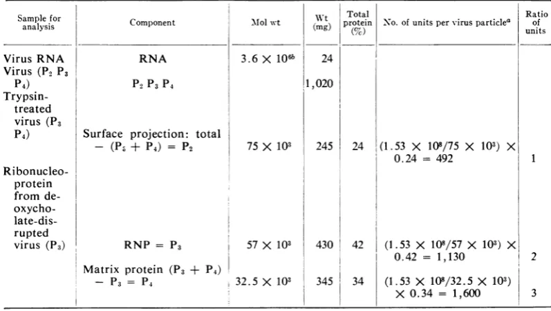
Vesicular stomatitisvirus contains single-stranded ribonucleic acid of molecular weight 3.6 X 10⁶and threemajor proteinswith molecularweightsof 75 x103,57 x 108, and 32.5 X 10³. The proteins have been shown tobe subunits of the surface
projections, ribonucleoprotein, and matrixprotein, respectively. From thesevalues andfrom estimates of the proportions of the individualproteins, it has been calcu-lated that the virus hasapproximately 500 surface projections, 1,100 protein units ontheribonucleoprotein strand, and 1,600 matrix proteinunits. Possible modelsof thevirus areproposed in which the proteins areinterrelated.
Vesicular stomatitis virus is a bullet-shaped, lipid-containing virus which measures approxi-mately 175 X 65 nm and which has well
de-fined surface projections. The virus ribonucleic acid (RNA) is generallyregarded as existing inthe
form of a highly organized helix of ribonucleo-protein (RNP), buttheexactlength oftheRNP strand and the precise form it takes within the
virus particle are not known. For example,
Simpson and Hauser (25) proposed a model for the virusinwhichtheRNP wasarrangedastwo concentric coils with a length of 7.3 ,um, from
which they calculated that the RNAhad a
mo-lecular weight of 7 X 10⁶. In contrast, Nakai
and Howatson (21) showed that the RNP
strands obtained by disruption ofthe virus with
sodium deoxycholate had an average length of 3.6
,m.
Onthebasisofthis value,theysuggested thattherewas onlyonecoil ofRNP in the virusparticle. Such a strand of RNP would contain RNA with a molecular weight of 3.4 X 10⁶ to 3.8 X 10⁶. This value is in fair agreement with
those calculated from sedimentation data (3, 13, 16, 20, 23, 26). The virus contains three
major proteins (18, 29), each of which is
associ-ated with a distinct structural unit (6). This paper reports on the proportions of the three
proteins, and, fromthe molecular weights of the RNA and of theindividual proteins, we suggest further details regarding the architecture of the virus.
MATERIALS AND METHODS
Virus.The Indiana serotype was used in all experi-ments. Unlabeled virus and virus labeled with 3H-uridine were grown in BHK-21 cell monolayers in Eagle's medium. When virus labeled with 14C-amino acids was required, Earle's medium was used. The viruswas purified by the method described previously
(2).
Preparation of virus substructures. Surface projec-tionswereremovedby incubating virus pellets in 0.04 Mphosphate, pH 7.6, with trypsin (1 mg/ml) for 15 minat37 C.The residual projection-free particleswere
isolatedby layeringonto 1 ml of30%sucrosein 0.04 M phosphate, pH7.6,andbycentrifuging intheSW39 rotorof theSpinco ultracentrifuge at30,000rev/min for 1 hr. Thepellet was drained well andsuspended in water. Virus skeletons, i.e., particles from which the surface projections and lipid coat had been
re-moved bytreating atroomtemperature withNonidet P-40 (2mg/ml finalconcentration), wereisolatedby centrifuging throughalayer of 30% sucrose in 0.04 M phosphate, pH 7.6. RNP was isolated in a similar manner after firsttreating the virus with0.5% sodium deoxycholate.
Estimation of RNA and protein. Virus RNA was
extracted from preparations of purified virusby mix-ing with 0.1% sodium dodecyl sulfate and shaking withphenol saturated with 0.1 M NaCl. Theseparated phenol layer was extracted with0.5 volume of water, and this aqueousextract wascombinedwith the
aque-ouslayerobtained in the firstphenolextraction. The combinedlayers were extracted twice with ether and then precipitated with 2 volumes of cold ethanol. After being stored for several hours at -20C, the RNA was separated by centrifuging at 15,000 X g for 1 hr and thenwas dissolved inasmallvolume of
water. Theamountof RNA in this solutionwas esti-matedby the orcinolmethod.
Proteinwasestimatedby the modified
Folin-Ciocal-teau method (19). Estimations were made on intact virus, i.e., virus which had beenincubated with trypsin
to remove thesurfaceprojections and RNP. Ineach case, equal volumes of thepreparationswerelayered
overacushion of 30% sucroseandwerecentrifuged in the SW39 rotorfor 1 hr at 30,000rev/min.
Esti-mateswerethenmadeonsamples of the resuspended pellets.
Electron microscopy. Samples ofvirus, RNP, and skeletons,isolated bysucrosegradientcentrifugation,
were mixed with bovine serum albumin to a final concentration of
0.05%1,
and fixedcrystallineoxliver 256on November 10, 2019 by guest
http://jvi.asm.org/
catalase was added (30). Drops of the samples were
applied to Formvar-carbon grids, negatively stained with
17%
potassium phosphotungstate at pH 7.0, orwith 1%O ammonium molybdate, pH 7.0, and
exam-ined in a Siemens Elmiskop I. Measurements were
madedirectly on thephotographic plates eitherbya
travelingmicroscopeorbyarecording
microdensitom-eter.
RESULTS
Relative proportions of the major structural
proteins ofthevirus. Apurified virus preparation containing 1,020 ,ug ofprotein/ml was disrupted
according tothe scheme in Table 1, and protein estimations were made on the fractions.
Esti-mations were made on virus, virus treated with
trypsin (to remove surface projections), and
isolated RNP. Thematerial used in each estima-tionwas derived from equal volumes ofthe
un-treatedviruspreparation. Theamountofprotein invirus (P2, P3, P4) minus theamountin trypsin-treated virus (P3, P4) gives the protein content
ofthe surface projections. Similarly, the amount
in trypsin-treated virus (P3, P4) minus that in theRNP (P3) gives P4. From the calculationsin Table 1, the percentage of the total virus
pro-tein in each subunit was used to calculate the molar ratios of each protein. These are very
close to those given recently by Helleiner and Wunner(9).
RNA of the virus. Aportion of thepreparation
of virus containing 1,020 Mg ofprotein/ml was
mixed with a trace amount of virus which had been grown in the presence of "4C-amino acids and 3H-uridine, and then was disrupted with
0.1%7,
sodium dodecyl sulfate. After shaking with phenol as described above, the aqueouslayer contained less than 2% of the 14C counts in the virus, and precipitation with ethanol
re-moved this small amount of14C from theRNA preparation. All the 3H radioactivity was
re-covered in the aqueous layer after sodium
dode-cyl sulfate-phenol treatment, and 85% of the
3H counts were present in the resuspended
ethanol precipitate. After allowing for this loss, the RNA present in the virus preparation was
estimated to be 24
Ag/ml,
an amount equal to 2.3 %Oofthevirusprotein.Electron microscopy. The measurements which
were made on electron micrographs of the vi-rusparticles, using crystals of catalaseasinternal
standards (30), are summarized in Table 2, where they are compared with those obtained
by Simpson and Hauser (25) and Nakai and
Howatson (21).
DISCUSSION
We have used the data presented above and that published by other workers (4, 6, 18, 20,
TABLE1. Calculationz ofthe nlumberofproteinzsubunits in one vesicularstomatitis virusparticle
Samplefor wt ToaRti
analysis Component Mlolwt (mg) protein(%c) N o. of units pervirusparticle' unitsof
VirusRNA RNA 3.6 X 10⁶b 24
Virus (P2 P3
P4) P2P3 P4 1,020
Trypsin-treated virus (P3
P4) Surface projection: total
-
(P3
+ P4) = P2 75 X 103 245 24 (1.53 X10⁸/75
X103)
X0.24 = 492 1
Ri bonucleo-protein from de- oxycho-
late-dis-rupted
virus (P3) RNP = P3 57 X 103 430 42 (1.53 X 10⁸/57 X 103) X
0.42 = 1,130 2
Matrix protein (P3 + P4)
- P3 = P4 32.5 X 10³ 345 34 (1.53 X 10⁸/32.5 X 103)
X 0.34 =
1,600
3aWeightofproteinin eachvirusparticle = 3.6 X 106 X (1,020/24) = 1.53 X 10⁸.
bThe useofadifferent molecularweightfor the RNA would alter thetotalweightof the
protein
but would havenoeffectontheratio of thepolypeptides.on November 10, 2019 by guest
http://jvi.asm.org/
[image:2.499.42.441.404.629.2]CARTWRIGHT ET AL.
TABLE2. Dimensions of vesicularstomatitis virusandsome ofitsconstituents
Dimension measured Presentwork Nakai andHowatson Simpsonand Hauser
(1968) (1966)
Overalllengthofvirus(excluding surface
projections)... 175 nm 173 nm 178 nm
Overall diameter of virus (excluding
sur-faceprojections... 65 nm 72 nm 80nm
Number ofstriations ofRNPhelix 35 34 35
Distance between striations... 4.5 nm 4.6nm 5nm
Diameter ofstriated structure... 40-49 nm 49 nm 50nm
Numberofproteinunits percoil of RNP.. 34-35
Size of protein units ofRNP 3by3by9nm 5 by 2 nm
di-ameter
29) to develop a model for the virus. To
calcu-late the number of proteinsubunits inthevirus, it is necessary to know their molecular weights
and the molecular weight of the RNA. Nakai and Howatson (21) calculated from
measure-ments ofthe strand length ofthe RNP that the RNA had a molecular weight of 3.6 X 10⁶, a
valuein good agreement with those obtained by sedimentation studies (3, 13, 16, 20, 23, 26).
The molecular weights of the protein subunits
were taken tobe 75 X 103 for the surface pro-jection, 57 X 103 for the RNP protein, and
32.5 X 10³ for the matrix protein. These are average values calculated from our own deter-minations (6) and from the values published in
the literature (4, 18, 20,29).
Using these values, our estimate of
approxi-mately 1,100 forthe number of protein units on
the RNP strand is similar to that obtained by
electron microscopy
(21).
The surface projec-tions account for 24% of the virus protein,equivalent
to a molecularweight
of 37 X 106and approximately 500 molecules of protein (Table 1). The surface projections are about 10.0 nm inlength and approximately 3.5 nm in
diameter, which suggests that each projection contains only one protein unit of molecular weight 75 X 103. This would mean that there
are approximately 500 surface projections, a figure in good agreement with the figure of 550
given recently for influenza virus (27), the PR-8
strain of which has a surface area about 12% lessthan thatofvesicularstomatitis virus.
The residue, after removing the surface
pro-jections and RNP, accounts for 34% of the
total protein ofthe virus. Ifthis protein is as-sumed to be entirely P4, then we estimate that
there are 1,600 units of the matrix protein in
each virus particle. However, several authors have shown by polyacrylamide gel electroph-oresisthat small amounts of alarge polypeptide
(Pi)
and a smaller polypeptide of molecularweight
ca. 30 x 103(20)
arepresent inthe virus.In our experiments, these polypeptides account for only a smallproportion ofthe total
radioac-tivity
in the protein of the virus. Thepolypep-tides would be found in theP4 fractionwhen the method of fractionation described above is
used. This would reduce the proportion of
P4
andcould meanthat thereare rather fewer than
1,600
matrix proteinunits in the virusparticle.
Our finding that the proportions ofthe three proteins of vesicular stomatitis virus are in the
approximate ratio of 1:2:3, together with the
fact that the matrix protein and RNP form a stable skeletonlike structure (5), led us to ex-amine thefeasibility ofthe proteins being struc-turally interrelated. The published electron
mi-crographs show that the RNP is helically ar-rangedin the virus with 35 turns spaced at
4.75-nm intervals (Table 2). We have no direct evi-dence concerning the arrangement of the matrix
protein subunits and the surfaceprojections, but
electron micrographs of the virus particles sug-gest that they are not helically arranged. The most probable arrangement would seem to be a hexagonal lattice and, in fact, several groups of workers have already found that viruses with surface projections and a lipid envelope often show hexagonal structures when viewed in the electron microscope. Hexagonal structures have been observed in influenza virus (e.g., 1, 8, 12, 22, 24), Rous sarcoma virus (7), monkey kidney vacuolating agent (17), and, probably more
significantly, in rabies virus (15) and inbroccoli
necroticyellows virus (10), both members of the rhabdovirus group.
In designing a model for the virus, the main problem is to relate thehelically arranged RNP to a hexagonal arrangement of the matrix pro-tein and surfaceprojections. Wehave considered two alternativemodels. Inthefirst, eachsurface projection is situated betweenthree matrix pro-tein subunits (Fig. 1) in such a way that the
258 J. VIROL.
on November 10, 2019 by guest
http://jvi.asm.org/
FIG. 1. Hexagonal lattice in which each surface
projectioni (X) issurrountdedby three matrixproteint
subunzits(E). Thesearearratngedtofitatubular struc-ture witht the distribution ofmatrix proteini subunzits
basedoni a92-subuniticosahedronicutacrossits
intier-latticeaxis. The distributioniofthesurface projections
is based on a 32-subuniit icosahedron cut across its
twofoldaxis.
matrix protein subunits of the tubular part of the particle and the surface projections are
ar-ranged as separate hexamers. This is in accord
withthepreviouslydescribedhexagonal
arrange-mentofproteinsubunits (e.g., 11, 28).
The rounded ends of tubular or bullet-shaped
particles can be formed by the use ofpentamer subunits instead of certain hexamers. Hull, Hills, and Markham (14) suggested that the
tubular portion can be considered as being based on a half-icosahedron, with the
arrange-mentofhexamersinthe tubularportion depend-ing on the axis across which the icosahedron is cut. From the dimensions of the virus particles and the numbers of surface projections and
matrix protein subunits per particle (Table 1), thesizes of the hexamers showninFig. 1 canbe
estimated as about 13.5 nm center-to-center for
the surface projection hexamers and about 7.5
nm center-to-center for the matrix protein
hexa-mers. These sizes suggest that the surface
pro-jection structure of this model might be based
on a 32-subunit icosahedron, and that of the
matrix proteinmight be based on a 92-subunit
icosahedron. If theinterrelationship between the
surface projections and matrix protein subunits
isasinFig. 1, thereare onlytwo possible
struc-tures for the tubular portion: (i) a matrix pro-tein layer based on an icosahedron cut across the twofold axis with the surface projection layer on an inter-lattice axis, and (ii) a matrix protein layer based on an inter-lattice axis with the surface projection layer on the twofold axis. However, a helically arranged RNP of the di-mensions given in Table 2 will not fit regularly with the matrix protein layer on either of these two structures.
In the second model, the units of matrix pro-tein are linked directly to the RNP to form a hexagonal array (Fig. 2) in which the matrix protein hexamers are linked. The placing of each surface projection in the center of a matrix
pro-tein hexamer (Fig. 2) gives the ratio of one surface projection to three matrix protein sub-units. A three-dimensional model incorporating these features (Fig. 3) shows that the matrix protein hexamersarein ahelix following that of the RNP coil. End caps for this model can be formed by the substitution of hexamers by pentamers, but they would not have the
icosa-hedral symmetry described for those in the first
model.
At present, there is insufficient evidence on vesicular stomatitis virus to allow us to decide
between these two models. Nevertheless, we think that some of the features of the models
..
+> 4 *~
FIG. 2. RNP(0) linked withahexagonalarrayof matrixprotein subunits tofit a tubularstructure. The surface projections (X) are placedat the centers of thematrixprotein hexamers.
259
on November 10, 2019 by guest
http://jvi.asm.org/
[image:4.499.91.186.81.293.2] [image:4.499.294.391.385.613.2]CARTWRIGHT ET AL.
JN L)IVi) J,N! 'IN RNp p T E
1A'R.X
F' OEFIN
i,i FROJEC T-JNS
FIG. 3. Virusparticle in which a three-,
relationship between the tlhree proteins
at
layerissuggested.
proposed in this paper could be applie
rhabdoviruses and even to other env ruses.
LITERATURE CITED
1. Almeida, J. D., and A. P. Waterson. 1967. Som
on the envelope ofaninfluenza virus. J.G&
46:107-110.
2. Brown, F.,B.Cartwright,and J.D. Almeida. 1
gensofvesicular stomatitis virus.1.Separation
genicity of three complement-fixinig compo munol. 96:537-545.
3. Brown, F., S. J.Martin, B. Cartwright, and J
Theribonucleic acids of the infective andin
ponentsofvesicular stomatitis virus. J. Gen.
486.
4. Burge, B.W., and A. S. Huang. 1970. Compa
brane proteinglycopeptides of Sindbis virus
stomatitisvirus.J.Virol. 6:176-182.
5. Cartwright, B., C. J. Smale, and F. Brown.
structureof vesicularstomatitis virus. J. Gen.
6. Cartwright,B., P. Talbot, and F. Brown. 1970
ofbiologicallyactivesub-unitsofvesicularst(
J. Gen. Virol.7:267-272.
7. Dourmashkin,R., and P. J.Simons. 1961. The
of Roussarcomavirus. J. Ultrastruct. Res. 5
8. Flewett,T. H., and K. Apostolov. 1967. Areti
inthe wall of influenza C virus. J. Gen. Vir
9. Helleiner,C.W., and W.H. Wunner. 1971. As
for counting 14C- and3H-proteins in polyac
Analyt.Biochem. 39:333-338.
10. Hills, G. J., and R. N. Campbell. 1968. M
broccoli necroticyellowsvirus.J.Ultrastruct
144.
Ei, 11. Hitchborn,J. H.,and G. J.Hills. 1968.Astudyoftubes
pro-ducedinplantsinfectedwithastrain of turnip yellowmosaic
virus. Virology.35:50-70.
12. Hoyle,L., R. W.Home, andA.P.Waterson.1962. The
struc-tureandcomposition ofthemyxoviruses.ItI. The interac-tionof influenza virus particles with cytoplasmicparticles derived from normal chorioallantoic membrane cells. Virology 17:533-542.
-'!;-'> 13. Huang, A. S., and R. R. Wagner. 1966. Comparative
sedi-RNF mentation coefficients of RNA extracted from plaque-forming anddefective particles ofvesicularstomatitisviruls.
J. Mol.Biol. 22:381-384.
14. Hull, R., G. J.Hills, andR.Markham. 1969. Studiesonalfalfa
mosaic virus.II. The structure of the virus components.
Virology 37:416-428.
15. Hummeler, K., H.Koprowski,and T. J.Wiktor. 1967.
Struc-ture and development of rabies virus in tissueculture. J.
Virol. 1:152-170.
16. Huppert, J., M. Rosenbergova, L. Gresland, and L. Harel.
1967. Properties of RNA from vesicularstomatitis virus,
p. 463-468. In J. S. Colter and W. Paranchych (ed.),
Molecular biology of viruses. Academic Press Inic., New
York.
17. Jordan,L.E.,G. Plummer,and H.D. Mayor. 1965. The fine
;;-, structureoffoamy virus.Virology25:156-159.
18. Kang,C. Y.,andL.Prevec. 1969. Proteinsofvesicular
stoma-dimenisional titis virus. 1.Polyacrylamide gel analysis ofviralantigens.
,id the lipid J.Virol. 3:404-413.
19. Lowry, 0. H., N. J. Rosebrough, A. L. Farr, and R. J.
Randall. 1951. Proteinmeiasuremerntwith theFolinphenol
reagent. J.Biol. Chem. 193:265-275.
bdto other 20. Mudd, J. A., and D. F. Summilers. 1970. Protein synthesisin
,eloped vi- vesicular stomatitis virus-infected HeLa cells. Virology 42:328-340.
21. Nakai, T., and A. F. Howatson. 1968.The finestructure of
vesicularstomatitis virus. Virology 35:268-281.
22. Nermut, M. V., and H. Frank. 1971. Fine structure of
in-leobservations fluenza A2 (Singapore) as revealed by negative staining,
en. Microbiol. freeze dryingandfreeze-etching. J. Gen. Virol. 10:37-51.
23. Petric, M., and L. Prevec. 197C.Vesicularstomatitis virus-a 966. Theanti- newinterfering particle, intracellular structures,and
virus-andimmuno- specific RNA. Virology 41:615-630.
nents. J. Im- 24. Reginster, M., and C-M. Calberg-Bacq. 1968. Further
ob-servations ontheeffects of caseinase Ccntheenvelopeof
1.Crick. 1967. influenza and Newcastle disease viruses. J. Ultrastruct.
terfering com- Res. 23:144-152.
Virol. 1:479- 25. Simpson, R. W., and R. E. Hauser. 1966. Structural
com-ponentsofvesicularstomatitis virus. Virology 29:654-667.
rison ofmem- 26. Stampfer, M., D. Baltimcre, andA. S. Huang. 1969.
Ribo-and vesicular nucleic acid synthesis of vesicular stomatitis virus. I.
Species ofiibonucleicacid found in Chinese hamsterovary
1969. Surface cells infected withplaque-formning and defective particles.
Virol. 5:1-10. J. Virol. 4:154-161.
The proteins 27.Tiffany, J. M., and H. A. Blough. 1970. Estimation ofthe
omatitisvirus. number of surface projections on myxo- and para
myxo-viruses. Virology41:392-394.
ultrastructure 28. Vergara,J., W.Longley, and J.D. Robertson. 1969.A
hexag-i:505-522. onal arrangement of sub-units in membrane of mouse cularstructure urinarybladder. J. Mol.Biol. 46:593-596.
ol. 1:297-304. 29. Wagner, R. R.,T. A.Schnaitman, andR.M.Snyder. 1969.
simple method vesicular
-rylamide gels. 3:395403.
lorphology of 30. Wrigley,N. G.1968. The latticespacingofcrystallinecatalase
Res.24:134- as aninternal standard length in electron microsccpy.J.
Ultrastruct.Res.24:454-464.