STUDIES ON TEMPERATURE INDUCED MUCOADHESIVE IN SITU GEL
FORMULATION OF RIZATRIPTAN BENZOATE FOR NASAL ADMINISTRATION
A Dissertation Submitted to
The Tamil Nadu Dr. M.G.R. Medical University Chennai - 600 032
In partial fulfillment for the award of Degree of MASTER OF PHARMACY
(Pharmaceutics) Submitted by PARAMESWARI. P Register No. 26116010 Under the Guidance of Mr. T. AYYAPPAN, M. Pharm.,
Assistant Professor, Department of Pharmaceutics.
ADHIPARASAKTHI COLLEGE OF PHARMACY
CERTIFICATE
This is to certify that the dissertation entitled “STUDIES ON
TEMPERATURE INDUCED MUCOADHESIVE IN SITU GEL
FORMULATION OF RIZATRIPTAN BENZOATE FOR NASAL
ADMINISTRATION” Submitted to The Tamil Nadu Dr. M.G.R. Medical University, Chennai, in partial fulfillment for the award of the Degree of the Master of
Pharmacy was carried out by PARAMESWARI.P (Register No.26116010) in the Department of Pharmaceutics under my direct guidance and supervision during the
academic year 2012-2013.
T. AYYAPPAN, M. Pharm.,
Assistant Professor,
Department of Pharmaceutics,
Place: Melmaruvathur Adhiparasakthi College of Pharmacy, Date: Melmaruvathur - 603 319.
CERTIFICATE
This is to certify that the dissertation entitled “STUDIES ON
TEMPERATURE INDUCED MUCOADHESIVE IN SITU GEL
FORMULATION OF RIZATRIPTAN BENZOATE FOR NASAL
ADMINISTRATION” is the bonafide research work carried out by
PARAMESWARI. P (Register No. 26116010) in the Department of Pharmaceutics,
Adhiparasakthi College of Pharmacy, Melmaruvathur which is affiliated to The
Tamil Nadu Dr. M.G.R. Medical University, Chennai, under the guidance of
Mr. T. AYYAPPAN,M. Pharm., Assistant Professor, Department of Pharmaceutics,
Adhiparasakthi College of Pharmacy, during the academic year 2012-2013.
Prof. Dr. T. VETRICHELVAN, M.Pharm., Ph.D.,
Principal,
My Heartfelt Dedication
To
My Beloved Parents
And
ACKNOWLEDGEMENT
First and foremost, I wish to express my deep sense of gratitude to his Holiness ARULTHIRU AMMA, President, ACMEC Trust, Melmaruvathurfor their ever growing Blessings in each step of the study.
With great respect and honor, I extend my thanks to THIRUMATHI
LAKSHMI BANGARU ADIGALAR, Vice President, ACMEC Trust,
Melmaruvathur for given me an opportunity and encouragement all the way in completing the study. Her excellence in providing skillful and compassionate spirit of unstinted support to the department for carrying out research work.
I got inward bound and brainwave to endure experimental investigations in novel drug delivery systems, to this extent; I concede my inmost special gratitude and thanks to Mr. T. AYYAPPAN, M.Pharm., Assistant Professor, Department of Pharmaceutics, Adhiparasakthi College of Pharmacy for the active guidance, valuable suggestions and a source of inspiration where the real treasure of my work.
I owe my sincere thanks with bounteous pleasure to
Prof. (Dr). T.VETRICHELVAN, M.Pharm., Ph.D., Principal, Adhiparasakthi
College of Pharmacy, without his encouragement and supervision it would have been absolutely impossible to bring out the work in this manner.
I have great pleasure in express my sincere heartfelt thanks to
Prof. K. SUNDARA MOORTHY, B.Sc., M.Pharm., Prof. Dr.S. SHANMUGAM,
M. Pharm., Ph.D., Professor and Mr. A. UMAR FARUKSHA, M.Pharm.,
My sincere thanks to our lab technicians Mrs. S. KARPAGAVALLI,
D.Pharm., B.B.A., and Mr. M. GOMATHI SHANKAR, D. Pharm., for their kind
help throughout this work.
I am very grateful to our Librarian Mr. SURESH, M.L.I.S., for his kind cooperation and help in providing all reference books and journals for the completion of this project.
My sincere appreciation is extended to pharmaceutics department for their assistance and cooperation. Furthermore, I would like to express my gratitude for the many people who have walked any forward step with me along this work journey, contributed directly and indirectly in this project.
I am thankful to all my class friends and my best friends M.SUJITHA and
M.ARCHANA for their support and suggestion during my work.
Finally, I would like to extend my heartfelt thanks to my parents
M.PARATHOBAN and P.VIJAYA , to hear my complaints, to help me, to share my
joy, to feel my pain, to contribute frequent prayers, to listen to my latest adventure, to advise me, to ensure that I have the best in life and to hope the best for me. I am certainly very fortunate to have such a wonderful family, without them it would have been impossible for me to achieve this success.
CONTENTS
CHAPTER TITLE PAGE No.
1. INTRODUCTION 1
2. LITERATURE SURVEY
2.1. Literature Review 52
2.2. Drug Profile 64
2.3. Polymers Profile 70
3. AIM AND OBJECTIVE 79
4. PLAN OF WORK 81
5. MATERIALS AND EQUIPMENTS
5.1. Materials used 83
5.2. Equipments used 84
6. PREFORMULATION STUDIES 85
7. FORMULATION OF MUCOADHESIVE NASAL IN
SITU GEL
90
8. EVALUATION OF MUCOADHESIVE NASAL IN
SITU GEL
8.1. Clarity 93
8.2. Measurement of Gelation Temperature (T1) 93
8.3. Measurement of Gel Melting Temperature (T2) 93
CHAPTER TITLE PAGE No.
8.5. Drug Content 94
8.6 Determination of Mucoadhesive Strength 94
8.7. Measurement of viscosity 97
8.8. In-vitro Drug Permeation Studies 97
8.9. Histopathological Study of Optimized Formulation 97
8.10. Kinetics of In-vitro Drug Permeation 98
8.11. Stability Studies 99
9. RESULTS AND DISCUSSION
9.1. Identification of Drug 102
9.2. Evaluation of Mucoadhesive Nasal In Situ Gels 118
9.2.1. Clarity 118
9.2.2. Measurement of Gelation Temperature (T1) 120
9.2.3. Measurement of Gel Melting Temperature (T2) 121
9.2.4. Determination of pH 124
9.2.5. Drug Content 125
9.2.6. Determination of Mucoadhesive Strength 127
9.2.7. Measurement of Viscosity 130
9.2.8. In-vitro Drug Permeation Studies 134
9.2.9. Histopathological Study of Optimized Formulation 143
CHAPTER TITLE PAGE No.
9.2.11. Stability Studies 149
10 SUMMARY AND CONCLUTION 155
12 FUTURE PROSPECTS 158
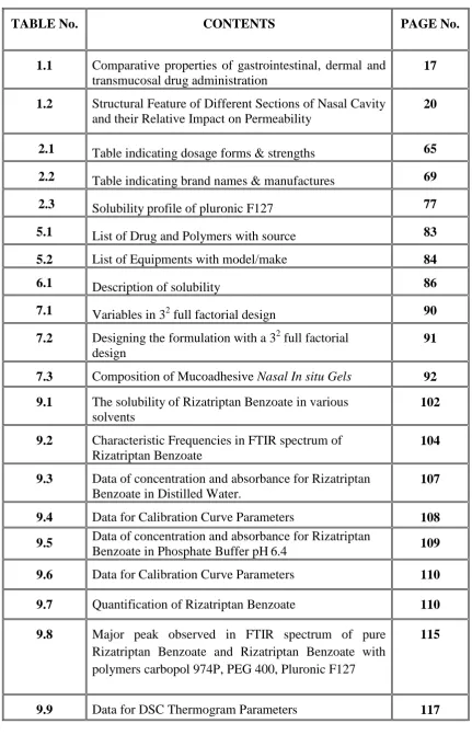
LIST OF TABLES
TABLE No. CONTENTS PAGE No.
1.1 Comparative properties of gastrointestinal, dermal and transmucosal drug administration
17
1.2 Structural Feature of Different Sections of Nasal Cavity and their Relative Impact on Permeability
20
2.1 Table indicating dosage forms & strengths 65
2.2 Table indicating brand names & manufactures 69
2.3 Solubility profile of pluronic F127 77
5.1 List of Drug and Polymers with source 83
5.2 List of Equipments with model/make 84
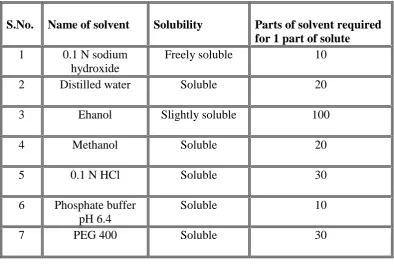
6.1 Description of solubility 86
7.1 Variables in 32
full factorial design 90
7.2 Designing the formulation with a 32 full factorial design
91
7.3 Composition of Mucoadhesive Nasal In situ Gels 92 9.1 The solubility of Rizatriptan Benzoate in various
solvents
102
9.2 Characteristic Frequencies in FTIR spectrum of Rizatriptan Benzoate
104
9.3 Data of concentration and absorbance for Rizatriptan Benzoate in Distilled Water.
107
9.4 Data for Calibration Curve Parameters 108
9.5 Data of concentration and absorbance for Rizatriptan
Benzoate in Phosphate Buffer pH6.4 109
9.6 Data for Calibration Curve Parameters 110
9.7 Quantification of Rizatriptan Benzoate 110
9.8 Major peak observed in FTIR spectrum of pure Rizatriptan Benzoate and Rizatriptan Benzoate with polymers carbopol 974P, PEG 400, Pluronic F127
115
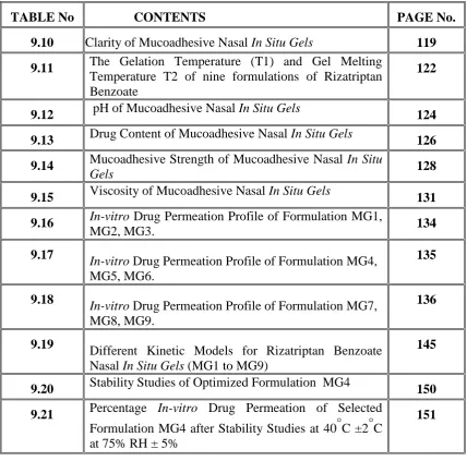
TABLE No CONTENTS PAGE No.
9.10
Clarity of Mucoadhesive Nasal In Situ Gels 119
9.11 The Gelation Temperature (T1) and Gel Melting
Temperature T2 of nine formulations of Rizatriptan Benzoate
122
9.12 pH of Mucoadhesive Nasal In Situ Gels 124
9.13 Drug Content of Mucoadhesive Nasal In Situ Gels 126
9.14 Mucoadhesive Strength of Mucoadhesive Nasal In Situ
Gels 128
9.15 Viscosity of Mucoadhesive Nasal In Situ Gels 131
9.16 In-vitro Drug Permeation Profile of Formulation MG1,
MG2, MG3. 134
9.17 In-vitro Drug Permeation Profile of Formulation MG4,
MG5, MG6.
135
9.18
In-vitro Drug Permeation Profile of Formulation MG7,
MG8, MG9.
136
9.19
Different Kinetic Models for Rizatriptan Benzoate Nasal In Situ Gels (MG1 to MG9)
145
9.20 Stability Studies of Optimized Formulation MG4 150
9.21 Percentage In-vitro Drug Permeation of Selected
Formulation MG4 after Stability Studies at 40°C ±2°C at 75% RH ± 5%
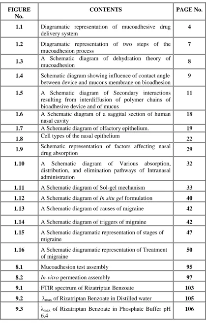
LIST OF FIGURES
FIGURE No.
CONTENTS PAGE No.
1.1 Diagramatic representation of mucoadhesive drug delivery system
4
1.2 Diagramatic representation of two steps of the mucoadhesion process
7
1.3 A Schematic diagram of dehydration theory of
mucoadhesion 8
1.4 Schematic diagram showing influence of contact angle between device and mucous membrane on bioadhesion
9
1.5 A Schematic diagram of Secondary interactions resulting from interdiffusion of polymer chains of bioadhesive device and of mucus
11
1.6 A Schematic diagram of a saggital section of human nasal cavity
18
1.7 A Schematic diagram of olfactory epithelium. 19
1.8 Cell types of the nasal epithelium 22
1.9 Schematic representation of factors affecting nasal
drug absorption 29
1.10 A Schematic diagram of Various absorption,
distribution, and elimination pathways of Intranasal administration
32
1.11 A Schematic diagram of Sol-gel mechanism 33
1.12 A Schematic diagram of In situ gel formulation 40
1.13 A Schematic diagram of causes of migraine 42
1.14 A Schematic diagram of triggers of migraine 42
1.15 A Schematic diagramatic representation of stages of
migraine
47
1.16 A Schematic diagramatic representation of Treatment
of migraine
50
8.1 Mucoadhesion test assembly 95
8.2 In-vitro permeation assembly 97
9.1 FTIR spectrum of Rizatriptan Benzoate 103
9.2 λmax of Rizatriptan Benzoate in Distilled water 105
9.3 λmax of Rizatriptan Benzoate in Phosphate Buffer pH
6.4
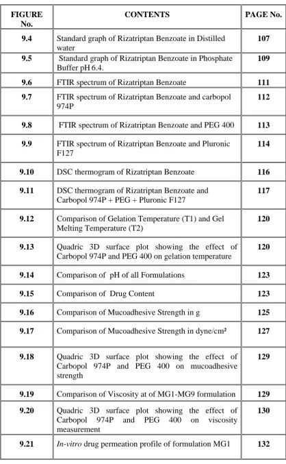
FIGURE No.
CONTENTS PAGE No.
9.4 Standard graph of Rizatriptan Benzoate in Distilled water
107
9.5 Standard graph of Rizatriptan Benzoate in Phosphate Buffer pH6.4.
109
9.6 FTIR spectrum of Rizatriptan Benzoate 111
9.7 FTIR spectrum of Rizatriptan Benzoate and carbopol 974P
112
9.8 FTIR spectrum of Rizatriptan Benzoate and PEG 400 113 9.9 FTIR spectrum of Rizatriptan Benzoate and Pluronic
F127
114
9.10 DSC thermogram of Rizatriptan Benzoate 116
9.11 DSC thermogram of Rizatriptan Benzoate and
Carbopol 974P + PEG + Pluronic F127
117
9.12 Comparison of Gelation Temperature (T1) and Gel
Melting Temperature (T2)
120
9.13 Quadric 3D surface plot showing the effect of
Carbopol 974P and PEG 400 on gelation temperature
120
9.14 Comparison of pH of all Formulations 123
9.15 Comparison of Drug Content 123
9.16 Comparison of Mucoadhesive Strength in g 125
9.17 Comparison of Mucoadhesive Strength in dyne/cm² 127
9.18 Quadric 3D surface plot showing the effect of
Carbopol 974P and PEG 400 on mucoadhesive strength
129
9.19 Comparison of Viscosity at of MG1-MG9 formulation 129
9.20 Quadric 3D surface plot showing the effect of
Carbopol 974P and PEG 400 on viscosity measurement
130
FIGURE No.
CONTENTS PAGE No.
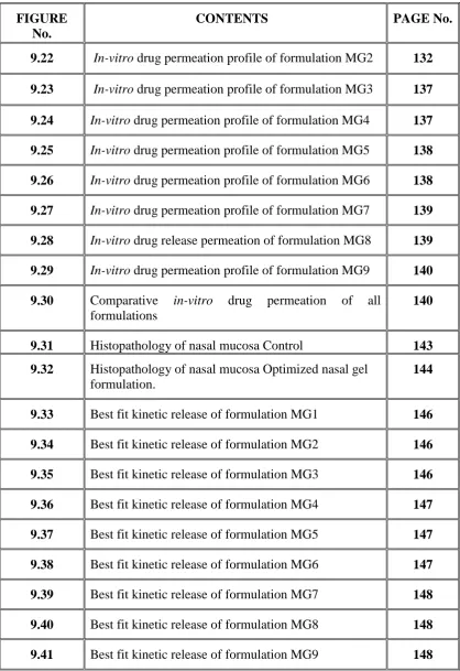
9.22 In-vitro drug permeation profile of formulation MG2 132
9.23 In-vitro drug permeation profile of formulation MG3 137
9.24 In-vitro drug permeation profile of formulation MG4 137
9.25 In-vitro drug permeation profile of formulation MG5 138
9.26 In-vitro drug permeation profile of formulation MG6 138
9.27 In-vitro drug permeation profile of formulation MG7 139
9.28 In-vitro drug release permeation of formulation MG8 139
9.29 In-vitro drug permeation profile of formulation MG9 140
9.30 Comparative in-vitro drug permeation of all
formulations
140
9.31 Histopathology of nasal mucosa Control 143
9.32 Histopathology of nasal mucosa Optimized nasal gel
formulation.
144
9.33 Best fit kinetic release of formulation MG1 146
9.34 Best fit kinetic release of formulation MG2 146
9.35 Best fit kinetic release of formulation MG3 146
9.36 Best fit kinetic release of formulation MG4 147
9.37 Best fit kinetic release of formulation MG5 147
9.38 Best fit kinetic release of formulation MG6 147
9.39 Best fit kinetic release of formulation MG7 148
9.40 Best fit kinetic release of formulation MG8 148
FIGURE No.
CONTENTS PAGE No.
9.42 Comparison of gelation and gel melting temperature
before and after stability studies at 40°C ±2°C at 75% RH ± 5% for optimized formulation MG4
150
9.43 Comparison of pH before and after stability studies at
40°C ±2°C at 75% RH ± 5% for optimized formulation MG4
150
9.44 Comparison of drug content of selected formulation
MG4after stability studies at 40°C ±2°C at 75% RH ± 5%
151
9.45 Percentage in-vitro drug permeation of selected
formulation MG4 after stability studies at 40°C ±2°C at 75% RH ± 5%
152
9.46 Comparison of in- vitro drug permeation before and
after stability studies at 40°C ±2°C at 75% RH ± 5% for optimized formulation MG4
ABBREVIATIONS
% ---- Percentage
°C ---- Degree Celsius
µg ---- Microgram
µg/mL ---- Microgram per milliliter
BBB ---- Blood brain barrier
cm ---- Centimeter
CNS ---- Central nervous system cPs ---- Centipoise
DSC ---- Differential Scanning Calorimetry edn ---- Edition
eg ---- Example
Eq ---- Equation
Fig ---- Figure
FTIR ---- Fourier Transform Infra Red Spectroscopy
gm ---- Grams
d/cm2 ---- Dyne per square centimeter
h ---- Hours
HCl ---- Hydrochloric acid
ICH ---- International Conference on Harmonization
[image:16.595.105.488.76.754.2]I.N. ---- Intranasal
ml ---- Milliliter
mg ---- Milligram
mg/mL ---- Milligram per milliliter
MDDS ---- Mucoadhesive drug delivery system
N ---- Normality
NDDS ---- Novel drug delivery system
nm ---- Nanometer
No. ---- Number
PEG 400 ---- Polyethlene glycol 400
PF127 ---- Pluronic flake127
pH ---- Negative logarithm of hydrogen ion
RB ---- Rizatriptan Benzoate
rpm ---- Revolutions per Minute
SD ---- Standard Deviation
S.No. ---- Serial Number
t ---- Time
UV ---- Ultra Violet
w/v ---- weight in volume
1. INTRODUCTION
(Panchal DR., et al., 2012)
Recently, controlled and sustained drug delivery has become the standard in modern Pharmaceutical design and an intensive research have been undertaken in achieving much better drug product effectiveness, reliability and safety. Over the past 30 years greater attention has been focused on development of controlled and sustained drug delivery systems.
The most desirable and convenient method of drug administration is the oral route because of their ease of administration. However, in many instances oral administration is not desirable when the drug undergoes significant degradation via first pass effect in liver. Hence, lack of systemic absorption through the gastrointestinal tract led to research on alternate routes of drug delivery such as parenteral, intramuscular, subcutaneous, intranasal, transdermal etc.
Intranasal (IN) administration is a needle free and hence an ideal alternative to the parenteral route for systemic drug delivery. Nasal mucosa consists of a rich vasculature and a highly permeable structure for systemic absorption. Drug administration through the nasal cavity is easy and convenient. Avoidance of first pass metabolism is the main advantage of nasal route of drug delivery.
1.1. Mucoadhesive Drug Delivery System:
(Flavia Chiva Carvalho, et al., 2010)
Bioadhesive is the term that describes the adhesion of a polymer to a biological substrate. More specifically, when adhesion is restricted to the mucous layer lining of the mucosal surface it is termed as mucoadhesion.
Mucoadhesive controlled release devices can improve the effectiveness of a drug by maintaining the drug concentration between the effective and toxic levels, inhibiting the dilution of drug in the body fluids, and allowing targeting and localization of a drug at a specific site.
Mucoadhesion also increases the intimacy and duration of contact between a drug containing polymer and a mucous surface. The combined effects of the direct drug absorption and decrease in excretion rate (due to prolonged residence time) allow for an increased bioavailability of the drug with a smaller dosage and less frequent administration.
1.1.1. Fundamentals of Bioadhesion:
Development of an adhesive bond between a polymer and biological membrane or its coating can be visualized as a two step process:
Initial contact between the two surfaces.
Formation of secondary bonds due to non-covalent interaction.
This process of bond formation attributed to surface (or surface coat) of the biological membrane surface of the adhesive and the interfacial layer between the two surfaces. Molecular events that take place in the interfacial layer depend on the properties of the polymer and membrane.
1.1.2. NEED OF MUCOADHESIVE DELIVERY
Fig 1.1: Diagramatic representation of mucoadhesive drug delivery system 1.1.3. Mechanism of Mucoadhesion:
The mucoadhesive must spread over the substrate to initiate close contact and increase surface contact, promoting the diffusion of its chains within the mucus. Attraction and repulsion forces arise and, for a mucoadhesive to be successful, the attraction forces must dominate. Each step can be facilitated by the nature of the dosage form and how it is administered. For example, a partially hydrated polymer can be adsorbed by the substrate because of the attraction by the surface water.
phenomenon which involves wetting, adsorption and interpenetration of polymer chains. Mucoadhesion has the following mechanism,
1. Intimate contact between a bioadhesive and a membrane (wetting or swelling phenomenon).
2. Penetration of the bioadhesive into the tissue or into the surface of the mucous membrane (interpenetration)
The mechanism of mucoadhesion is generally divided in two steps,
The contact stage
The contact stage
The contact stage:
(osmotic pressure, electrostatic repulsion, etc.) and attractive forces (van der Waals forces and electrostatic attraction). Therefore, the particle must overcome this repulsive barrier).
The consolidation step:
In the consolidation step (Figure 1.2), the mucoadhesive materials are activated by the presence of moisture. Moisture plasticizes the system, allowing the mucoadhe-sive molecules to break free and to link up by weak van der Waals and hydrogen bonds.
Essentially, there are two theories explaining the consolidation step: the diffusion theory and the dehydration theory.
Fig 1.3: A Schematic diagram of dehydration theory of mucoadhesion
Mucoadhesion Theories: ( Flavia Chiva Carvalho1., et al., 2010)
Although the chemical and physical basis of mucoadhesion are not yet well understood, there are six classical theories adapted from studies on the performance of several materials and polymer-polymer adhesion which explain the phenomenon.
Electronic theory
Electronic theory is based on the premise that both mucoadhesive and biological materials possess opposing electrical charges. Thus, when both materials come into contact, they transfer electrons leading to the building of a double electronic layer at the interface, where the attractive forces within this electronic double layer determines the mucoadhesive strength.
Adsorption theory
phenomenon because, although they are individually weak, a great number of interactions can result in an intense global adhesion .
Wetting theory
The wetting theory applies to liquid systems which present affinity to the surface in order to spread over it. This affinity can be found by using measuring techniques such as the contact angle. The general rule states that the lower the contact angle then the greater the affinity (Figure 1.4). The contact angle should be equal or close to zero to provide adequate spreadability.
Fig 1.4: Schematic diagram showing influence of contact angle between device and
mucous membrane on bioadhesion.
The spreadability coefficient, SAB, can be calculated from the difference between the surface energies γB and γA and the interfacial energy γAB, as indicated in equation (1).
S AB = γ B – γ A- γ AB (1)
needed to separate the two phases.
WA = γ A+ γ B – AB (2)
Diffusion theory
Diffusion theory describes the interpenetration of both polymer and mucin chains to a sufficient depth to create a semi-permanent adhesive bond (Figure 4). It is believed that the adhesion force increases with the degree of penetration of the polymer chains . This penetration rate depends on the diffusion coefficient, flexibility and nature of the mucoadhesive chains, mobility and contact time. According to the literature, the depth of interpenetration required to produce an efficient bioadhesive bond lies in the range 0.2-0.5 μm. This interpenetration depth of polymer and mucin chains can be estimated by equation 3:
l = (tDb) ½ (3)
where t is the contact time, and Db is the diffusion coefficient of the mucoadhesive material in the mucus. The adhesion strength for a polymer is reached when the depth of penetration is approximately equivalent to the polymer chain size.
Fig 1.5: A Schematic diagram of Secondary interactions resulting from
interdiffusion of polymer chains of bioadhesive device and of mucus
Fracture theory
This is perhaps the most-used theory in studies on the mechanical measurement of mucoadhesion. It analyses the force required to separate two surfaces after adhesion is established. This force, sm, is frequently calculated in tests of resistance to rupture by the ratio of the maximal detachment force, Fm, and the total surface area, A0, involved in the adhesive interaction (equation 4):
S m = Fm/Ao (4)
In a single component uniform system, the fracture force, sj, which is equivalent to the maximal rupture tensile strength, sm, is proportional to the fracture energy (gc), for Young’s module (E) and to the critical breaking length (c) for the fracture site, as described in equation 5:
Sf ~ [gcE/c]1/2 (5) Fracture energy (gc) can be obtained from the reversible adhesion work, Wr
work, Wi (work of plastic deformation provoked by the removal of a proof tip until the disruption of the adhesive bond), and both values are expressed as units of fracture surface (Af).
gc = Wr +W1 (6)
The elastic module of the system (E) is related to the stress (s) and to the shear (e) by Hooke’s law:
E = [σ/ε] ε ->0 = [F/A0/ΔI/l0] Δl ->0 (7)
In equation 7, the stress is the ratio between force (F) and area (A0), and shear is given by the ratio between the variation of system thickness (Dl) and the original thickness (l0).
Mechanical theory
Mechanical theory considers adhesion to be due to the filling of the irregularities on a rough surface by a mucoadhesive liquid. Moreover, such roughness increases the interfacial area available to interactions thereby aiding dissipating energy and can be considered the most important phenomenon of the process.
It is unlikely that the mucoadhesion process is the same for all cases and therefore it cannot be described by a single theory. In fact, all theories are relevant to identify the important process variables.
The mechanisms governing mucoadhesion are also determined by the intrinsic properties of the formulation and by the environment in which it is applied . Intrinsic factors of the polymer are related to its molecular weight, concentration and chain flexibility. For linear polymers, mucoadhesion increases with molecular weight, but the same relationship does not hold for non-linear polymers. It has been shown that more concentrated mucoadhesive dispersions are retained on the mucous membrane for longer periods, as in the case of systems formed by in situ gelification. After application, such systems spread easily, since they present rheological properties of a liquid, but gelify as they come into contact the absorption site, thus preventing their rapid removal. Chain flexibility is critical to consolidate the interpenetration between formulation and mucus.
interpenetration. Super-hydration of the system can lead to build up of mucilage without adhesion. The thickness of the mucus layer can vary from 50 to 450 μm in the stomach to less than 1μm in the oral cavity. Other physiological variations can also
occur with diseases.
None of these mechanisms or theories alone can explain the mucoadhesion which occurs in an array of different situations. However, the understanding of these mechanisms in each instance can help toward the development of new mucoadhesive products .
1.1.3. Common Sites of Application for Mucoadhesive Drug Delivery Platform:
Mucoadhesive formulations have been widely used for their targeted and controlled release delivery to many mucosal membrane based organelles. Such formulations may deliver active ingredient for local systemic effect, while bioavailability limiting effects such as enzymatic or hepatic degradation can be avoided or minimized.
In each case of these mucosal routes, mucus characteristics and functions are different. By this definition, the mucosal routes for drug delivery are:
Buccal drug delivery system
Ophthalmic drug delivery system
Vaginal drug delivery system
Nasal drug delivery system
Buccal drug delivery
Additionally, buccal drug delivery can be promptly terminated in cases of toxicity through the removal of dosage form thereby offering a safe and easy method of drug utilization. Various polymers such as sodium carboxymethylcellulose, hydroxypropylcellulose and polycarbophil are used for delivery of peptides, protein and polysaccharides by this routes have been examined.Although gel and ointments are the most patient convenient tablets, patches and films have also been examined. Furthermore buccal drug delivery is associated with high patient compliance, low levels of irritation and offers significant ease of administration.
Ophthalmic drug delivery
The delivery of therapeutic agents to the eye may be achieved using various types of dosage forms including liquid drops, gels, ointments and solid ocular inserts (both degradable and nondegradable). Another interesting delivery system is in situ gelling polymer that undergoes a phase transition after application. Mucoadhesive polymers would be expected only to attach to conjunctival mucus in vivo. Additionally limited bioavailability has been experienced in vivo for carbomer and polycarbophil, as a result of the high swelling capacity of such polymers in the neutral pH environment of the eye. Maintenance of a low viscosity in such systems through pH regulation in the range 4–5 is not acceptable as it may result in patient unease and mild lacrimation, both of which will have an effect on treatment success. User acceptance and compliance may subsequently be limited by physical and psychological barriers surrounding such dosage forms.
Vaginal drug delivery systems
first-damage, and infection commonly observed for parenteral drug delivery routes of administration. While the vagina provides a promising site for systemic drug delivery because of its large surface area, rich blood supply and high permeability, poor retention due to the self-cleansing action of the vaginal tract is often problematic. However, residence times within the vagina tend to be much higher than at other absorption sites such as the rectum or intestinal mucosa. Another important consideration is the change in the vaginal membrane during the menstrual cycle and post-menopausal period. Typical bioadhesive polymers that have been in vaginal formulations include polycarbophil, hydroxypropylcellulose and polyacrylic acid.
Nasal drug delivery
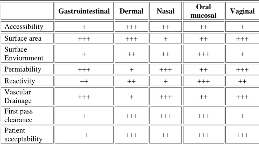
Table 1.1: Comparative properties of gastrointestinal, dermal and transmucosal drug administration:
+ Poor, + + Good, + + + Excellent
1.2. Nasal Drug Delivery System:
The administration of drugs via nose is not a novel approach for drug delivery. In ancient days, nasal drug delivery was used for the systemic administration of psychotherapeutic compounds and other similar substances. But in modern pharmaceutics, nasal drug delivery is considered as a route of choice for local effect rather than systemic effect. Delivery of drugs via nose for maintenance therapy of nasal allergy, sinusitis, nasal congestion, and nasal infections.
In recent years, research has established that the nasal route is a safe and acceptable alternative to the parenteral administration of drugs. The nasal route has
Gastrointestinal Dermal Nasal Oral
mucosal Vaginal
Accessibility + +++ ++ ++ +
Surface area +++ +++ + ++ +++
Surface
Enviornment + ++ ++ +++ +
Permiability +++ + +++ ++ +++
Reactivity ++ ++ + +++ ++
Vascular
Drainage +++ + +++ ++ +++
First pass
clearance + +++ +++ +++ +
Patient
The greater permeability of nasal mucosa with large surface area affords a rapid onset of therapeutic effect. The low metabolic environment of nose has potential to overcome the limitations of oral route and duplicate the benefit of intravenous administration.
1.2.1. Anatomy and Physiology of Nose: (Amol., et al., 2011)
The nose is divided into two nasal cavities via the septum. The volume of nasal cavity is approximately15 ml with a surface area of around 150 cm².
Fig 1.6: A Schematic representation of a sagittal section of human nasal cavity
Schematic representation of a sagittal section of human nasal cavity showing the nasal vestibule , Atrium , respiratory region: inferior turbinate, middle turbinate and the superior turbinate, the olfactory region and nasopharynx .
The Vestibule:
The Respiratory Region:
It contains three nasal turbinates, the superior, middle and inferior which project from the lateral wall of each half of the nasal cavity. The presence of these turbinates creates a turbulent airflow through the nasal passages, which ensures better contact between the inhaled air and the mucosal surface. The respiratory epithelial cells are covered with cilia and microvilli, which increase the surface area available for the absorption of drugs.
The Olfactory Region:
It is situated in the roof of the nasal cavity (15cm²). The olfactory tissue is often yellow in colour, in contrast to the surrounding pink tissue.
Table 1.2: Structural Feature of Different Sections of Nasal Cavity and their Relative Impact on Permeability.
Region Structural Features Structural Features
Nasal vestibule Nasal hairs (vibrissae) Epithelial cells are stratified, squamous and keratinized Sebaceous glands present
Least permeable because of the presence of Keratinized cells
Atrium Transepithelial region Stratified squamouscells present anteriorly and pseudo stratified cells with microvilli present posteriorly
Less permeable as it has small surface area and stratified cells are present anteriorly
Respiratory region (inferior turbinate middle turbinate superior turbinate)
Pseudo stratified ciliated columnar cells with microvilli (300 per cell),large surface area
Receives maximum nasal secretions becauseof the presence of
seromucus glands, nasolacrimal duct and goblet cells
Richly supplied with blood for heating and humidification of inspired air, presence of paranasal sinuses
Most permeable regionbecause of large surfacearea and rich vasculature
Olfactory region Specialized ciliated olfactory nerve cells for smell perception
Receives ophthalmic and maxillary divisions of trigeminal nerve
Direct access to cerebrospinal fluid
Nasopharynx Upper part contains ciliated cells and lower part contains squamous epithelium
1.2.2.Advantages of Nasal Drug Delivery System: (Rahishuddin., et al., 2011)
Drug degradation that is observed in the gastrointestinal tract is absent.
Hepatic first pass metabolism is avoided.
Rapid drug absorption and quick onset of action can be achieved.
The bioavailability of larger drug molecules can be improved by means of
absorption enhancer or other approach.
The nasal bioavailability for smaller drug molecules is good.
Drugs that are orally not absorbed can be delivered to the systemic circulation
by nasal drug delivery.
Drugs possessing poor stability in G.I.T. fluids are given by nasal route.
Polar compounds exhibiting poor oral absorption may be particularly suited for
this route of delivery.
Convenient for those on long term therapy, when compared with parenteral
medication.
1.2.3. Limitations of Nasal Drug Delivery System:
Absorption surface area is less when compared to GIT.
Once the drug administered cannot be removed.
Nasal irritation
1.2.4.Mechanism of Nasal Absorption: (Rahisuddin., et al., 2010)
(i.e. pH, temperature, etc.). So many absorption mechanisms were established earlier but only two mechanisms have been predominantly used, such as:
First mechanism
It involves an aqueous route of transport, which is also known as the paracellular route but slow and passive. There is an inverse log-log correlation between intranasal absorption and the molecular weight of water-soluble compounds. The molecular weight greater than 1000 Daltons having drugs shows poor bioavailability
Second mechanism
It involves transport through a lipoidal route and it is also known as the transcellular process. It is responsible for the transport of lipophilic drugs that show a rate dependency on their lipophilicity. Drug also cross cell membranes by an active transport route via carrier-mediated means or transport through the opening of tight junctions .
For examples: Chitosan, a natural biopolymer from shellfish, opens tight
junctions between epithelial cells to facilitate drug transport.
Figure
The cell type of nasal epithelium showing ciliated cell (A), non-ciliated cell(B), goblet cells(C), gel mucus layer (D), sol layer (E), basal cell (F) and basement membrane (G).
1.2.5. Barriers to Nasal Absorption: ( Swamya N.G.N, et al., 2012)
Nasal drug delivery system is considered has a profitable route for the formulation scientist because it has easy and simple formulation strategies. Intranasally administered drug products therapeutic efficacy and toxicities are influenced by number of factors.
Following factors are the barriers to the absorption of drugs through nasal cavity.
Low bioavailability
Bioavailability of polar drugs is generally low about 10% for low molecular weight drugs and not above 1% for peptides such as calcitonin and insuin.The most important factor limiting the nasal absorption of polar drugs and especially large molecular weight polar drugs such as peptides and proteins is the low membrane permeability. Larger peptides and proteins are able to pass the nasal membrane using an endocytotic transport process but only in low amounts.
Mucociliary clearance
formulations is an approach to overcome the rapid mucociliary clearance. The clearance may also be reduced by depositing the formulation in the anterior and less ciliated part of the nasal cavity thus leading to improved absorption.
Enzymatic degradation
Another contributing, but often less considered factor to the low bioavailability of peptides and proteins across the nasal mucosa is the possibility of an enzymatic degradation of the molecule in the lumen of the nasal cavity or during passage through the epithelial barrier. Both these sites contain exopeptidases such as mono and diamino peptidases that can cleave peptides at their N and C termini and endopeptidases such as serine and cysteine, which can attack internal peptide bonds .
The use of enzyme inhibitors and/or saturation of enzymes may be the approaches to overcome this barrier.
1.2.6.Factors Influencing Nasal Drug Absorption: (Panchal D. R., et al., 2012)
Several factors affect the systemic bioavailability of drugs which are administered through the nasal route. The factors influencing nasal drug absorption are described as follows.
Physiochemical properties of drug.
Molecular size.
Lipophilic-hydrophilic balance.
Enzymatic degradation in nasal cavity.
Nasal Effect
Membrane permeability.
Mucociliary clearance
Cold, rhinitis.
Delivery Effect
Formulation (Concentration, pH, osmolarity)
Delivery effects
Drugs distribution and deposition.
Viscosity
Physiochemical properties of drug
Molecular size
The molecular size of the drug influence absorption of the drug through the nasal route. The lipophilic drugs have direct relationship between the MW and drug permeation whereas water- soluble compounds depict an inverse relationship. The rate of permeation is highly sensitive to molecular size for compounds with MW ≥ 300 Daltons.
Lipophilic-hydrophilic balance
In case of peptides and proteins are having low bioavailability across the nasal cavity, so these drugs may have possibility to undergo enzymatic degradation of the drug molecule in the lumen of the nasal cavity or during passage through the epithelial barrier. These both sites are having exopeptidases and endopeptidases, exopeptidases are monoaminopeptidases and diaminopeptidases. These are having capability to cleave peptides at their N and C termini and endopeptidases such as serine and cysteine, which can attack internal peptide bonds.
Nasal effect factors
Membrane permeability
Nasal membrane permeability is the most important factor, which affect the absorption of the drug through the nasal route. The water soluble drugs and particularly large molecular weight drugs like peptides and proteins are having the low membrane permeability. So the compounds like peptides and proteins are mainly absorbed through the endocytotic transport process in low amounts. Water-soluble high molecular weight drugs cross the nasal mucosa mainly by passive diffusion through the aqueous pores (i.e. tight junctions).
Environmental pH
transcellular route, whereas the more lipophilic ionized form passes through the aqueous paracellular route.
Mucociliary clearance
Mucociliary clearance is a one of the functions of the upper respiratory tract is to prevent noxious substances (allergens, bacteria, viruses, toxins etc.) from reaching the lungs. When such materials adhere to, or dissolve in, the mucus lining of the nasal cavity, they are transported towards the nasopharynx for eventual discharge into the gastrointestinal tract . Clearance of this mucus and the adsorbed/dissolved substances into the GIT is called the MCC. This clearance mechanism influence the absorption process due to the dissolved drugs in the nasal cavity are discharge by the both the mucus and the cilia, which is the motor of the MCC and the mucus transport rate is 6 mm/min. It is of utmost importance that the MCC is not impaired in order to prevent lower respiratory tract infections.
Cold, rhinitis
Rhinitis is a most frequently associated common disease, it influence the bioavailability of the drug. It is mainly classified into allergic rhinitis and common, the symptoms are hyper secretion, itching and sneezing mainly caused by the viruses, bacteria or irritants. Allergic rhinitis is the allergic airway disease, which affects 10% of population. It is caused by chronic or acute inflammation of the mucous membrane of the nose. These conditions affect the absorption of drug through the mucus membrane due the inflammation.
Delivery effect factors
clearance, drug structure can be used to advantage to improve absorption.
Formulation (Concentration, pH, Osmolarity)
The pH of the formulation and nasal surface, can affect a drugs permeation. To avoid nasal irritation, the pH of the nasal formulation should be adjusted to 4.5-6.5 because lysozyme is found in nasal secretions, which is responsible for destroyin certain bacteria at acidic pH. Under alkaline conditions, lysozyme is inactivated and the tissue is susceptible to microbial infection. In addition to avoiding irritation, it results in obtaining efficient drug permeation and prevents the growth of bacteria.
Concentration gradient plays very important role in the absorption /permeation process of drug through the nasal membrane due to nasal mucosal damage. Examples for this are nasal absorption of L-Tyrosine was shown to increase with drug concentration in nasal perfusion experiments. Another is absorption of salicylic acid was found to decline with concentration. This decline is likely due to nasal mucosa damage by the permanent.
The osmolarity of the dosage form affects the nasal absorption of the drug; it was studied in the rats by using model drug. The sodium chloride concentration of the formulation affects the nasal absorption. The maximum absorption was achieved by 0.462 M sodium chloride concentration; the higher concentration not only causes increased bioavailability but also leads to the toxicity to the nasal epithelium.
Drugs distribution and deposition
mainly depends on the site of disposition. The anterior portion of the nose provides a prolonged nasal residential time for disposition of formulation, it enhances the absorption of the drug. And the posterior chamber of nasal cavity will use for the deposition of dosage form, it is eliminated by the mucociliary clearance process and hence shows low bioavailability. The site of disposition and distribution of the dosage forms are mainly depends on delivery device, mode of administration, physicochemical properties of drug molecule.
Viscosity
A higher viscosity of the formulation increases contact time between the drug and the nasal mucosa thereby increasing the time for permeation. At the same time, highly viscous formulations interfere with the normal functions like ciliary beating or mucociliary clearance and thus alter the permeability of drugs.
1.2.7. Nasal Drug Delivery Formulations:
The nasal drug formulations are,
Nasal Drops
Nasal Sprays
Nasal Gels
Nasal Powders
Microemulsions
Mucoadhesives
Nasal Drops:
Nasal drops are one of the most simple and convenient systems developed for nasal delivery. Nasal drops contain therapeutically active ingredients dissolved in solutions or mixtures of excipients (for example, preservatives, viscosity modifiers, emulsifiers and buffering agents). Usually they are administered to a dropper. The main disadvantage of this system is the lack of dose precision.
Nasal Sprays:
Nasal Gels:
Nasal gels are highly viscous, thickened solutions or suspensions. The advantages of the nasal gel include the reduction of postnasal drip due to high viscosity, lowering of the taste impact due reduced swallowing, less anterior leakage of the formulation, reduced irritation by using soothing/emollient excipient and target delivery to the mucosa for better absorption.
Nasal Powders:
This dosage form may be developed if the drug lacks stability in the solution and suspension dosage forms. The other advantages of the nasal powder dosage form are the absence of preservatives and superior stability of the formulation. However, the suitability of the powder formulation is dependent on the solubility, particle size, aerodynamic properties and nasal irritancy of the nasal drug and/or excipients. An intranasal powder form of glucagon was reported to have improved the metabolic status and fatty liver in patients with pancreatectomy.
Microemulsions:
Mucoadhesives:
Mucoadhesive formulations using polymers such as carbopol, chitosan and poloxamers have been prolong the duration of contact between the nasal mucosa and the formulation.
Fig 1.10: A Schematic diagram of Various absorption, distribution, and elimination
pathways of Intranasal administration.
1.3. In situ Gelling System: (Parekh Hejal B., et.al., 2012)
Controlled and sustained drug delivery has become the necessity in modern pharmaceutical design and an intensive research have been undertaken in achieving much better drug product effectiveness, reliability and safety. This problem has been solved by In situ drug delivery system.
In situ forming polymeric formulations are drug delivery systems that are in sol form before administration in the body, but once administered, undergo gelation in situ, to form a gel.
Fig 1.11: A Schematic diagram of Sol-gel mechanism
1.3.1. Advantages of In situ gels:
A drug can prolong the drug contact at the site of administration due to its
rheological and mucoadhesive properties as compared to an aqueous solution.
The gels also possess a broad application spectrum and can be applied in
The gel formulations can be used to enhance the local and systemic exposure
of potential lead compounds, which ideal to establish animal models for various conditions quickly and cost efficiently.
Ease of administration and reduced frequency of administration.
Improved patient compliance and comfort.
In situ gel formulations offers an interesting alternative for achieving systemic
drug effects of parenteral routes, which can be inconvenient of oral route, which can result in unacceptably low bioavailability and passes the hepatic first pass metabolism in particular of proteins and peptides.
It makes the production less complex and hence lowers the investment and
manufacturing cost.
1.3.2. Classification of In Situ Gel Formulation:
Based on Route of Administration:
In situ polymeric system for oral administration.
In situ polymeric system for ocular delivery.
In situ polymeric systems for rectal and vaginal delivery.
In situ polymeric system forming injectable drug delivery system.
In situ polymeric systems forming nasal drug delivery system.
1.3.3. Importance of In Situ Gelling System:
The major importance is the possibilities of administrating accurate
& reproducible quantities compared to already formed gel.
In situ forming polymeric delivery system such as ease of
Poor bioavailability & therapeutic response exhibited by conventional
ophthalmic solution due to rapid precorneal elimination of drug may be overcome by use of gel system that are instilled as drops into eye & undergoes a sol-gel transition from instilled dose.
Liquid dosage form that can sustain drug release & remain in contact
with cornea of eye for extended period of time is ideal.
Reduced systemic absorption of drug drained through the nasolacrimal
duct may result in some undesirable side effects.
1.3.4. Ideal Characteristics of Polymer:
A polymer used to in situ gels should have following characteristics,
It should be biocompatible.
It should be capable of adherence to mucus.
It should have pseudo plastic behaviour.
It should be good tolerance & optical activity.
It should influence the tear behavior.
The polymer should be capable of decrease the viscosity with
increasing shear rate there by offering lowered viscosity during blinking & stability of the tear film during fixation.
1.4. Mucoadhesive In Situ Gels as Nasal Drug Delivery Systems:
as the large surface area, highly vascularized epithelium, porous endothelial membrane, and the avoidance of first-pass metabolism.
Nasal drug delivery can also provide a route of entry to the brain that circumvents the blood–brain barrier because the olfactory receptor cells are in direct contact with the central nervous system.
In Situ is a Latin word which means in position.
1.4.1. Advantages of Mucoadhesive In situ Gels as Nasal Drug Delivery System:
Avoids degradation of drug in gastrointestinal tract resulting from acidic or
enzymatic degradation.
Avoids degradation of drug resulting from hepatic first-pass metabolism.
Results in rapid absorption and onset of action.
Results in higher bioavailability thus uses lower doses of drug.
Easily accessible, non-invasive route.
Self-medication is possible through this route.
Direct transport into systemic circulation and CNS is possible Offers lower risk
of overdose.
Does not have any complex formulation requirement.
1.4.2. In situ forming hydrogels:
There are three broadly defined mechanisms used for triggering the in situ gel
formation of biomaterials.
In situ gel formation based on physiological stimuli
(Eg., temperature and pH)
In situ gel formation based on chemical reactions
1.4.2.1. In situ gel formation based on physiological stimuli Thermally triggered systems:
Eg., temperature
Temperature-sensitive hydrogels are probably the most commonly studied class of environment-sensitive polymer systems in drug delivery research. The use of biomaterial whose transitions from sol to gel is triggered by increase in temperature provides an attractive way to approach in situ gel formation. The ideal critical temperature range for such system is ambient and physiologic temperature, such that clinical manipulation is facilitated and no external source of heat other than that of body is required for triggering gelation.
Three main strategies exist in engineering of thermo responsive sol-gel polymeric system. For convenience, temperature-sensitive hydrogels are classified into negatively thermo sensitive, positively thermo sensitive and thermally reversible gels.
Critical solution temperature (LCST) and contract upon heating above the LCST. Polymers with low critical solution temperature (LCST) transition between ambient and physiologic temperature are used for this purpose. One of the most extensively investigated polymers that exhibit useful LCST transition is poly (N isopropylacrylamide) (PNIPAAm). PNIPAAm is a water soluble polymer at its low
LCST, but hydrophobic above LCST, which result on precipitation of PNIPAAm from the solution at the LCST.
positive temperature sensitive hydrogel has an upper critical solution temperature (UCST) such a hydrogel contracts upon cooling below the UCST. Polymer networks of poly (acrylic acid) (PAA) and polyacrylamide (PAAm) or poly (acrylamide-co-butyl methacrylate) have positive temperature dependence of swelling. The most commonly used thermo reversible gels are those prepared from poly (ethylene oxide)-b-poly (propylene oxide)-oxide)-b-poly (ethylene oxide) (Pluronics®, Tetronics®, poloxamer).
pH triggered systems
Another formation of in situ gel based on physiologic stimuli is formation of gel induced by pH changes. All the pH-sensitive polymers contain pendant acidic or basic groups that either accept or release protons in response to changes in environmental pH. The polymers with a large number of ionizable groups are known as polyelectrolytes. Swelling of hydrogel increases as the external pH increases in the case of weakly acidic (anionic) groups, but decreases if polymer contains weakly basic (cationic) groups. The majority of anionic pH-sensitive polymers are based on PAA (Carbopol®, carbomer) or its derivatives.
1.4.2.2. In situ gel formation based on chemical reactions (Eg., chemical and photo-initiated polymerization).
Chemical reactions that results in situ gelation may involve precipitation of inorganic solids from supersaturated ionic solutions, enzymatic processes, and photo-initiated processes.
Ionic cross linking
Polymers may undergo phase transition in presence of various ions. Some of the polysaccharides fall into the class of ion-sensitive ones.While k-carrageenan forms rigid, brittle gels in replacement of small amount of K+, i-carrageenan forms elastic gels mainly in the presence of Ca2+. Gellan gum available commercially as Gelrite® is an anionic polysaccharide that undergoes in situ gelling in the presence of mono- and divalent cations, including Ca2+, Mg2+, K+ and Na+. Gelation of the low-methoxypectins can be caused by divalent cations, especially Ca2+. Likewise, alginic acid undergoes gelation in presence of divalent/ polyvalent cations eg. Ca2+ due to the interaction with guluronic acid block in alginate chains.
Photo-polymerization
Photo-polymerization is commonly used for in situ gel formation of biomaterials. A solution of monomers or reactive macromers and initiator can be injected into a tissue site and the application of electromagnetic radiation
Fig 1.12: A Schematic diagram of In situ gel formulation
1.5. Migraine: (Viram., et al., 2012)
Migraine is a physiologic condition, in which a person suffers from tremendous headache. Generally, this headache affects only one side of the head and body. Migraine is a neurological syndrome characterized by altered bodily perceptions, headaches and nausea. Physiologically the migraine headache is a neurological condition more common to women than to men.
In such people, the blood flow in the brain muscles drops, as a result of too much load, squeezing the arteries. When the person suddenly relaxes, these tight brain muscles expand, stretching the blood vessel walls. The blood pumped with each heartbeat, then pushes the vessels further, causes immense pain. Though the exact cause of migraine has not been identified. There are a number of factors that can trigger the severe headache. The typical headache is unilateral and pulsating, lasting from 4 to 72 hours.
enlarges that is located on the outside of the skull just under the skin of the temple (temporal artery). This causes a release of chemicals that cause inflammation, pain and further enlargement of the artery.
A migraine is a severe painful headache that is often preceded or accompanied by sensory warning signs such as flashes of light, blind spots, tingling in the arms and legs, nausea, vomiting, and increased sensitivity to light and sound. A migraine headache causes the sympathetic nervous system to respond with feelings of nausea, diarrhea, and vomiting. This response also delays the emptying of the stomach in to the small intestine (affecting food absorption), decreases blood circulation (leading to cold hands and feet) and increases sensitivity to light and sound.
1.5.1. Causes of Migraine: (http://www.medicalnewstoday.com/articles)
Some people who suffer from migraines can clearly identify triggers or factors that cause the headaches, but many cannot. Potential migraine triggers include:
Allergies and allergic reactions
Bright lights, loud noise and certain odors and perfumes
Physical or emotional stress
Changes in sleep patterns or irregular sleep
Smoking or exposure to smoke
Skipping meals or fasting Alcohol
Menstrual cycle fluctuations, birth control pills hormone fluctuations during menopause onset.
Fig 1.13: A Schematic diagram of causes of migraine
1.5.2. Symptoms of Migraine:
Symptoms of migraine can occur a while before the headache, immediately before the headache, during the headache, and after the headache. Although not all migraines are same, typical symptoms are include:
Moderate to severe pain, usually confined to one side of the head, but switching in sucessessive migraines.
Pulsing and throbbing head pain
Increasing pain during physical activity
Inability to perform regular activities due to pain
Nausea, vomiting and increased sensitivity to light and sound
1.5.3. Stages of migraine: (http://www.nhs.uk/conditions/migraine)
There are five distinct stages to a migraine, although not everyone goes through all these stages,
Prodromal (Pre-headache) stage:
Some people experience change in moode, energy levels, behavior and appetite, and sometimes aches and pains several hours or days before an attack. Sometimes also referred to as the “premonitory stage” this phase can begin hours or even days prior to the onset of the acute pain phase. It may be characterized by sleepiness, irritability, and other change in mood or any number of diverse symptoms specific to the individual sufferer. About half of all migraine patients report consistent awareness of a prodrome stage. They ‘just know’ when a migraine is coming.
Aura:
Some people experience in a sensation, or aura, just before their migraine starts. Symptoms of aura include flashes of light or blind spots, difficulty focusing, and seeing things as if you are looking through a broken mirror. The aura usually develops over 5-20 minutes and lasts less than 60 minutes. The aura is not harmful and results in no permanent damage. But for someone who has never had a migraine, or never had an aura, the experience can be a very frightening one.
The aura usually disappears before the start of headache pain, which usually follows the aura by a few minutes to an hour. Infrequently the aura may continue during the time of headache pain. Even more infrequently the headache pain may never arrive. When an aura is not followed by a headache, it is called a migraine equivalent, acephalic migraine or silent migraine.
Headache Stage or Acute Pain Stage:
This is usually a pulsating or throbbing pain on one side of the head. The characteristic feature of migraine disease is, of course, the migraine headache. But even the term, “migraine headache” is, if not entirely misleading, at least deficient.
The headache of migraine generally exhibits at least some of the following characteristics:
Throbbing, pounding, or pulsating pain
Often on just one side of the head, or begins on one side before spreading to both sides
Pain may alternate sides from one attack to the next
Made worse with mild activity, such as walking or bending
Most intense pain is often concentrated around the temple(s) – on the side of the forehead
Commonly lasts from 4 to 72 hours in adults, 30 minutes to 48 hours in children
Nausea is reported by almost 90% of migraine sufferers Vomiting may occur with up to 35% of migraine attacks
Resolution Stage:
Most attacks gradually fade away. Some people find the headache stops suddenly after they have been sick. Sleep have been relieves the symptoms.
Postdromal or Recovery phase:
That averages 24-48 hours and post-headache impairment lasting an additional 24-48 hours, the total duration of migraine impairment, per attack, can be from three to six days. Of course some attacks are more or less severe, and the extent to which migraine impairs functioning is obviously greatest during the acute pain stage. Nonetheless, considering that a person with moderate migraine disease may experience two to four attacks per month, it is often the case that there are more days with migraine impairment than there are without migraine impairment.
There are several types of Migraine:
Migraine with Aura is when there is a warning sign, known as aura, before
the migraine begins. Warning sounds may include visual problem (such as flashing lights) and stiffness in the neck, shoulders or limbs.
Migraine without Aura
Migraine without Headache also known as silent migraine is when an aura or
Fig 1.15: A Schematic diagramatic representation of stages of migraine
1.5.4. Diagnosis of Migraine:
The International Headache Society recommends the following criteria to diagnose migraines without aura. This stands for:
5 or more attacks
4 hours to 3 days in duration
At least 2 of unilateral location, pulsating quality, moderate to severe pain aggravation by or avoidance of routine physical activity
At least 1 additional symptoms such as nausea, vomiting, sensitivity to light, sensitivity to sound.