Int. J. Electrochem. Sci., 9 (2014) 148 - 160
International Journal of
ELECTROCHEMICAL
SCIENCE
www.electrochemsci.org
Determination of Acetaminophen in the Presence of Ascorbic
Acid Using a Glassy Carbon Electrode Modified with
Poly(Caffeic acid)
Hayati Filik*, Asiye Aslıhan Avan, Sevda Aydar, Gamze Çetintaş
Istanbul University, Faculty of Engineering, Department of Chemistry, 34320 Avcılar Istanbul, Turkey *
E-mail: [email protected]
Received: 5 July 2013 / Accepted: 19 September 2013 / Published: 1 November 2013
A stable electroactive thin film of poly(caffeic acid) was deposited on the surface of a glassy carbon electrode by potentiostatic technique in an aqueous solution containing caffeic acid. The electrochemical behaviors of acetaminophen on poly(caffeic acid) modified glassy carbon electrodes (poly(CA)-GCEs) were investigated by cyclic voltammetry and square-wave voltammetry (SWV). The results obtained that the poly(CA)-modified electrode was exhibited excellent electrochemical activity on the oxidation of acetaminophen. The calibration curve for APAP was shown two linear segments: the first linear segment increases from 0.2 to 1.0 and second linear segment increases up to 10 µM. Using the first range of this calibration plot, a detection limit of 0.026 µM is obtained for acetaminophen using square wave voltammetry. The poly(CA) electrode is selective not only for the detection of acetaminophen in the presence of ascorbic acid, dopamine, p-aminophenol and uric acid but also selective for the simultaneous determination of these four species present in a mixture. Finally, the proposed method was successfully applied to analysis of acetaminophen in pharmaceutical tablets.
Keywords: Acetaminophen, Paracetamol, Voltammetry, Poly(caffeic acid), Pharmaceutical Analysis.
1. INTRODUCTION
hepatotoxicity and acute liver failure when taken in large amounts [2]. In therapeutic use, acetaminophen is often found associated with ascorbic acid and/or other pharmacologically and biologically active compounds [3,4]. In the electrochemical investigations of APAP in clinical preparations, presence of some potentially interfering constituents (i.e., electroactive species), especially ascorbic acid (AA), is considered as a significant problem in the accuracy of the determinations. Ascorbic acid (vitamin C) is involved, amongst many things, in maintaining a healthy immune system. Since body fluids contain high concentrations of ascorbic acid (AA), the determination of APAP in body fluids based on the electrochemical oxidation of APAP is difficult. Ascorbic acid at higher concentration in biological samples can lead to false negative results in a number of urine tests. This compound generally shows overlapping signals on the surface of most chemically modified electrodes, which limit the analytical applicability of the sensors. Development of a method that could determine acetaminophen in pharmaceutical and clinical preparations without any interference from the other ingredient is of great importance.
Acetaminophen (APAP) is an electroactive compound and voltammetric mechanistic studies for the electrode processes of the acetaminophen/N-acetyl-p-quinoneimine (APAP/NAPQI) redox system are presented[5]. The electrochemical oxidation of acetaminophen (APAP) has been studied by cyclic voltammetry on bare glassy carbon (GC) electrodes [6,7]. Nematollahi et al. demonstrated the electrochemical oxidation of APAP in various pHs using cyclic voltammetry and controlled-potential coulometry. The rate constants were estimated by comparing the experimental cyclic voltammetric responses with the digital simulated results. The results indicated that electrochemically generated NAPQI participates in different type reactions based on solution’s pH [8].
The use of conventional material electrodes usually requires high potentials for the electro-oxidation of target compounds or suffers fouling of electrode surface by the products generated from electrochemical reactions; consequently, the achieved detection limits are relatively high. In order to eliminate these drawbacks, various approaches using different modified electrodes have been proposed [9-38]. The use of electropolymerization to prepare modified electrodes is a practical approach to the direct formation of films on small or irregularly shaped electrodes, especially microelectrodes or microarray electrodes[39,40]. Electropolymerization can be carried out in three ways: potentiostatic (constant potential) method, galvanostatic (constant current) method and potentiodynamic (potential scanning or cyclic voltammetric) method. As compared with the cyclic voltammetric electropolymerization, the constant-potential electropolymerization performed under a stable electrode potential exhibits some superiority because it can effectively hamper the side reactions and yield polymers with regular structure, high electroactivity, and purity [41,42].
preconcentration of APAP on a glassy carbon electrode modified with poly(CA), as well as the selective determination of APAP in urine free from interference from ascorbic acid. A electroactive thin film of poly(CA) was deposited on the surface of a glassy carbon electrode by using the electrodeposition technique in an aqueous solution containing caffeic acid (CA).
2. EXPERIMENTAL
2.1. Apparatus
The voltammetric experiments were performed in an electrochemical assemble with a platinum wire as the counter electrode, a glassy carbon electrode (3 mm diameter) as working electrode and Ag/AgCl reference electrode. Cyclic voltammetry (CV) experiments were carried out with a Gamry Reference 600 potentiostat (Gamry, USA). All experiments were performed at room temperature (25oC). Before each experiment, the working electrode was polished with slurry containing 0.3 µm and then 0.05 µm sized aluminum oxide particles for 5 min. After each treatment the electrode was washed and ultrasonicated in distilled water for 5 min to remove retained aluminum oxide particles on the electrode surface. The pH values of the solutions were measured by a Hanna HI 221 pH-meter using the full range of 0-14. Various buffers were prepared using 0.1 M H3PO4, 0.1 M NaOH, 0.1 M NaH2PO4 and 0.1 M Na2HPO4 and 0.1 M HCl. All buffers were prepared with distilled water.
2.2. Reagents and materials
All chemicals used were of analytical-reagent grade, and distilled water was used throughout. All the experiments were carried out at room temperature. Acetaminophen and p-aminophenol were purchased from Sigma (St. Louis, MO, USA), and they were all used as received. Aqueous stock solution of 1.0×10⁻² mol L−1 acetaminophen (APAP) was used for further preparation of final solutions. Standard stock solutions (1.0×10⁻² mol L−1) of the compounds (p-aminophenol, dopamine, ascorbic acid, caffeine) were prepared by dissolving appropriate amounts in ethanol. The solutions were protected from light and stored at 4°C. Before use, all sample solutions were prepared by appropriate dilutions to the desired concentration with distilled water. Caffeic acid, 3,4-dihydroxycinnamic acid, were purchased from Merck and it was used as received. All potentials reported were versus the Ag/AgCl. All of the measurements were carried out without removing dissolved oxygen.
2.4. Sample preparation
to 100-mL flask and diluted with phosphate buffer (pH=7.0) so that the concentration of APAP lies in the range of the calibration plot, and then appropriate amounts of these diluted samples (10 mL) were transferred to the electrochemical cell for the determination of APAP using SWV.
2.5. Experimental procedure
A three-electrode system was used, including a poly(CA)-modified electrode as the working electrode, a platinum wire electrode as a counter electrode, and Ag/AgCl as a reference electrode. About 10 mL of the electrolyte solution containing an appropriate amount of APAP standard solution or sample was added to the electrochemical cell. The electrode was then immersed into the supporting electrolyte (i.e, PBS) and measured. SWV experiments were performed. Scan parameters were square-wave mode, step: 8 mV, amplitude: 100 mV, frequency: 50 Hz. Cyclic voltammetric experiments were performed with a scan rate of 100 mV s−1 unless otherwise stated.
3. RESULTS AND DISCUSSION
3.1. Characteristics of the poly(caffeic acid) film
The electropolymerization of caffeic acid was carried out in phosphate buffer solution(PBS), as reported recently [50,51]. Before each experiment, the glassy carbon electrode was first polished with 0.05-μm alumina in a water slurry using a polishing cloth and rinsed with 1:1 HNO3, acetone and water, respectively.
[image:4.596.73.516.493.682.2]
A GCE electrode was placed in a pH 7.0 phosphate buffer solution containing 0.2 mmol caffeic acid and modified by deposition at the potential of +2.0 V for 30 s. Then, the poly(CA)-modified glassy carbon electrode was prepared. After this step, the electrode was washed with distilled water and transferred into a supporting electrolyte phosphate buffer pH 7.0 solution, where cyclic voltammograms were recorded. The CVs of the poly(caffeic acid)-modified glassy carbon electrode at various scan rates in phosphate buffer (pH 7.0) in the potential range of − 0.5 to 0.5 V (vs. Ag/AgCl) are shown in Fig. 1. As can be seen from Fig. 1 anodic and cathodic peak potentials are observed at 241 mV and 202 mV, respectively. This demonstrated that the polymer film was formed on the GC electrode. The peak currents were directly proportional to the potential scan rates. The relationship between peak current and potential scan rate was found to be linearly with v, which indicates that the charge-transport process within the film was adsorption-controlled.
3.2. Electrochemical behavior of APAP
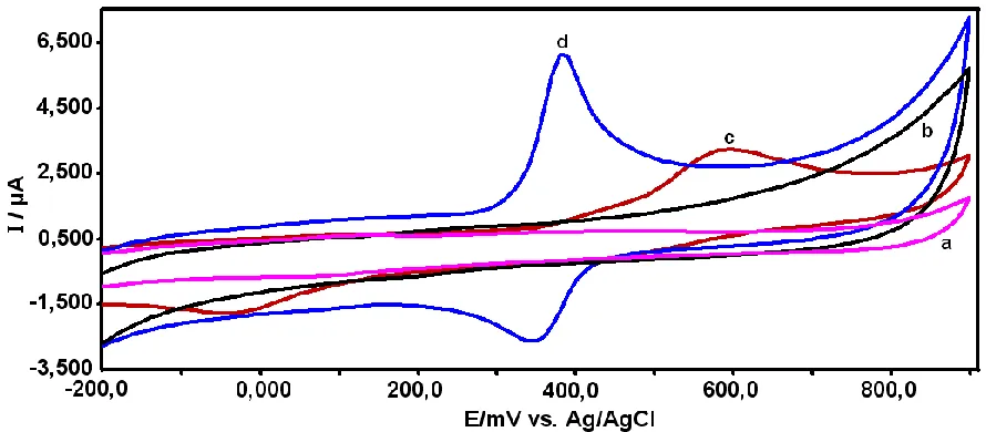
[image:5.596.76.521.427.622.2]The electrochemical behavior of APAP on poly(CA)-modified GCE and bare GCE were investigated by cyclic voltammetry (CV). At the bare GCE (Fig. 2, curve c), a pair of relatively weak redox peak currents corresponding to the electrochemical redox of APAP was observed at an anodic peak potential (Epa) of 638 mV and a cathodic peak potential (Epc) of -41 mV. The peak-to-peak potential separation (ΔEp=Epa-Epc) was as large as 597 mV (Fig. 2 curve c).
Figure 2. CVs of APAP on the bare GCE and poly(CA)-modified GCE in 0.1 M PBS (pH 7.0) at scan rate of 100 mV s-1. (a) and (b) represent the absence of 0.1 mM APAP, (c) and (d) represent the presence of 0.1 mM APAP.
rate kinetics occur on poly(CA)-modified GCE. Compared with the unmodified GCE, the redox peak currents obtained on poly(CA)-modified GCE were much higher and the ΔEp was much lower, which are clear evidences of good electrocatalytic activity of poly(CA) toward APAP. In this experiment, the net values of oxidation peak current of APAP obtained at the poly(CA)/GCE (6.0 µA) is about 2.0 times higher than that at the GCE (3.0 µA). The electrochemistry of poly(CA)-modified GCE was performed as control experiment (curve c), and no obvious redox peaks were appeared. As can be seen in Fig. 2 (curve a and b) the background current of poly(CA)-modified electrode(Fig 2 curve b) is much larger than that of the unmodified GCE (Fig 2 curve a), indicating high specific capacitance of poly(CA) films on electrode.
3.3. Effect of scan rate
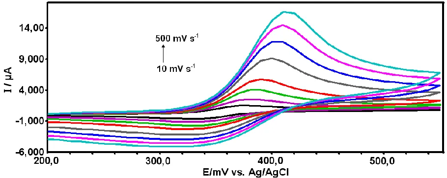
The effect of the potential scan rate on the peak current of APAP was investigated. Fig. 3 shows the CVs of the poly(CA)-modified glassy carbon electrode at various scan rates (v) (at all scan rates at 25 oC) obtained in 0.1 mol L-1 PBS (pH 7.0) containing 0.1 mM APAP. As shown in Fig. 3, the anodic and cathodic peak currents increased with increasing the potential scan rate. With increasing the potential scan rate, the oxidation peak potential shifted positively and the reduction peak potential shifted negatively. As can be seen (Fig. 4), the redox peak currents were found linearly proportional to the scan rate ranging from 10 to 500 mV s-1.
Figure 3. CVs of 0.1 mM APAP on poly(CA)-modified electrode at different scan rates from 10 to 500 mV s−1 (10, 25, 50, 100, 200, 300, 400 and 500), respectively. Inset: is the plot of the peak current of APAP versus scan rate.
The linear regression equations are Ipa (µA)= 0.0321 v (mV s-1) + 1.9978 (R= 0.9954) and Ipc (µA)= -0.0087 v (mV s-1
[image:6.596.71.526.429.611.2][image:7.596.179.403.155.293.2]
- 0.2829 (R=0.9939), respectively. This indicated that the modified electrode reaction of APAP is a adsorption-controlled process. On the other hand, the anodic potentials shifted positively with the increase of scan rate, indicating the quasi-reversible nature of the electrode reaction.
Figure 4. The plot of the peak current of APAP vs. scan rate.
3.4. Effect of pH
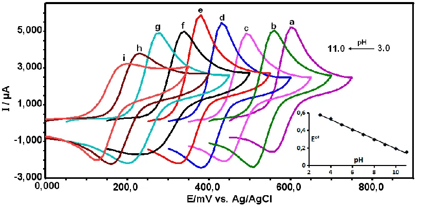
The influence of solution pH value on the redox reaction of acetaminophen at the Poly(CA) modified GCE was studied in the pH range from 3 to 11. It was observed that the oxidation potential decreased when the pH of the APAP solution increased. As shown in Fig. 5, a negative shift of both the cathodic and anodic peak potentials occurs when the pH value is increased and the redox peak currents of APAP reached a maximum at a pH value of 7.0; thus, pH 7.0 phosphate buffer solution was chosen as the supporting electrolyte.
[image:7.596.96.506.511.713.2]
When pH was higher than 7.0, the peak current of APAP decreased gruadally with the increasing of pH. This indicates that for pH greater than 7.0, APAP is converted to the phenoxide ion. Fig. 5 displays the average variation of the anodic and cathodic peak potentials for the APAP oxidation with pH at the poly(CA)-modified GCE. This average could be considered approximately as the standard (formal) potential (E°′). Fig. 5 (inset) shows E°' as a function of pH with the linear equation as: E0' (V) = -0.055 pH + 0.8122 (R = 0.9933). The slope value of -55 mV pH−1 is close to the theoretical value of -59 mV pH−1 according to Nernst equation, suggesting equal numbers of proton and electron are involved in the redox reaction. Thus, at the poly(CA)-modified electrode, the electrochemical reaction of APAP is a two-proton coupled two-electron process.
3.5. Voltammetric determination of APAP
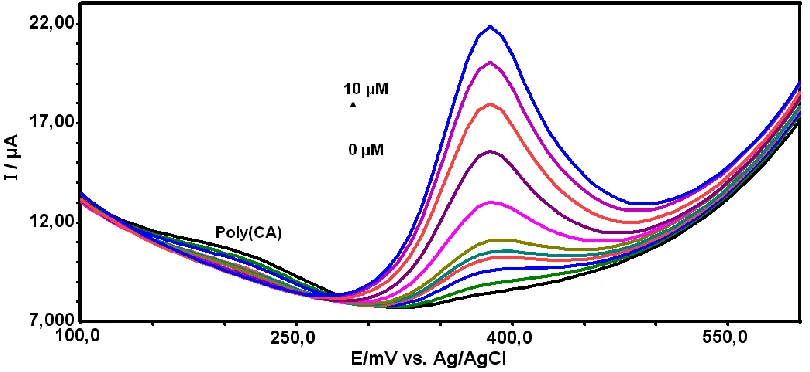
[image:8.596.94.499.541.727.2]In order to decrease the detection limit of APAP using the proposed method, the square wave mode was utilized at the poly(CA)-modified electrode. Fig. 6 depicted the SWV curves of different concentration of APAP at poly(CA)-modified electrode. Using the SWV method, the SWV peak current increased linearly with APAP concentration with very good correlation coefficients. The calibration curve for APAP shows (Fig. 7) two linear segments: the first linear segment increases from 0.2 to 1.0 µM with linear regression equation of Ip (µA)=2.7305 C (µM) + 0.2355 (R=0.9980), and second linear segment increases up to 10 µM with linear regression equation of Ip (µA)=1.13 C (µM) + 2.738 (R=0.9950). The limit of detection, defined as CL=3Sy/x/b [55] (where Sy/x is the standard deviation of y-residuals and b is the slope of the calibration plot) was 0.026 µM. The detection limit for the determination of acetaminophen at the proposed modified electrode was lower than in those of some previously published studies [9-38]. The detection limit and linear range for detection of APAP were compared with previously published methods in Table 1. Under the optimized conditions, the repeatability of the proposed method expressed as relative standard deviations (RSDs, n =5) were found to be 3.5 and 3.0 % for the concentration of 0.5 and 5.0 µM, respectively.
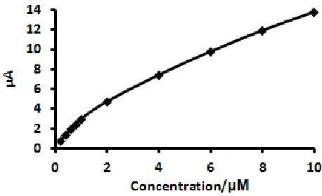
Figure 7. The plot of peak current vs. APAP concentration.
3.6. Interference experiments
The current response of the poly(CA)-modified electrodes in the presence of some interferent compounds was also examined. The electrochemical behaviors of the coexisting electroactive species, which often cause serious interference with the determination of APAP (1.0 µM), such as ascorbic acid, uric acid, dopamin and p-aminophenol (4-AP), were investigated by using SWV. The oxidation potential of each interferent ascorbic acid (Epa = 67 mV), dopamin (Epa=203 mV), uric acid (Epa =349 mV) and p-aminophenol (4-AP) (Epa =135 mV) at the poly(CA) film-coated electrode was found to be more negative than that of APAP (Epa =385 mV). The results showed that 20-fold excess of UA and 100 fold excess of DA and 4-AP did not interfere with the measurement of 1.0 µM APAP. Therefore, it could be possible for selective detection of APAP in the presence of AA, dopamine, uric acid and p-aminophenol but also selective for the simultaneous determination of these four species present in a mixture. On the other hand, no interference has been found when including up to 1000 µM of glucose, salicylic acid and caffeine.
3.7. Determination of APAP in the presence of AA
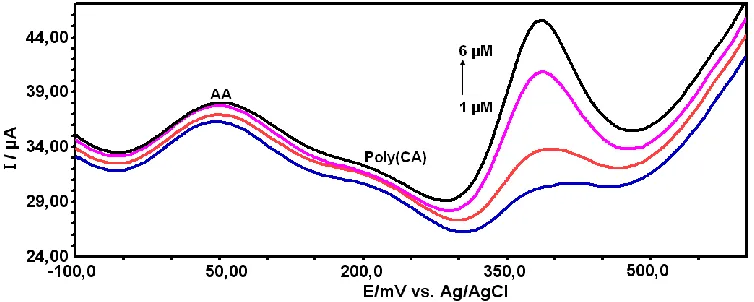
Figure 8. SWVs of APAP and AA at the poly(CA)-modified electrode in 0.1 M PBS (pH 7.0). [APAP] was changed and [AA] was kept constant (i.e., [AA]= 500 µM). [APAP]; (a) 1,0, (b) 2.0, (c) 4.0 and 6.0 µM APAP.
3.8. Repeatability and stability
The repeatability and stability of this assay were also investigated. The reproducibility of the method was evaluated by measuring on ten solutions of 1.0 µM of APAP in phosphate buffer (pH 7.0) leading to a relative standard deviation of 3.7 %. The experimental results indicated that the as-prepared poly(CA)-modified electrode has a good repeatability. The poly(CA)- modified electrode exhibited a high stability whenever it was placed in a dry state or in the phosphate buffer solution. No loss of electroactivity of the electrode was found for the continuous cyclical sweep for 10 h. The poly(CA)-modified electrode was also not deteriorated even for as long as two weeks.
3.9. Applications
[image:10.596.109.486.75.226.2]The content of APAP was determined by standard addition method, and the results shown in Table 2. The results obtained by poly(CA) modified GCE are in good agreement with the declared acetaminophen content and we have compared the electrochemical determination of APAP in commercial drugs with HPLC method [56]. The recoveries (97%-100%) indicate that the accuracy and repeatability of this method are very good. According to experimental results, it is very clear that this new method has great potential for practical APAP sample analysis.
Table 1. Comparison of the electrochemical behavior of poly(CA)/GCE for APAP with some of the previously reported electrodes.
Modified Electrode
Linear range (mol L-1)
Detection limit
(mol L-1) References
SWNT-DCP 1.0×10⁻⁷-2.0×10⁻⁵ 4.0×10⁻⁸ [9]
NanoTiO2/poly(AY) 1.2×10⁻⁵-1.2×10⁻⁴ 2.0×10⁻⁶ [10]
[image:10.596.98.498.686.761.2]
Cu(II)-conducting polymer complex
2.0×10⁻⁵-5.0×10⁻³ 5.0×10⁻⁶ [12]
Graphene 1.0×10⁻⁷-2.0×10⁻⁶ 3.2×10⁻⁸ [13]
Nafion/TiO2-Graphene 1.0×10⁻⁶-1.0×10
−4
2.1×10⁻⁷ [14]
Ionic liquid electrode 1.0×10⁻⁶-2.0×10⁻³ 3.0×10⁻⁷ [15]
Graphene/Nafion 4.0×10⁻⁷-1.0×10⁻⁶ 2.5×10⁻⁸ [16]
MWCNT-ACS 5.0×10⁻⁸-2.0×10⁻⁶ 5.0×10⁻⁸ [17]
Palladium nanoclusters-coated polyfuran
5.0×10⁻⁷-1.0×10⁻⁵ 7.64×10⁻⁸ [18]
FIA-Carbon film resistor 8.0×10⁻⁷ -5.0×10⁻⁴ 1.36×10⁻⁷ [19]
Polypyrrole- modified pencil 5.0×10⁻⁷ -5.0×10⁻⁴ 7.9×10⁻⁷ [20]
Micro-crystalline natural graphite–polystyrene
composite
2.0×10⁻⁶-1.0×10⁻⁴ 3.4×10⁻⁸ [21
Renewable glassy carbon annular band electrode
6.6×10⁻⁶
-6.6×10⁻⁵ 1.32×10⁻⁷ [22]
Nanogold modified indium tin oxide (ITO)
2.0×10⁻⁷
-1.5×10⁻³ 1.8×10⁻⁷ [23]
Fullerene /GCE 5.0×10⁻⁵-1.5×10⁻³ 5.0×10⁻⁵ [24]
N (3,4dihydroxyphenethyl)3,5dinitrobenzamide
-MWCNT/CPE
1.5×10⁻⁵-2.7×10⁻⁴ 1.0×10⁻⁵ [25]
Nano-TiO2/GCE 1.2×10
-5
-1.2×10⁻⁴ 2.0×10⁻⁶ [26]
Poly(3,4-
ethylenedioxythiophene)-/SPE
4.0×10⁻⁶-4.0×10⁻⁴ 1.39×10⁻⁶ [27]
ZrO2/CPE 1.0×10
-6
-2.5×10⁻³ 9.12×10⁻⁷ [28]
C-Ni/GCE 2.0×10⁻⁶-2.3×10⁻⁵ 6.0×10⁻⁷ [29]
Multiwall carbon nanotubes /GCE
5.0×10⁻⁶-3.0×10⁻⁵ 3.0×10⁻⁷ [30]
Poly(Patton and Reeder’s reagent) /MCPE
7.0×10⁻⁷-1.0×10⁻⁵ 5.3×10⁻⁷ [31]
Ionic liquid /CNTPE 1.0×10⁻⁶-6.0×10⁻⁵ 5.0×10⁻⁷ [32]
Functionalized MWCNTs /GE
2.5×10⁻⁵-4.0×10⁻⁴ 5.0×10⁻⁷ [33]
Poly(taurin)-MWCNT/GCE 1.0×10⁻⁶-1.0×10⁻⁴ 5.0×10⁻⁷ [34]
CoOx/CCE 5.0×10⁻⁶-3.5×10⁻⁵ 3.7×10⁻⁷ [35]
Chitosan-MWCNT/GCE 1.0×10⁻⁶-1.545×10⁻⁴ 1.0×10⁻⁷ [36]
polypyrrole/aszophloxine/Au 2.0×10⁻⁷-1.0×10⁻⁴ 8.0×10⁻⁸ [37]
Poly (calconcarboxylic acid)/ GCE
1.0×10⁻⁷-1.0×10⁻⁵ 1.0×10⁻⁸ [38]
[image:11.596.66.533.623.766.2]Poly(caffeic acid) 2.0×10⁻⁷-1.0×10⁻⁵ 2.6×10⁻⁸ This work
Table 2. Determination of acetaminophen in pharmaceutical samples.
Commercial drugs Labeled (mg unite-1)
Added (mg unite-1)
Found / Recovery (%)
Reference method (mg unite-1)
4. CONCLUSIONS
The poly(CA)-modified electrode, which can be prepared by electropolymerization of CA, possesses an excellent electrocatalytic activity for APAP oxidation in neutral aqueous media. The prepared poly(CA)/GCE exhibited best performance as a sensor for the determination of trace APAP based on the square-wave voltammetry (SWV). The poly(CA)-modified electrode exhibited an excellent sensitivity and selectivity towards APAP even in the presence of 500-fold excess of AA. The above mentioned method can be employed for the detection of APAP, ascorbic acid, p-aminophenol, dopamine and uric acid simultaneously without interference of each other. The poly(CA) film coated electrode exhibited a stable and sensitive response to APAP in the presence of interferents. This method could be an alternative method for the analysis of APAP in the future with its high sensitivity, stability, good reproducibility and it being devoid of interference. The fabrication of poly(CA) film on GC electrode surface is very simple and less time consuming (ca. 30 s).
ACKNOWLEDGEMENTS
We gratefully acknowledge Istanbul University Scientific Research Fund (Project Nos. BYP-20075, UDP-26215 and UDP-34672) for financial support.
References
1. G. G. Graham, K. F. Scott, R. O. Day, Drug. Saf., 28 (2005) 227.
2. N. Wangfuengkanagul, O. Chailapakul, J. Pharm. Biomed. Anal., 28 (2002) 841.
3. J. Guzy, Z. Chovanova, M. Marekova, Z. Chavkova, V. Tomečkova, G. Mojžišová, J. Kušnir, Biologia Bratislava, 59 (2004) 399.
4. H. R. B. Raghavendran, A. Sathivel, T. Devaki, J. Health Sci., 50 (2004) 42.
5. P. Fanjul-Bolado, P. J. Lamas-Ardisana, D. Hernández-Santos, A. Costa-García, Anal. Chim. Acta, 638 (2009) 133.
6. P. T. Kissinger, D. A. Roston, J. J. Van Benschoten, J. Y. Lewis and W. R. Heineman, J. Chem. Educ., 60 (1983) 772.
7. D. J. Miner, J. R. Rice, R. M. Riggin and P. T. Kissinger, Anal. Chem.,53 (1981) 2258.
8. D. Nematollahi, H. Shayani-Jam, M. Alimoradi, S. Niroomand, Electrochim. Acta, 54 (2009) 7407. 9. [9] D. Sun, H. Zhang, Microchim. Acta, 158 (2007) 131.
10.S. A. Kumar, C. F. Tang, S. M. Chen, Talanta, 76 (2008) 997. 11.M. Li, L. Jing, Electrochim. Acta, 52 (2007) 3250.
12.M. Boopathi, M.-S.Won, Y.-B. Shim, Anal. Chim. Acta, 512 (2004) 191. 13.X. Kang, J. Wang, H. Wu, J. Liu, I. A. Aksay, Y. Lin, Talanta, 81 (2010) 754. 14.Y. Fan, J.-H. Liu, H.-T. Lu, Q. Zhang, Coll. Surf. B: Biointerface, 85 (2011) 289. 15.X. S. Guan, H. Zhang, J. Zheng, Anal. Bioanal. Chem., 391 (2008) 1049.
16.H. Filik, G. Çetintaş, A. A. Avan, S. N. Koç, İ. Boz, Int. J. Electrochem. Sci., 8 (2013) 5724. 17.T.-L. Lu, Y.-C. Tsai, Sens. Actuators B, 153 (2011) 439.
18.N. F. Atta, M. F. El-Kady, A. Galal, Sen. Actuators B., 141 (2009) 566.
19.F.S. Felix, C.M.A. Brett, L. Angnes, J. Pharm. Biomed. Anal., 43 (2007) 1622. 20.L. Özcan, Y. Şahin, Sens. Actuators B: Chem., 127 (2007) 362.
22.Bogusław Bas´, A. Bugajna, M. Jakubowska, W. Reczynśki, A. Smalec, Electrochim. Acta, 99 (2013)190.
23.R. N. Goyal, V. K. Gupta, M. Oyama, N. Bachheti, Electrochem. Commun., 7 (2005) 803. 24.R.N. Goyal, S.P. Singh, Electrochim. Acta, 51 (2006) 3008.
25.A.A. Ensafi, H. Karimi-Maleh, S. Mallakpour, M. Hatami, Sens. Actuators B, 155 (2011) 464. 26.S.A. Kumar, C.F. Tang, S.M. Chen, Talanta, 76 (2008) 997.
27.W.Y. Su, S.H. Cheng, Electroanal., 22 (2010) 707.
28.M. Mazloum-Ardakani, H. Beitollahi, M.K. Amini, F. Mirkhalaf, M. Abdollahi-Alibeik, Sens. Actuators B, 151 (2010) 243.
29.S.F. Wang, F. Xie, R.F. Hu, Sens. Actuators B, 123 (2007) 495.
30.Z. A. Alothman, N. Bukhari, S.M. Wabaidur, S. Haider, Sens. Actuators B, 146 (2010) 314. 31.T. Thomas, R.J. Mascarenhas, F. Cotta, K.S. Guha, B.E.K. Swamy, P. Martis, Colloids Surf. B:
Biointerfaces, 101 (2013) 91.
32.T. Tavana, M.A. Khalilzadeh, H. Karimi-Maleh, A.A. Ensafi, H. Beitollahi, D.Zareyee, J. Mol. Liq., 168 (2012) 69.
33.R. Manjunatha, D.H. Nagaraju, G.S. Suresh, J.S. Melo, S.F. D’Souza, T.V. Venkatesha, Electrochim. Acta, 56 (2011) 6619.
34.Q. Wan, X. Wang, F. Yu, X. Wang, N. Yang, J. Appl. Electrochem., 39 (2009) 785. 35.H. Razmi, E. Habibi, Electrochim. Acta, 55 (2010) 8731.
36.A. Babaei, M. Afrasiabi, M. Babazadeh, Electroanal., 22 (2010)1743. 37.M.B. Gholivand, M. Amiri, J. Electroanal. Chem., 676 (2012) 53.
38.A.L. Liu, K. Wang, W. Chen, F. Gao, Y.S. Chai, X.H. Lin, Y.Z. Chen, X.H. Xia, Electrochim. Acta, 63 (2012) 161.
39.S. A. Emr, A. M. Yacynych, Electroanal., 7 (1995) 913.
40.Q. Gao, X. Cui, F. Yang, Y. Ma, X. Yang, Biosens. Bioelectron., 19 (2003) 277. 41.X.-G. Li, M.R. Huang, W. Duan, Y.L. Yang, Chem. Rev., 102 (2002) 2925.
42.X.-G. Li, W. Duan, M.-R. Huang, L. N.J. Rodriguez, React. Funct. Polym., 62 (2005)261. 43.S. K. Trabelsi, N. B. Tahar, R. Abdelhedi, Electrochim. Acta, 49 (2004) 1647.
44.O. Makhotkina, P. A. Kilmartin, J. Electroanal. Chem., 633 (2009) 165.
45.W. R. Sousa, C. da Rocha, C. L. Cardoso, D. H. S. Silva, M. V. B. Zanoni, J. Food Comp. Anal., 17 (2004) 619.
46.C. C. Zeng, C. –F. Liu, J. Zeng, R. -G Zhong, J. Electroanal. Chem., 608 (2007) 85.
47.A. B. Moghaddam, M. R. Ganjali, R. Dinarvand, P. Norouzi, A. A. Saboury, A. A. Moosavi-Movahedi, Biophys. Chem., 128 (2007) 30.
48.R. Amadalli, A. De Battisti, D. V. Girenko, S. V. Kovalyov, A. B. Velichenko, Electrochim. Acta, 46 (2000) 341.
49.C. Giacomelli, K. Ckless, D. Galato, F. S. Miranda, J. Braz. Chem. Soc., 13 (2002) 332. 50.N. B. Li, W. Ren, H. Q. Luo,J. Solid State Electrochem., 12 (2008) 693.
51.N. B. Li, W. Ren, H. Q. Luo, Electroanal., 19 (2007) 1496.
52.H. Hotta, M. Ueda, S. Nagano, Y. Tsujino, J. Koyama, T. Osakai, Anal. Biochem., 303 (2002) 66. 53.R. Arakawa, M. Yamaguchi, H. Hotta, T. Osakai, T. Kimoto, J. Am. Soc. Mass Spectrom., 15
(2004) 1228.
54.Y. Li, S.-M. Chen, Int. J. Electrochem. Sci., 7 (2012)2175.
55.J. C. Miller, J. N. Miller, Statistics for Analytical Chemistry. Ellis Horwood Series, PTR Prentice Hall, London (1993).