R E S E A R C H A R T I C L E
Open Access
Differences in fungi present in induced sputum
samples from asthma patients and non-atopic
controls: a community based case control study
Hugo Cornelis van Woerden
1*, Clive Gregory
1*, Richard Brown
2, Julian Roberto Marchesi
2,
Bastiaan Hoogendoorn
1and Ian Price Matthews
1Abstract
Background:There is emerging evidence for the presence of an extensive microbiota in human lungs. It is not known whether variations in the prevalence of species of microbiota in the lungs may have aetiological significance in respiratory conditions such as asthma. The aim of the study was to undertake semi-quantitative analysis of the differences in fungal species in pooled sputum samples from asthma patients and controls.
Methods:Induced sputum samples were collected in a case control study of asthma patients and control subjects drawn from the community in Wandsworth, London. Samples from both groups were pooled and then tested for eukaryotes. DNA was amplified using standard PCR techniques, followed by pyrosequencing and comparison of reads to databases of known sequences to determine in a semi-quantitative way the percentage of DNA from known species in each of the two pooled samples.
Results:A total of 136 fungal species were identified in the induced sputum samples, with 90 species more common in asthma patients and 46 species more common in control subjects.Psathyrella candolleana, Malassezia pachydermatis, Termitomyces clypeatusandGrifola sordulentashowed a higher percentage of reads in the sputum of asthma patients andEremothecium sinecaudum, Systenostrema alba, Cladosporium cladosporioidesand
Vanderwaltozyma polysporashowed a higher percentage of reads in the sputum of control subjects. A statistically significant difference in the pattern of fungi that were present in the respective samples was demonstrated using the Phylogenetic (P) test (P < 0.0001).
Conclusion:This study is novel in providing evidence for the widespread nature of fungi in the sputum of healthy and asthmatic individuals. Differences in the pattern of fungi present in asthma patients and controls merit further investigation. Of particular interest was the presence ofMalassezia pachydermatis, which is known to be associated with atopic dermatitis.
Keywords:Asthma, Sputum, Fungi, Case–control study
Background
The human lung has a surface area of around 50 m2[1]
and is in contact with more than 15,000 litres of air each day [2]. At each breath around 5,000 particles of dust are inhaled [3]. On average, the dust in the earth’s at-mosphere contains 10,000 to 100,000 organisms per gram of dust [4], some of which is in the‘respirable dust’
fraction, consisting of particles smaller than 5μm [5]. This extensive exposure to the environment means that the lungs are a common portal for infection by viruses, bacteria, fungi, protozoa and other infectious agents.
Historically, healthy lungs were believed to be free of
bacteria and that during infection organisms “gain a
foothold in the normally sterile lung tissue” [6]. How-ever, there is increasing evidence that microbiota are present even in healthy lungs [7]. This finding raises the possibility of a potential overlap between pathogenic and commensal microbiota in the respiratory tract.
* Correspondence:[email protected];[email protected] 1
Institute of Primary Care & Public Health, Cardiff University School of Medicine, Neuadd Meirionnydd, Heath Park, Cardiff CF14 4YS, UK Full list of author information is available at the end of the article
There is relatively little literature examining microbiota in human lungs. Tunney et al. [8] showed that approxi-mately 50% of healthy individuals harbour between 1000 and 10,000 culturable anaerobic bacteria per ml of induced sputum A range of microbial species were also found in induced sputum at low numbers in another study which examined sputum from healthy subjects [9]. One previous study has been identified which used metage-nomic culture independent gemetage-nomic techniques and demonstrated that microbial communities in asthmatic airways were disordered, with pathogenic Proteobacteria
more frequently found in the bronchi of asthmatics patients than in controls [10].
The current study further examines the role of atypical microbiota in respiratory disease. The study used mo-lecular techniques to identify eukaryote species that were present in induced sputum samples taken from asthma patients and controls, living in Wandsworth, London. The aim of the study was to undertake semi-quantitative analysis of the differences in fungal species present in pooled sputum samples from asthma patients and controls.
Methods Study population
The study protocol was approved by Camden and Isling-ton community local research ethics committee (ref 08/ H0722/540). All patients participating in the study sup-plied informed consent. This case control study from which the induced sputum samples were drawn has pre-viously been described. Further information on the char-acteristics of the subjects in this study has been provided in that paper [11]. In summary, participants were resi-dents of Wandsworth, London, and were primarily iden-tified from the patient registers of two GP practices. Asthma patients were defined as those individuals who had a current diagnosis of asthma, for example, by being on the GP practice asthma register. Most of the asthma patients were on inhaled corticosteroids. Non-atopic controls were defined as individuals who on questioning did not report having current or previous asthma, ec-zema or hay fever. All participants competed a published questionnaire [12] to assess the risk of mould in the home. The questionnaire contained four questions: Is there any visible mould growth on your house? Is there any odour of mould or cellar-like musty air in your house? Is there any moisture stains in your house? Is there any water/moisture damage in your house?
Sputum collection and DNA extraction
Participants inhaled isotonic saline via an ultrasonic nebu-liser. Globules of sputum were coughed up into petri dishes, spread on microscope slides and stained for
micro-scopic examination. Approximately 5 mm2areas were
excised from each microscope slide. The samples were combined to yield two pooled samples for subsequent DNA extraction and PCR: asthma patients and control subjects. (A sample from one asthma patient was inadvert-ently included in the control set). DNA was subsequinadvert-ently extracted using the Zymo research pinpoint system (Zymo Research, Irvine, Ca) in accordance with manufacturer’s instructions. The samples were taken from 30 asthma patients and 13 non-atopic control subjects involved in the case control study.
Pyrosequencing of extracted DNA and statistical analysis Extracted DNA was amplified using a PCR protocol for the partial 18S rRNA gene using the primer pair (Euk1a
(5’ CTG GTT GAT CCT GCC AG 3’) and Euk516r (5’
ACC AGA CTT GCC CTC C 3’)) in accordance with
previously described protocols [13,14]. The two pooled extract amplicons, from asthma patients and from con-trols, were sequenced using a 454 pyrosequencer by Re-search and Testing Inc, Lubbock, Texas, USA. DNA sequences were compared to the SILVA database of known eukaryotic 18S rRNA gene sequences to deter-mine in a semi-quantitative way the proportional distri-bution in each of the two samples.
The difference between the pattern of fungal species in each of the two pooled samples was compared using Unifrac [15,16]. This online software uses phylogenetic in-formation to test whether or not two environments are sig-nificantly different. The software estimates the similarity between communities by measuring the number of changes that would be required to explain the differences in the dis-tribution of sequences between the two environments.
Results
Study population and presence of mould in the home Patients had a mean age of 41.6 years (SD 14.9, range 18–65 years) and control participants a mean age of 35.7 years (SD 12.8, range 24– 58 years). The patient group was 40% male and the control group was 46% male.
A positive answer to at least one of the four questions regarding possible mould in the home was recorded in 30% (9/30) asthma patients and 15.4% (2/13) of control subjects. Due to the small sample size, this relatively large difference was not statistically significant.
Analysis of pyrosequencing data
The differences based on the percent of total DNA reads of in the pooled samples from asthma patients and non-atopic controls are shown in Tables 1 and 2. A statisti-cally significant difference in the pattern of fungi that were present in the respective samples was demon-strated using the Phylogenetic (P) test (P <0.0001).
Table 1 Fungi that were more common in asthma patients than in control participants (difference in percent of DNA reads in descending order)
Fungal species Control
participants
Asthma patients
Difference
Psathyrella candolleana 0.000 27.294 27.294
Malassezia pachydermatis
0.000 21.651 21.651
Termitomyces clypeatus 0.000 7.071 7.071
Grifola sordulenta 0.000 4.489 4.489
Pycnoporus sp 0.000 2.938 2.938
Phlebiopsis gigantean 0.000 2.932 2.932
Dichostereum pallescens 0.000 2.746 2.746
Peniophorella praetermissa
0.168 2.617 2.449
Aspergillus zonatus 0.016 1.959 1.942
Acanthophysium cerussatum
0.000 1.825 1.825
Pleurotus ostreatus 0.000 1.615 1.615
Candelabrochaete africana
0.016 1.556 1.540
Basidiobolus ranarum 0.000 1.510 1.510
Tapinella atrotomentosa 0.000 1.487 1.487
Pleurocybella porrigens 0.000 1.399 1.399
Debaryomyces hansenii 0.000 1.294 1.294
Collybia tuberosa 0.000 1.282 1.282
Galerina atkinsoniana 0.000 1.230 1.230
Punctularia strigosozonata
0.000 1.148 1.148
Elderia arenivaga 0.000 1.143 1.143
Pseudoarmillariella ectypoides
0.000 0.834 0.834
Pulcherricium caeruleum 0.000 0.665 0.665
Tilletia goloskokovii 0.000 0.618 0.618
Cerrena sp 0.000 0.600 0.600
Serpula lacrymans 0.000 0.571 0.571
Bondarcevomyces taxi 0.000 0.554 0.554
Resinicium bicolor 0.000 0.519 0.519
Cortinarius sodagnitus 0.000 0.414 0.414
Trichaptum abietinum 0.000 0.315 0.315
Chamaeota sinica 0.000 0.262 0.262
Peziza vesiculosa 0.000 0.239 0.239
Pterula echo 0.000 0.204 0.204
Laccocephalum mylittae 0.000 0.198 0.198
Coprinopsis cinerea 0.000 0.198 0.198
Exidiopsis calcea 0.000 0.187 0.187
Dioszegia fristingensis 0.000 0.157 0.157
Inonotus baumii 0.060 0.210 0.150
Hydnochaete olivacea 0.000 0.134 0.134
Derxomyces boekhoutii 0.000 0.128 0.128
Xeromphalina campanella
0.000 0.117 0.117
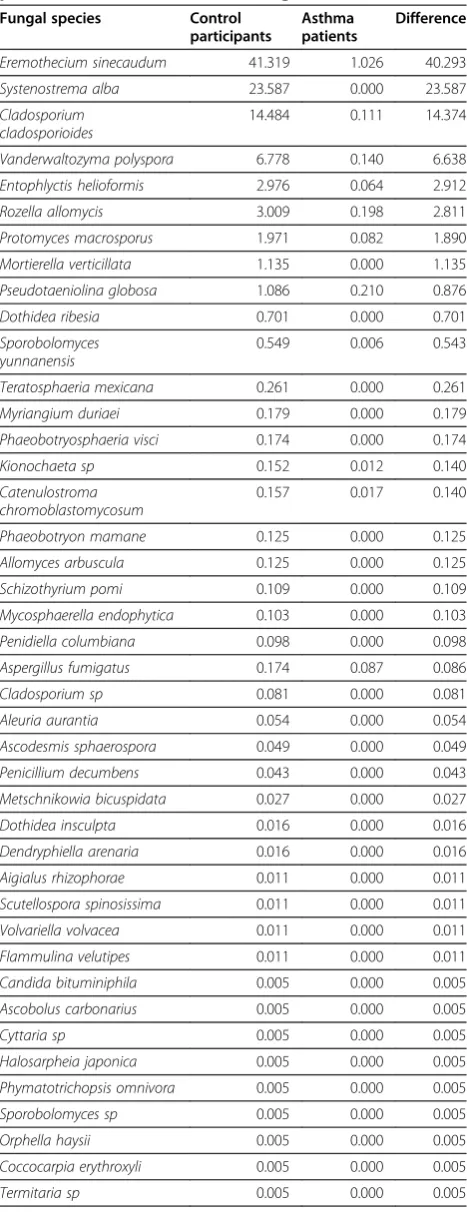
Table 1 Fungi that were more common in asthma patients than in control participants (difference in percent of DNA reads in descending order)(Continued)
Pulchromyces fimicola 0.000 0.111 0.111
Aspergillus oryzae 0.000 0.099 0.099
Entoloma prunuloides 0.000 0.099 0.099
Dioszegia zsoltii 0.000 0.093 0.093
Basidiobolus haptosporus 0.000 0.087 0.087
Saccharomycopsis fibuligera
0.000 0.087 0.087
Galiella rufa 0.000 0.082 0.082
Derxomyces simaoensis 0.000 0.070 0.070
Mycoclelandia arenacea 0.000 0.064 0.064
Steccherinum fimbriatum 0.000 0.064 0.064
Austropaxillus sp 0.000 0.058 0.058
Meyerozyma guilliermondii
0.000 0.058 0.058
Volvariella caesiotincta 0.000 0.058 0.058
Galerina marginata 0.000 0.052 0.052
Occultifur externus 0.000 0.052 0.052
Hericium americanum 0.000 0.047 0.047
Penicillium commune 0.000 0.041 0.041
Volvopluteus earlei 0.000 0.041 0.041
Gymnopus dryophilus 0.000 0.029 0.029
Aspergillus terreus 0.000 0.023 0.023
Tritirachium sp 0.000 0.023 0.023
Gloiocephala aquatic 0.000 0.023 0.023
Tricholoma matsutake 0.000 0.023 0.023
Exidia uvapsassa 0.000 0.017 0.017
Rhodocollybia maculate 0.000 0.017 0.017
Lentinus sp 0.000 0.017 0.017
Teratosphaeria acidotherma
0.076 0.093 0.017
Taphrina deformans 0.000 0.012 0.012
Antrodia vaillantii 0.000 0.012 0.012
Aspergillus penicillioides 0.000 0.012 0.012
Passalora vaginae 0.000 0.012 0.012
Malassezia furfur 0.000 0.012 0.012
Candida sp 0.000 0.012 0.012
Piromyces sp 0.000 0.012 0.012
Paxillus vernalis 0.000 0.012 0.012
Derxomyces mrakii 0.000 0.012 0.012
Lasiodiplodia gonubiensis 0.000 0.012 0.012
Teratosphaeria ohnowa 0.005 0.012 0.006
Diversispora celata 0.000 0.006 0.006
Geomyces destructans 0.000 0.006 0.006
Coprinopsis sp 0.000 0.006 0.006
Chlamydosauromyces punctatus
0.000 0.006 0.006
in asthma patients and 46 species more common in con-trol subjects, based on the percent of total DNA reads (see Figure 1).Psathyrella candolleana, Malassezia pachy-dermatis, Termitomyces clypeatus and Grifola sordulenta
were particularly prevalent in the sputum of asthma
patients and Eremothecium sinecaudum, Systenostrema
alba, Cladosporium cladosporioidesand Vanderwaltozyma polysporawere particularly prevalent in the sputum of con-trol subjects. No other eukaryote species were identified in the sputum samples.
Discussion
The range of fungal species present in both asthma patients and control subjects was larger than expected. There were also clear differences in the pattern of fun-gal species between asthma patients and control
sub-jects. The fungi Malassezia pachydermatis, was found
in patients with asthma and not the control group. This organism has a known association with atopic conditions including atopic dermatitis [17]. However, there were no other obvious associations were identi-fied in the published literature between asthma and the other fungi found in the pooled samples from the asthma patients. Two of the fungi most commonly
found in the sputum of asthma patients (Termitomyces
clypeatus and Psathyrella candolleana) represent mem-bers of the basidiomycete family [18]. The latter has been found in indoor dust [19] and one can speculate that fungal spores may have been inhaled within the home. It is possible that most of the fungi identified could have come from a single individual, or a small number of individuals, whose samples were heavily colonised by fungi.
Except for Cladosporium, the species identified in
[image:4.595.305.539.120.727.2]induced sputum are not commonly found in air samples examined using standard culture techniques [20]. Ana-lysis of air samples using molecular techniques may demonstrate that these species are commonly present in the air, but this research has not been undertaken so far. Three out of four species detected in the sputum of asthma patients were from the macromycetes group (commonly known as mushrooms). Although asthma is
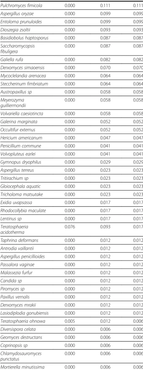
Table 2 Fungi that were more common in control participants than in asthma patients (difference in percent of DNA reads in descending order)
Fungal species Control
participants
Asthma patients
Difference
Eremothecium sinecaudum 41.319 1.026 40.293
Systenostrema alba 23.587 0.000 23.587
Cladosporium cladosporioides
14.484 0.111 14.374
Vanderwaltozyma polyspora 6.778 0.140 6.638
Entophlyctis helioformis 2.976 0.064 2.912
Rozella allomycis 3.009 0.198 2.811
Protomyces macrosporus 1.971 0.082 1.890
Mortierella verticillata 1.135 0.000 1.135
Pseudotaeniolina globosa 1.086 0.210 0.876
Dothidea ribesia 0.701 0.000 0.701
Sporobolomyces yunnanensis
0.549 0.006 0.543
Teratosphaeria mexicana 0.261 0.000 0.261
Myriangium duriaei 0.179 0.000 0.179
Phaeobotryosphaeria visci 0.174 0.000 0.174
Kionochaeta sp 0.152 0.012 0.140
Catenulostroma chromoblastomycosum
0.157 0.017 0.140
Phaeobotryon mamane 0.125 0.000 0.125
Allomyces arbuscula 0.125 0.000 0.125
Schizothyrium pomi 0.109 0.000 0.109
Mycosphaerella endophytica 0.103 0.000 0.103
Penidiella columbiana 0.098 0.000 0.098
Aspergillus fumigatus 0.174 0.087 0.086
Cladosporium sp 0.081 0.000 0.081
Aleuria aurantia 0.054 0.000 0.054
Ascodesmis sphaerospora 0.049 0.000 0.049
Penicillium decumbens 0.043 0.000 0.043
Metschnikowia bicuspidata 0.027 0.000 0.027
Dothidea insculpta 0.016 0.000 0.016
Dendryphiella arenaria 0.016 0.000 0.016
Aigialus rhizophorae 0.011 0.000 0.011
Scutellospora spinosissima 0.011 0.000 0.011
Volvariella volvacea 0.011 0.000 0.011
Flammulina velutipes 0.011 0.000 0.011
Candida bituminiphila 0.005 0.000 0.005
Ascobolus carbonarius 0.005 0.000 0.005
Cyttaria sp 0.005 0.000 0.005
Halosarpheia japonica 0.005 0.000 0.005
Phymatotrichopsis omnivora 0.005 0.000 0.005
Sporobolomyces sp 0.005 0.000 0.005
Orphella haysii 0.005 0.000 0.005
Coccocarpia erythroxyli 0.005 0.000 0.005
Termitaria sp 0.005 0.000 0.005
Table 1 Fungi that were more common in asthma patients than in control participants (difference in percent of DNA reads in descending order)(Continued)
Saccobolus dilutellus 0.000 0.006 0.006
Thanatephorus fusisporus 0.000 0.006 0.006
Boletellus shichianus 0.000 0.006 0.006
Puccinia poarum 0.000 0.006 0.006
Mallocybe dulcamara 0.000 0.006 0.006
Coniophora marmorata 0.000 0.006 0.006
[image:4.595.57.291.124.220.2]associated with damp environments that are affected by mould growth, we are unaware of any study that has identified an association between macromycetes and asthma. Future studies should consider analysing air samples from the homes of participants using molecular techniques, so as to take into account the presence of fungi in the ambient environment of participants.
We used universal primers for the eukaryotic 18S rRNA gene and were surprised that no eukaryotes other than fungi were identified in cases or controls. We have consid-ered a number of potential reasons why this may be the case and the most likely was that levels of non-fungal eukaryotic DNA, present in the samples, was below the limits of detection. The PCR primers were chosen after considerable deliberation and a probeCheck test showed that they were universal and matchedHomo sapiens’18S rRNA gene 100% [21]. However other potential reasons include: a genuine absence of other eukaryotes and
unintended removal of DNA from other eukaryotes as part of the processing of the samples.
Individual level analysis of samples was considered, but rejected as it was anticipated that, if the samples from each individual were analysed separately, the num-ber of eukaryotes in each sample would be below the threshold of detection. Samples for this study were therefore pooled to maximise the number of copies of each species in the pooled samples and consequently maximise the probability of detecting all the species that were present.
The study has a number of weaknesses. The sputum was not fresh when it was examined and although every effort was made to prevent contamination of samples by spores in the air, this is a possibility. The sample size is small and therefore may not be representative of asthma patients. Unfortunately, information on pets was not col-lected in this study and therefore could not be correlated with the presence of absence of particular fungi. It is possible to speculate that the presence of a pet (particu-larly a dog) in the subjects' house might be associated
with the presence of Malassezia pachydermatis in the
sputum of the research subjects, as this organism has been identified as a commensal on the skin of dogs and have could contaminated air in the homes of some of the research participants.
The potential significance of these fungi is unclear. There is tentative emerging evidence that microbiota
45 35 25 15 5 5 15 25 35 45
Eremothecium sinecaudum
Protomyces macrosporus Cladosporium cladosporioides
Systenostrema alba
Pseudotaeniolina globosa Vanderwaltozyma polyspora
Entophlyctis helioformis Mortierella verticillata
Rozella allomycis Candelabrochaete africana
Aspergillus zonatus Peniophorella praetermissa Malassezia pachydermatis Acanthophysium cerussatum Basidiobolus ranarum Dichostereum pallescens Collybia tuberosa Debaryomyces hansenii Phlebiopsis gigantea Grifola sordulenta Psathyrella candolleana Tapinella atrotomentosa Elderia arenivaga Termitomyces clypeatus Pleurotus ostreatus Galerina atkinsoniana Pycnoporus sp Pleurocybella porrigens Punctularia strigosozonata
Other <1%
[image:5.595.55.291.124.189.2]% reads in C % reads in A
[image:5.595.59.540.441.705.2]Figure 1Graph showing the percentage of pyrosequencing reads for fungal species identified in the Asthma patient (A) or control participant (C) samples (species identified for reads greater than 1%).
Table 2 Fungi that were more common in control participants than in asthma patients (difference in percent of DNA reads in descending order)(Continued)
Candida glabrata 0.005 0.000 0.005
Schizosaccharomyces japonicus
0.005 0.000 0.005
Cladochytrium sp 0.005 0.000 0.005
may form part of a complex causal web that results in disease, for example, by their effects on the immune sys-tem, without becoming pathogenic in the classical sense. For example, microbial compounds present in sputum may play a role as adjuvant factors and encourage a Th2-biased allergic response [22,23].
Conclusion
This study provides emerging evidence for the wide-spread presence of fungi in the sputum of asthma patients and control subjects. Significant differences have been identified in the pattern of fungi present in asthma patients and control subjects drawn from the same community. Although this method demonstrates the possibility of using microscopy samples, further in-vestigation is warranted which applies these techniques to fresh sputum samples. This method may in itself be applicable to analysis of historical samples and may in turn prove of interest in evaluating the microbiome of the lung demographically and between generations.
Competing interests
The authors declare that they have no competing interests.
Authors’contributions
HCVW obtained the initial funding, designed the case control study, obtained ethical and NHS R&D permission and wrote the first draft of the paper. CG, RG and JM undertook analysis of the sputum samples. All authors have contributed to, read and approved the final manuscript.
Acknowledgments
Funding was by Wandsworth PCT and Cardiff University. We are grateful to a range of colleagues who have contributed to the previously reported study based on this case control study and to the two GP practices that provided access to patients.
The authors would like to acknowledge the assistance of Research and Testing Inc, Lubbock, Texas, USA in carrying out the final stage pyrosequencing.
Author details
1Institute of Primary Care & Public Health, Cardiff University School of
Medicine, Neuadd Meirionnydd, Heath Park, Cardiff CF14 4YS, UK.2Cardiff School of Bioscience, Main Building, Museum Avenue, Cardiff CF10 3AT, UK.
Received: 10 August 2012 Accepted: 28 January 2013 Published: 5 February 2013
References
1. Hasleton PS:The internal surface area of the adult human lung.J Anat
1972,112(Pt 3):391–400.
2. Bartsch P, Collignon A, Weber G, Robaye G, Delbrouck JM, Roelandts I, Yujie J:
Distribution of metals in human lung: analysis by particle induced x-ray emission.Arch Environ Health1982,37(2):111–117.
3. Bunnell J:Editorial.Epidemio-Ecology News2002,1(2):1.
4. Griffin DW, Kellogg CA, Garrison VH, Holmes C, Shinn EA:The movement of soil and sediment in Earth's atmosphere: microbiology and ecosystem health.Epidemio-Ecology News2002,1(2):1–6.
5. World Health Organisation:Hazard prevention and control in the work environment: airborne dust. Geneva: WHO; 1999.
6. Scott JAG, Hall AJ:The value and complications of percutaneous transthoracic lung aspiration for the etiologic diagnosis of community-acquired pneumonia*.Chest1999,116(6):1716–1732.
7. Rogers GB, Carroll MP, Serisier DJ, Hockey PM, Jones G, Kehagia V, Connett GJ, Bruce KD:Use of 16S rRNA gene profiling by terminal restriction fragment length polymorphism analysis to compare bacterial communities in
sputum and mouthwash samples from patients with cystic fibrosis. J Clin Microbiol2006,44(7):2601–2604.
8. Tunney MM, Field TR, Moriarty TF, Patrick S, Doering G, Muhlebach MS, Wolfgang M, Boucher R, Gilpin DF, McDowell A:Detection of anaerobic bacteria in high numbers in sputum from patients with cystic fibrosis. Am J Respir Crit Care Med2008,177:995–1001.
9. Rogers GB, Carroll MP, Serisier DJ, Hockey PM, Kehagia V, Jones GR, Bruce KD:
Bacterial activity in cystic fibrosis lung infections.Respir Res2005,6(1):49. 10. Hilty M, Burke C, Pedro H, Cardenas P, Bush A, Bossley C, Davies J, Ervine A,
Poulter L, Pachter L,et al:Disordered microbial communities in asthmatic airways.PLoS One2010,5(1):e8578.
11. van Woerden HC, Ratier-Cruz A, Aleshinloye OB, Martinez-Giron R, Gregory C, Matthews IP:Association between protozoa in sputum and asthma: a case– control study.Respir Med2011,105(6):877–884.
12. Pirhonen I, Nevalainen A, Husman T, Pekkanen J:Home dampness, moulds and their influence on respiratory infections and symptoms in adults in Finland.Eur Respir J1996,9(12):2618–2622.
13. Amann RI, Krumholz L, Stahl DA:Fluorescent-oligonucleotide probing of whole cells for determinative, phylogenetic, and environmental studies in microbiology.J Bacteriol1990,172(2):762–770.
14. Sogin ML, Gunderson JH:Structural diversity of eukaryotic small subunit ribosomal RNAs. Evolutionary implications.Ann N Y Acad Sci1987,
503:125–139.
15. Lozupone C, Hamady M, Knight R:UniFrac–an online tool for comparing microbial community diversity in a phylogenetic context.BMC Bioinformatics
2006,7:371.
16. Martin AP:Phylogenetic approaches for describing and comparing the diversity of microbial communities.Appl Environ Microbiol2002,
68(8):3673–3682.
17. Gaitanis G, Magiatis P, Hantschke M, Bassukas ID, Velegraki A:The Malassezia genus in skin and systemic diseases.Clin Microbiol Rev2012,25(1):106–141. 18. Vasutov M, Urban A:Phylogenetic studies in Psathyrella focusing on
sections Pennatae and Spadiceae–new evidence for the paraphyly of the genus.Mycol Res2008,112(10):1153–1164.
19. Pitkaranta M, Meklin T, Hyvarinen A, Paulin L, Auvinen P, Nevalainen A, Rintala H:Analysis of fungal flora in indoor dust by ribosomal DNA sequence analysis, quantitative PCR, and culture.Appl Environ Microbiol
2008,74(1):233–244.
20. Burr ML, Matthews IP, Arthur RA, Watson HL, Gregory CJ, Dunstan FD, Palmer SR:Effects on patients with asthma of eradicating visible indoor mould: a randomised controlled trial.Thorax2007,62(9):767–772. 21. Quast C, Pruesse E, Yilmaz P, Gerken J, Schweer T, Yarza P, Peplies J, Glockner FO:
The SILVA ribosomal RNA gene database project: improved data processing and web-based tools.Nucleic Acids Res2013,41(D1):D590–596.
22. Jacquet A:The role of the house dust mite-induced innate immunity in development of allergic response.Int Arch Allergy Immunol2010,
155(2):95–105.
23. Jacquet A:Interactions of airway epithelium with protease allergens in the allergic response.Clin Exp Allergy2011,41(3):305–311.
doi:10.1186/1471-2334-13-69
Cite this article as:van Woerdenet al.:Differences in fungi present in induced sputum samples from asthma patients and non-atopic controls: a community based case control study.BMC Infectious Diseases 201313:69.
Submit your next manuscript to BioMed Central and take full advantage of:
• Convenient online submission
• Thorough peer review
• No space constraints or color figure charges
• Immediate publication on acceptance
• Inclusion in PubMed, CAS, Scopus and Google Scholar
• Research which is freely available for redistribution