This is a repository copy of
Comparing Fatigue when Using Large Horizontal and Vertical
Multi-Touch Interaction Displays
.
White Rose Research Online URL for this paper:
http://eprints.whiterose.ac.uk/87614/
Version: Submitted Version
Proceedings Paper:
Al-Megren, Shiroq, Kharrufa, Ahmed, Hook, Jonathan David
orcid.org/0000-0002-0588-7013 et al. (3 more authors) (2015) Comparing Fatigue when
Using Large Horizontal and Vertical Multi-Touch Interaction Displays. In: Human-Computer
Interaction – INTERACT 2015:15th IFIP TC 13 International Conference, Bamberg,
Germany, September 14-18, 2015, Proceedings, Part IV. The 15th IFIP TC.13 International
Conference on Human-Computer Interaction (INTERACT '15), 14-18 Sep 2015
SPRINGER-VERLAG BERLIN , pp. 156-164.
https://doi.org/10.1007/978-3-319-22723-8_13
[email protected] https://eprints.whiterose.ac.uk/ Reuse
Items deposited in White Rose Research Online are protected by copyright, with all rights reserved unless indicated otherwise. They may be downloaded and/or printed for private study, or other acts as permitted by national copyright laws. The publisher or other rights holders may allow further reproduction and re-use of the full text version. This is indicated by the licence information on the White Rose Research Online record for the item.
Takedown
If you consider content in White Rose Research Online to be in breach of UK law, please notify us by
Comparing Fatigue when Using Large Horizontal and
Vertical Multi-Touch Interaction Displays
Shiroq Al-Megren1, Ahmed Kharrufa2, Jonathan Hook3, Amey Holden2, Selina Sutton2, and Patrick Olivier2
1 School of Computing, University of Leeds, Leeds, United Kingdom [email protected]
2
Culture Lab, Newcastle University, Newcastle Upon Tyne, United Kingdom {ahmed.kharrufa, selina.sutton, patrick.olivier}@ncl.ac.uk
3
Department of Theatre, Film and Television, University of York, York, United Kingdom [email protected]
Abstract. We report on a user study that compared muscle fatigue experienced when using a large multi-touch display in horizontal and vertical configurations over a one-hour period. Muscle fatigue is recognized as the reduction in a mus-cleÕs capacity to generate force or power output and was measured objectively and subjectively before and after a puzzle-solving task. While subjective measures showed a significant level of overall arm muscle fatigue after the task for both configurations, objective measures showed a significant level of mus-cle fatigue on the middle deltoids and the non-dominant extensor digitorum for the vertical configuration only. We discuss the design implications of these findings and suggest relevant future areas of investigation.
Keywords. Large displays; interaction; tabletops; fatigue; ergonomics
1
!
Introduction
boards only). Other studies, such as [11, 12], have reported informal and conflicting insights for longitudinal usage of large interactive displays.
We present a study that measured muscle fatigue objectively and subjectively for both horizontal and vertical large interactive displays using an unconstrained task, which required users to perform gestures commonly used in multi-touch interaction. Our results suggest that when designing multi-touch applications that will be used for extended periods, designs that consider fatigue should, in general, favor horizontal over vertical displays, and in both cases should aim at locating interactions at closely accessible locations on the display whenever possible. Our results also suggest that designers should pay close attention to objective measures of fatigue when evaluating the appropriateness of their designs to usersÕ physiology, rather than relying on sub-jective measures alone.
2
!
Study Design
We performed the study on a Microsoft PixelSense multi-touch tabletop with a 30Ó rear projected display (21Ó high, 27Ó deep, and 42.5Ó wide). The task was to complete a series of 25-piece puzzles, using the tabletopÕs Puzzle application, which was adapted to include scaling of pieces. Pieces could be translated, scaled and rotated, or an integrated combination of two or more of these actions, using one or more fingers, uni- or bi-manually. Initially, the pieces of each puzzle were displayed at random orientations, sizes and locations. Two pieces could only connect to form one larger piece if they were a similar size (within 20%) and orientation (within 30¡). Once completed the puzzle was replaced with another. This exercise was repeated for one-hour. The puzzles were displayed in a random order and subjects completed five puz-zles on average. This task was chosen because it required participants to complete common types of interactions used when performing different tasks using large inter-active displays, along with intermittent periods of no activity.
3
!
Procedure
Surface Electromyography (SEMG) was used to monitor three muscles on each arm before, during and after the task. These muscles are: middle deltoid, bicep brachii, and extensor digitorum muscles (see Figure 1a). Lozano et al. [7] suggested these muscles are appropriate sites for measuring gestural interaction and fatigue on a multi-touch tablet when using SEMG. The deltoid placement was altered, from anterior to middle, to consider both shoulder abduction and flexion. To locate the placement of the SEMG sensor for each muscle, a specific location on the arm was palpated as the subject performed a movement activating the target muscle. The skin was then shaved, wiped with alcohol and the sensors placed parallel to their prospective muscle at 2cm apart. We followed CriswellÕs guidelines [13] to ensure correct placement.
the task as the possible interaction techniques were described verbally. They were then asked to wait for the start and the end of the session to be announced. During the session, ten electromyograms (EMG) were recorded for muscle activity analysis. Participants were all males to limit interferences due to anatomical differences [15].
3.1! Experiment 1: Horizontal Configuration
[image:4.595.131.467.285.381.2]Eighteen males, who were familiar with multi-touch technology and were free of musculoskeletal disorders, took part. The mean age was 26.4 (±4.6). Three were left-handed. The tabletop was raised to a height of 26Ó using wooden panels to allow com-fort and participants sat on a chair 18Ó in height (Figure 1a).
Fig. 1. The experimental set up for the horizontal (a) and vertical (b) configurations
3.2! Experiment 2: Vertical Configuration
A second participant group of eighteen males took part; all were right-handed, famil-iar with multi-touch technology and free of musculoskeletal disorder, with an average age of 28.8 (±4.8). The tabletop was placed on its side on top of a desk and partici-pants were sat on a chair 18Ó in height (Figure 1b).
4
!
Objective Measures
Muscle fatigue has objectively been detected non-invasively using sonomyography, near-infrared spectroscopy, mechanomyogram, and SEMG. Recent research has also proposed a novel approach, ÒConsumed EnduranceÓ [16], which does not necessitate the employment of specialized equipment. A survey carried out by Al-Mulla et al. [17] found SEMG to be the most suited measurement for the detection and quantifica-tion of fatigue. Previous applicaquantifica-tions of SEMG for the assessment of fatigue include interacting with software on a traditional setup [18] and a multi-touch table [6, 7]. Accordingly, SEMG was used to detect and measure physiological changes to skeletal muscles due to contraction. Processed EMG data can provide information about local-ized fatigue and force.
short period. To do this our subjects were asked to remain seated and maintain a pos-ture for 10 seconds while holding a 2.5kg weight. The pospos-tures were; i) Middle del-toid: arm elevated at 90¡ in the frontal plane; ii) Bicep brachii: arm held close to the body while elevating the forearm at 90¡ in the sagittal plane; iii) Extensor digitorum: the forearm resting on a desk with the wrist resting on the edge of the table. A MVIC is a baseline and so future readings may be greater than 100%.
Localized Fatigue. For fatigue indexing, we adopted the median power frequency (MPF) of the MVIC in the time domain as a reference point [20]. Essentially, a de-creasing MPF signal indicates that muscle fatigue is inde-creasing. Previous research corroborates the reliability and consistency of this method of analysis (e.g. [21]) un-like using the amplitude in the time domain, where the literature reports significant contradictions (e.g. [7], [21]).
Force. ÔForceÕ quantifies a muscleÕs electrical activity during contraction and is described as a percentage of a MVIC. To extract force information we integrated a rectified EMG, a long-established technique due to their linear relationship [22].
The SEMG used was ZeroWire, a wireless system with six surface channels, with BiosenseÕs bio-logic disposable press-stud electrodes (Ag-AgCI). This system oper-ates using light autonomous signal processing and power transmission units, each weighing 10gm. Each channel provides a bandwidth of 10-1000 Hz for a signal sam-pled at 2000 sample/sec. The transmitters wirelessly transfer the signals captured with the electrodes to the main unit, which is directly connected to a computer running the ZeroWire software suite. This minimizes the restriction of a userÕs movements. Matlab and Microsoft Excel were used to process the EMGs and analyze the results.
4.1! Localized Fatigue Analysis
The 10-second MVIC collected before and after the task was divided into 10 seg-ments with a 50% overlap. This led to 3000 data points for each segment from the original 2000. The last segment was excluded from the analysis because of the over-lap. Each segment was then rectified and passed through a low pass fourth-order But-terworth filter with a cut-off frequency of 500Hz. A fast Fourier transform was then performed to calculate the power spectrum of each segment from which the MPF is obtained. Matlab was used to process the EMG using functions provided by the Sig-nal Processing Toolbox (DSP) and the Biomechanics et al. Toolbox (BEAT) [23].
For normalization (to eliminate variations between the subjects such as age and muscle mass) the data recorded before the task was averaged and used as a reference value for the data collected after the task. These values were averaged to produce singular values representing the MPF of each muscle. A paired t-test was then carried out between the two data sets.
4.2! Localized Fatigue Results
Horizontal. While some muscles did show a decrease of MPF indicating some level
respectively, while the dominant and non-dominant bicep brachii showed a decrease to 88% and 95% respectively. Moreover, while the dominant middle deltoid also showed evidence of significant increase of MPF indicating decreased fatigue (119% and t(17)=1.74, p=0.01), no change was noted for the non-dominant middle deltoid.
Vertical. The middle deltoid showed a significant decrease of MPF indicating in-creased fatigue (63% and t(17)=1.74, p=0.003 for the dominant, and 71%, t(17)=1.74, p=0.02 for the non-dominant side). As for the extensor digitorum only the non-dominant side showed a significant decrease of MPF indicating increased fatigue (75% and t(17)=1.74, p=0.01) with no noted decrease for the dominant hand. The biceps brachii showed non-significant decrease of MPF for the non-dominant hand to 79% and no-decrease for the dominant hand.
4.3! Force Analysis
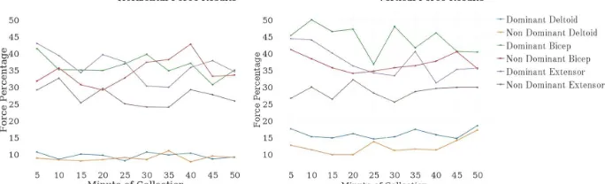
[image:6.595.126.468.407.511.2]Ten 1-minute EMGs were collected during the experimental task at 5-minute inter-vals. Each EMG was passed through a low-pass fourth-order Butterworth filter with a cut-off frequency of 500Hz. The averaged root mean square was then calculated using BEAT, which was then averaged. Normalization was carried out using the averaged MVICs collected before the start of the task. The normalized values were then aver-aged to represent the muscle activity as a percentage of the MVIC (see Figure 2). A one-way repeated measure analysis of variance (ANOVA) was then carried out.
Fig. 2 Force results for 1-minute readings collected every 5 minutes for both configurations
4.4! Force Results
Horizontal. A one-way repeated ANOVA for the 10 measures showed that only the
dominant extensor digitorum showed significant inconsistency of muscle activity throughout the task (a change of 13% and F(17, 9)=3.09, p=0.002). No significant evidence was found against the other muscles proving their activity to be consistent. Nevertheless, the non-dominant extensor digitorum showed an increase and decrease of up to 8% and the bicep brachii showed a change of up to 10% and 14% for the dominant and non-dominant sides respectively. The middle deltoidsÕ activity proved relatively stable with activations ranging from 8-11% for both sides.
digi-torum (a change of 13% and F(17, 9)=2.85, p=0.004). No significant evidence was found against the other muscles proving their activity to be consistent. Nevertheless, both biceps brachii showed an increase and decrease in activation of up to 13%, while both middle deltoid muscles showed relatively consistent activation of up to 18%.
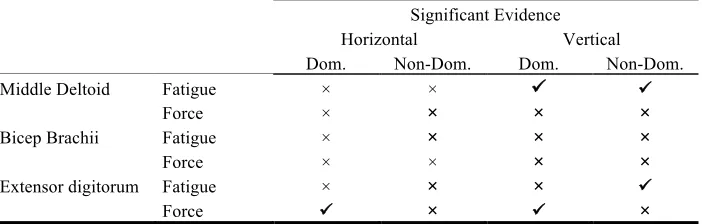
[image:7.595.125.476.262.374.2]Table 1 marks the muscles that showed significant evidence of increase in level of fatigue after the trial session for both configurations. It also marks significant incon-sistencies in activation of force during task interactions.
Table 1. Objective measures summary of significant increase in fatigue and force inconsistency
Significant Evidence
Horizontal Vertical
Dom. Non-Dom. Dom. Non-Dom.
Middle Deltoid Fatigue ! ! ! !
Force ! ! ! !
Bicep Brachii Fatigue ! ! ! !
Force ! ! ! !
Extensor digitorum Fatigue ! ! ! !
Force ! ! ! !
5
!
Subjective Measures
When examining fatigue, subjective ratings are commonly used to assess perceived physical exertion and supplement objective measures. We used the Borg CR100 scale [14], which is a fine graded ratio scaling method that estimates the level of exertion and determines the ratio relationship between perceptual responses. The scale con-tains subjective dynamic ranges with values ranging from 0 to 120 annotated with verbal anchors ranging from Ònothing at allÓ to Òabsolute maximumÓ. This scale was chosen as the most appropriate after critical assessment and comparison of four other scales. The subjects were asked to rate their perceived muscle exertion after the MVIC had been recorded before and after the experiment.
[image:7.595.120.475.651.692.2]The average of the scores collected was analyzed for statistical significance using a paired t-test. For the horizontal configuration, the averaged value of the ratings was significantly greater after the task (t(17)=1.74, p=0.0003). Similarly for the vertical configuration, the averaged value was significantly greater after the task (t(17)=1.74, p=0.0005). This indicates an overall increased perceived level of fatigue for both configurations (see Table 2).
Table 2. Subjective mean and standard deviation results for the horizontal and vertical confi-gurations
Configuration Before After
Horizontal 3.97 (±7.96) 15.44 (±14.93)
6
!
Discussion and Conclusion
The main aim of the study was to compare the muscle fatigue experienced when using large multi-touch interactive displays in horizontal and vertical configurations, over a one-hour period.
Our findings from the objective measures provide statistically significant evidence for the presence of potentially damaging fatigue for the middle deltoids for the verti-cal case only and not the horizontal. This can be due to the elevation required of the arms when interacting with the vertical display (i.e. gorilla-arm effect [24]) and the employment of larger force activation percentages. The presence of fatigue was also noted for the non-dominant extensor digitorum despite consistent activation of force. This can be due to the regular use of the non-dominant muscle (either uni- or bi-manually) in the vertical configuration, as can be derived from the significantly in-consistent activation of the dominant extensor digitorum. The inin-consistent activation of dominant extensor digitorum suggests that the users were alternating between dom-inant and non-domdom-inant sides. Unlike the objective measures, the results of the sub-jective measures showed that the subjects perceived the presence of fatigue in both the vertical and horizontal configurations with statistical significance. This emphasiz-es the unreliability of a personÕs perception of fatigue, where objective remphasiz-esults found significant levels of muscle fatigue that can be damaging only in the vertical case.
These results have clear implications on interaction design for large interactive displays. Vertical interactive displays, while suitable for intermittent use over short periods of time, are not as suitable for frequent longer use Ð our study showed pres-ence of fatigue for a one-hour task, but it might occur in shorter durations. Prolonged activities leading to muscle fatigue, which has previously been reported anecdotally, have the potential to lead to musculoskeletal disorders [1, 2]. For tasks that require frequent interactions in the range of one or more hours, designers should choose a horizontal display where possible to minimize damage to the musculoskeletal system, modify the design of interaction techniques to reduce the need for continuous interac-tion, or to change the location of distant interactions when possible to more accessible locations on the display. Moreover, subjective measures showed a significant level of fatigue for both horizontal and vertical cases, indicating that for either case, designers should locate points of interaction in spaces easily accessible to the user whenever possible. As a simple example, for large displays, the use of contextual commands that are located based on usersÕ interaction location is recommended over the tradi-tional desktop model of showing commands on upper or side toolbars and menus. However, identifying the display space that helps reduce fatigue, whether on horizon-tal or vertical displays, is the subject of further investigation.
the display and only two angles of interaction. In the future, we plan to assess the fatigability of other muscles (e.g. capitis muscles) at additional angles of interaction.
References
1. V¿llestad, N.K.: Measurement of human muscle fatigue. J Neurosci Methods 74,219-227 (1997)
2. Wahlstršm, J.: Ergonomics, musculoskeletal disorders and computer work. Occ Med 55, 168-176 (2005)
3. Meyer, S., Cohen, O., Nilsen, E.: Device comparisons for goal-directed drawing tasks. CHI'95 Conf Companion, pp. 251-252. ACM, Boston (1994)
4. Sears, A., Shneiderman, B.: High precision touchscreens: design strategies and comparisons with a mouse. Int J Man-Mach Stud 34,593-613 (1991)
5. Barriera-Viruet, H., Sobeih, T.M., Daraiseh, N., Salem, S.: Questionnaires vs observational and direct measurements: a systematic review. TIES'06 7,261-284 (2006)
6. Young, J.G., Trudeau, M.B., Odell, D., Marinelli, K., Dennerlein, J.T.: Wrist and shoulder posture and muscle activity during touch-screen tablet use. WORK 45,59-71 (2013)
7. Lozano, C., Jindrich, D., Kahol, K.: The impact on musculoskeletal system during multitouch tablet interactions. ACM CHI'11, pp. 825-828. ACM, Vancouver (2011)
8. Ichino, J., Isoda, K., Hanai, A., Ueda, T.: Effects of the display angle in museums on user's cognition, behavior, and subjective responses. CHI'13, pp. 2979-2988. ACM, Paris (2013) 9. Muller-Tomfelde, C., Wessels, A., Schremmer, C.: Tilted tabletops: In between horizontal and vertical workspaces. TABLETOP'08 pp. 49-56 (2008)
10. Zerpa, C., Lopez, N., Przysucha, E., Sanzo, P.: The effect of common teaching tools on upper extremity muscle activity. Education 4,160-166 (2014)
11. Morris, M.R., Brush, A.J.B., Meyers, B.R.: A field study of knowledge workers'; use of interactive horizontal displays. TABLETOP'08 pp. 105-112 (2008)
12. Wigdor, D., Perm, G., Ryall, K., Esenther, A., Chia, S.: Living with a tabletop: Analysis and observations of long term office use of a multi-touch table. TABLETOP'07, pp. 60-67 (2007)
13. Criswell, E.: Cram's Introduction to Surface Electromyography. Jones and Jones (2011) 14. Borg, G., Borg, E.: A new generation for scaling methods: Level-anchored ratio scaling. Psychologica 28,15-45 (2001)
15. Cioni, R., Giannini, F., Paradiso, C., Battistini, N., Navona, C., Starita, A.: Sex differences in surface EMG interference pattern power spectrum. J Appl Physiol 77,2163-2168 (1994) 16. Hincapi-Ramos, J.D., Guo, X., Moghadasian, P., Irani, P.: Consumed endurance: a metric to quantify arm fatigue of mid-air interactions. CHI'14, pp. 1063-1072. ACM, Toronto (2014) 17. Al-Mulla, M.R., Sepulveda, F., Colley, M.: A review of non-invasive techniques to detect and predict localised muscle fatigue. Sensors 11,3545-3594 (2011)
18. Peres, S.C., Nguyen, V., Kortum, P.T., Akladios, M., Wood, S.B., Muddimer, A.: Software ergonomics: relating subjective and objective measures. CHI'09 EA, pp. 3949-3954. ACM, Boston (2009)
19. Sherwood, L.: Human Physiology: From Cells to Systems. Cengage Learning (2012) 20. Phinyomark, A., Thongpanja, S., Hu, H., Phukpattaranont, P., Limsakul, C.: The Usefulness of Mean and Median Frequencies in Electromyography Analysis (2012)
21. Murata, A., Ishihara, H.: Evaluation of shoulder muscular fatigue induced during mouse operation in a VDT task. IEICE - Trans Inf Syst E88-D,223-229 (2005)
22. Lawrence, J.H., De Luca, C.J.: Myoelectric signal versus force relationship in different human muscles. J Appl Physiol Respir Environ Exerc Physiol 54,1653-1659 (1983)
23. NISMAT http://www.nismat.org/software/beat/doc/beat.html