Size, Composition,
and Structure
of the
Deoxyribonucleic
Acid
of
Herpes
Simplex
Virus
Subtypes
1 and 2
ELLIOTT D. KIEFF, STEVEN L. BACHENHEIMER, AND BERNARD ROIZMAN Department of Microbiology, The University of Chicago, Chicago, Illinois 60637
Receivedforpublication 29 April 1971
Studies of the size, composition, and structure of the deoxyribonucleic acid (DNA) of the F and G prototypes of herpes simplex virus (HSV) subtypes 1 and2
(HSV-1 and HSV-2) showed the following. (i) As previously reported by Good-heart et al. HSV-1 and HSV-2 DNA have a buoyant density of 1.726 and 1.728 g/cm3, corresponding to 67 and 69guanine i cytosine moles per cent, respectively. The differencein guanine plus cytosinecontent of the DNAspecieswasconfirmed by the finding of a 1 C difference in Tm. (ii) The DNA frompurified virus on
co-centrifugationwith T4 DNA in neutral sucrose density gradients sedimentedat55S, corresponding to 99 ± 5 million daltons in molecular weight. HSV-1 and HSV-2 DNAcould not be differentiated withrespect to size. (iii) Cosedimentation of al-kali-denatured DNA frompurified virus with T4 DNA on alkaline sucrose
den-sity gradients consistently yielded several bands of single-stranded HSV DNA
ranging from fragments 7 x 10⁶ daltons to intact strands 48 X 106 daltons in
molecularweight.
Naturally occurring herpes simplex viruses (HSV) differ ina numberofbiological, physical, andimmunological properties (5, 7, 8, 10,
17-19,
20, 27). As determined in neutralization tests, most[butnot all (31)] fresh isolates fall into two
subtype groups designated as 1 (HSV-1) and 2(HSV-2).
This report concerns the properties of HSV
deoxyribonucleic acid (DNA). Previous reports
have estimated the molecular weight of HSV-1 DNAto be 68 x 106 (24) to 100 x 106daltons
(2) and to contain 65 to 68 per cent guanine plus cytosine(10, 11,20,22,23).HSV-2 DNA was
reported (10) to contain 70.4 moles per cent
guaninepluscytosine, andnothingwas known of
its sizeorrelatednessto HSV-1 DNA. In the
ex-periments described in this paper, we have di-rectlycompared thesize, composition,and
struc-tureofHSV-1and HSV-2 DNA.
MATERIALS AND METHODS
Solutions. Virus buffer consisted of0.15 M NaCl
and 0.02 Mtris(hydroxymethyl)aminomethane
(Tris)-hydrochloride, pH 7.5. Neutral DNA buffer consisted of1 MNaCl, 0.001M ethylenediaminetetraacetic acid
(EDTA), and 0.05 M Tris-hydrochloride, pH 7.5. Alkaline DNA buffer consisted of 0.8 M NaCl, 0.3 M NaOH, and 0.001 M EDTA. Reticulocyte standard buffer (RSB) consisted of 0.01 M sodium chloride,
0.001 M magnesium chloride, 0.01 M Tris, pH 7.5. Standardsaline citrate (1 X SSC) consisted of 0.15 M sodiumchloride, 0.015 M sodium citrate. SDS buffer
consisted of 0.1 MNaCl, 0.001 MEDTA, 0.01 MTris,
pH7.4.
Cells. HEp-2 and Vero cells were originally ob-tained from FlowLaboratories, Rockville, Md., and were grown in minimal essential medium
supple-mented with 10% calfserum.
Virus and infection of cells. The properties of the Fand G prototypes of HSV-1 and HSV-2 have been
previously described(7). The procedures used for in-fection of HEp-2 and Vero cells with HSV-1 and HSV-2 werethose described previouslyforinfection
of HEp-2 cells with HSV-1 (7).
Labeling of cells with radioactive isotopes. HEp-2
cells infected with 20 plaque-forming units (PFU) of HSV-1 or HSV-2 percell werelabeled between 4 and 20 hr postinfectionwith 10,Ciof3H-thymidine
or0.1 ,uCiof14C-thymidine (Schwarz-Mann, Orange-burg, N.Y.) per ml of medium of mixture 199 lacking
thymidine and supplemented with 1% dialyzed calf serum.
Purification ofvirus: preparation of nucleocapsids. Cellsinfected with HSV-1orHSV-2 were harvested by
scraping and centrifuged at 800 X g in a PR-2
re-frigeratedcentrifuge for 10 min at 4 C. The cell pellet was washed with phosphate-buffered saline (PBS) and resuspended in RSB containing 0.5% Nonidet P-40 (Shell Chemical Co., New York, N.Y.) for 10 minat4C. Thecells were then homogenized with six 125
on November 11, 2019 by guest
http://jvi.asm.org/
strokes of a tight fitting Dounce homogenizer, and the nuclei were removed by centrifugation of the ho-mogenate at 800 X gfor 10min at 4 C in the PR-2
centrifuge. The cytoplasm was layered onto 37-ml
linear 10 to 50% (w/w) sucrose density gradients, prepared in virus buffer and centrifuged for 1 hr at 25,000 rev/minin theSW27 rotor at5C.
Purification of virus: preparation of envelopedvirus. Thepreparation of enveloped HSV-1 followeda
pro-cedureof Spear andRoizman(manuscript in prepara-tion). Briefly, cells infected with HSV-1 were har-vested and washed as described above. The cell pellet wasresuspendedin 0.01 MTris-hydrochloride (pH 7.4) for 10 min at 4 C,homogenized with four strokes of a
tight fitting Dounce homogenizer, and adjusted to 0.25 M sucrose by theaddition of one-seventh volume of2M sucrose in 0.01 MTris-hydrochloride (pH 7.4). Thenucleiwereremoved bycentrifugation for 10 min at 800 X g in a PR-2 refrigerated centrifuge. The cytoplasm was layered onto 17-ml linear dextran 10 gradients (1.04 to 1.09 g/cm3) and centrifuged for 1hrat25,000rev/min and 5 C in a SW 25.3 rotor.
Determination of relative sedimentation rates of HSV-1 and HSV-2 DNA: preparation of 14C-labeled T4 DNA. T4 phage prepared in E. coli and labeled
with 14C-uridinewasthe kind gift of Robert Hasel-korn. The phage was resuspended in 0.5% sodium dodecyl sulfate (Matheson Scientific Co., Elk Grove
Village, Ill.) and 2%sarkosyl NL97 (Geigy Chemical Co., Ardsley, N.Y.) in neutral DNA buffer and
gently rolled with phenol at 60 C for 2 min. The
phenolphase was removed, and the DNA wasgently
rolled with chloroform-isoamyl alcohol (2%, v/v)
untilthe aqueous phase wasclear.
3H-thymidine-labeled simian virus 40 form I DNA
(obtained from S. Kit and D. M. Dubbs) and
3H-thymidine-labeled E. coli plasmid DNA (obtained from N. Cozarelli) were used to determine therelative
sedimentation constant of the purified T4 DNA in
alkalinesucrosegradients.
Sedimentation in neutral sucrose gradients.
3H-thymidine-labeled HSV-1 or HSV-2 nucleocapsids were disruptedby exposure to0.5% sodium dodecyl sulfate, 2% sarkosyl inneutral DNAbuffer at60 C for 2 min; mixed with "4C-labeled T4 DNA; poured onto 12-ml linear 10to30% (w/w)sucrosegradients
prepared in neutral DNA buffer; and sedimentedfor 3.5 hrat40,000 rev/min in an SW41 rotor at20 C.
Fractions (0.3ml) werecollectedfrom the top of the
gradients, and the DNA wasprecipitated ontofilters (HAWP 25-mm; Millipore Filter Corp., Bedford, Mass.) with5%trichloroacetic acid byusingherring
spermDNA (Sigma Chemical Co., St. Louis, Mo.) as a carrier. The filters were dried and assayed for
radioactivityinaPackard Tri-Carbliquid scintillation
spectrometer.
Sedimentation in alkaline sucrose gradients.
3H-thymidine-labeled HSV-1 or HSV-2 nucleocapsids
weremixed with14C-labeledT4phageandresuspended
in0.5% sodiumdodecyl sulfate and 2% sarkosyl in alkaline DNA buffer. The DNAspecieswere poured
onto 12-ml linear 10to 30% sucrose gradients pre-pared inalkalineDNAbuffer and sedimented for 3.5 hrat40,000rev/min in an SW41 rotorat20 C.
Theprocedures for thecollection of sucrose density gradient fractions, precipitation of the DNA, and assayofradioactivitywere asdescribed above.
Extraction and sedimentation of viral DNA from whole cytoplasmic lysate and from purified enveloped virus.HEp-2cells wereinfected with 40 PFU of HSV-1 percell andlabeled from 4 hrpostinfection with 10
M&CiofSH-thymidineper ml ofmedium. At 16 hr post-infection, the cells were harvested, allowed to swell in 0.01 MTris (pH 7.4) and 2.5 mm EDTA, and then
Dounce-homogenized as previously described. A
sampleof thecytoplasmwaslayered on adextran 10 gradient and centrifuged to obtain enveloped virus. The DNA was extracted fromenveloped virions and
cosedimented with T4 DNA as described above. Another sample of the cytoplasm was dissolved in 0.5%sodiumdodecyl sulfate, 2%sarkosylinneutral DNAbufferat60 C for2min;mixed with 14C-labeled T4 DNA; andcosedimented in neutral sucrose density gradients. Finally, a sample of cytoplasm and a sample ofpurifiedenvelopedvirusharvestedfromthe dextran 10gradient were each mixed with T4 phage labeled with 14C in the DNA; solubilized in 0.5% sodium dodecyl sulfate,2%sarkosyl in alkalineDNAbuffer;
andcentrifugedinalkalinesucrosedensity gradients. Allcentrifugationsweredoneasdescribedabove.
Purification of viral DNA for isopycnic banding and thermaldenaturation. Purifiedherpessimplex nucleo-capsids were resuspended at 60 C in neutral DNA buffer containing 0.5% sodium dodecyl sulfate and 2% sarkosyl, gently poured onto 12-ml linear 10 to
30% sucrose gradients, and centrifuged in an SW41
rotor for 3.5 hr at 40,000 rev/min and 20 C.
Frac-tions (0.3ml) werecollected throughaflowcell
con-tinuously monitored for absorbance at 260 nm. The
fractionscontaining intact HSVDNAweredialyzed against 0.1X SSC, digested with 50 ttg of heat
in-activated pancreatic ribonuclease (Schwarz-Mann, Orangeburg, N.Y.) per ml, adjusted to 1.5X SSC,
and further digested with 50 ug ofribonuclease per
ml.Sodiumdodecyl sulfate wasaddedtomakeafinal concentration of0.5%O, and theproteinwasextracted
by rolling the DNA with phenol and chloroform-isoamyl alcohol. The DNA was then dialyzed four times for 24 hragainst 100volumes of 0.1X SSCto removetracesofphenol.
Isopycnicbanding of HSV DNA in cesium chloride.
PurifiedHSV-1 orHSV-2 DNAwasmixedwith2Mug
of SPO DNA (gift of K. Bott), dialyzed against
cesium chlorideat adensityof1.715g/cm3in 0.01 M
Tris-hydrochloride (pH 7.4), and centrifuged in the
Spinco model E analytical centrifugeat 44,700 rev/
minand 25 C for 20 hr.Ultraviolet (UV) absorption
photographs were scanned with the Joyce Loebel microdensitometer.
Melting temperature of HSV DNA.PurifiedHSV-1 DNA and HSV-2 DNA in0.1X SSCweredialyzed
against0.1 X SSC in thesameflasktoeliminate pos-sible artifacts atributable to differences in salt
con-centration. The thermal denaturation
profiles
of the two DNAspeciesweredeterminedsimultaneously
in 0.5-mlTeflon-stoppered quartzcuvettes witha 1-cmlight path(HellmaCellsInc.,Jamaica, N.Y.) by
using
aGilfordrecordingspectrophotometerequipped
withon November 11, 2019 by guest
http://jvi.asm.org/
a linear temperature programmer and internal
thermistor (16).
RESULTS
Viralpreparations: naked nucleocapsids.
Noni-det P-40-extracted cytoplasm of 20 hr-infected HEp-2orVero cellsyielded (2) prominentbands oncentrifugation througha10to50% (w/w) su-crose density gradient. Electron microscopy of
negatively stained preparations showed that the top band contained amixed population of
par-tially disrupted, intact, empty, and full nucleo-capsids. The bottom band contained predomi-nantly full nucleocapsids contaminated with
smallamountsof membranedebris.
The bottom band from the sucrose gradients was used as the source of DNA from
nucleo-capsid preparations.
Enveloped virus preparations. The cytoplasm
ofHSV-1-infected cellsoncentrifugation through
a dextran 10 density gradient asdescribed above
yielded a single band in the middle of the tube. Electron microscopic analysis indicated that the band contained fully enveloped virus imperme-able to phosphotungstic acid and wasfree from partially disaggregated virions. This finding
con-firmsnumerous other observations that the
cyto-plasm of cells infected with HSV-1 virus contains predominantly enveloped nucleocapsids (28).
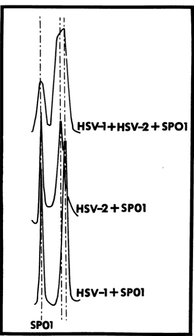
Isopycnic bandingof HSV-1 and HSV-2 DNA in neutralCsCI. Thedensity of HSV-1 and HSV-2 DNA in neutral CsCl was determined in the
Spinco model E centrifuge with SPOl as an
in-ternalmarkerasdescribed above. Theresultsare
shown inFig. 1. The densities of HSV-1 DNA andHSV-2 DNAcalculated accordingto Szybal-ski (30) with the value of 1.742 g/cm3 for the density of SPOl DNA are 1.726 i 0.0005 and 1.728 0.0005g/cm3, respectively.
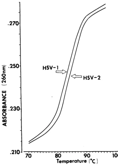
Thermal denaturation of HSV-1 and HSV-2 DNA. The UV absorption-thermal denaturation profile of HSV-1 and HSV-2 DNA was deter-mined in0.1x SSCasdescribed above. The re-sults shown in Fig. 2 indicate that the melting
temperatures of HSV-1 and HSV-2 DNA in
0.1X SSCare82and 83 C,respectively.
Determination of relative sedimentation rates
andmolecular weight of HSV-1 and HSV-2 DNA: sedimentation of native DNA. The sedimentation
rateofHSV-1andHSV-2 DNAin neutralsucrose
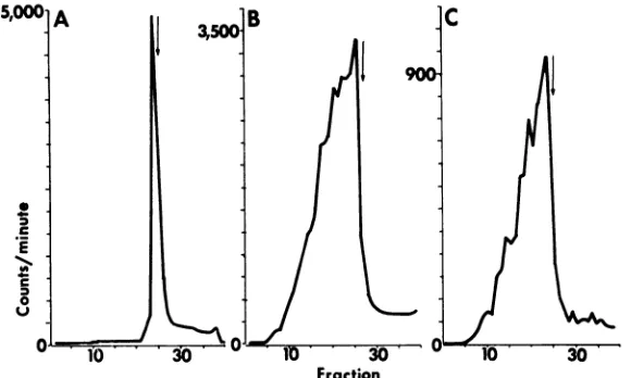
density gradientswas compared withthat ofT4 DNA as described above. Theresults shown in Fig. 3 indicate the following.
(i) Both HSV-1 and HSV-2 DNA extracted from nucleocapsids with sodium dodecyl sulfate and sarkosyl and centrifuged in neutral sucrose
density gradients formasinglesharpbandslightly above that ofT4 DNA (Fig. 3A, B). Identical
FIG. 1. Buoyant density determination of HSV-1
DNA, HSV-2 DNA, and ofan artificialmixture of
HSV-1 and HSV-2 DNAdetermined bycentrifugation
withSPOIDNA inamodel E analytical centrifugeas
described in Materials andMethods. The UV
absorp-tion photograph was scanned with a Joyce Loebel
microdensitometer.
results were obtained when HSV nucleocapsids were mixed with T4 phage and coextracted by
gently rolling with phenol and chloroform-iso-amylalcohol.
(ii) T4 DNA used in this study was intact
double-stranded DNA by the following criteria. T4DNAformedasingle discrete band in neutral sucrosegradients. Alkali denaturation of the T4 DNA yielded a single discrete band in alkaline
sucrose gradients. Cosedimentation of T4 DNA
with simian virsus 40form IDNAand Escherchia
coli plasmid DNA in alkaline sucrose gradients demonstrated that the sedimentation rate of the denaturated T4 DNA was identical to that pre-viously reported (3, 14).
(iii) The ratio of the distances sedimented by HSV-1 and HSV-2 DNAto that sedimented by
I
HSI
1+HSV-2+SPO1
Ii
HSV-+SPO1
l
ISW+
HJX|SV-
+
SPOI
SPO
on November 11, 2019 by guest
http://jvi.asm.org/
[image:3.484.240.433.59.392.2]T4 DNA was 0.96 ± 0.02. The sedimentation
coefficientof HSV-1 DNA and HSV-2 DNA in
neutralsucrosedensity gradients derived from the relationship of Burgi and Hershey (4), (d1/d2) =
(sl/s2),
was 55S. The molecular weight of HSVDNA wasdetermined from the equation Sl/S2 =
(M1/M2)0.38 (4, 9). With the value of 110 X 106
daltonsfor the molecularweight of T4 DNA (1, 6, 26), the molecular weight of HSV DNA was
calculatedtobe 99 ±+- 5 X 10⁶daltons.
.270--E .250 C
0
C'4
Lu z 4c
g .230
0
co
U,
HSV-(<Kzz
HSV-270 80 90
[image:4.484.57.248.176.445.2]Temperature
(1C)
FIG. 2. UVabsorbance-thermal denaturationprofile
of HSV-J and HSV-2 DNA in O.I X SSC, determined asdescribed in Materials and Methods.
(iv) Cosedimentation of HSV-1 and HSV-2 DNA in neutral sucrose gradients (Fig. 3C)
in-dicated that the sedimentation coefficients of these DNA species cannot be differentiated by zonecentrifugation.
Sedimentation of denatured DNA. The sedi-mentation rate of denatured HSV-1 and HSV-2 DNA was compared to that of denatured T4 DNAin alkaline sucrose density gradients as
de-scribed above. Figure 4 shows the distribution of DNA in the gradients. The results indicate the
following.
(i) HSV-1 and HSV-2 DNA formed multiple
bands inalkaline sucrose density gradients. The
formation ofmultiple bands isnot anartifactof
theprocedures used in thepurification of the
vi-rus orin the extractionand thesedimentationof
DNA.The DNAextracted fromenveloped HSV-1
showed a similar distribution inalkaline sucrose
gradients. The distribution of DNA bands was unaffected by the presence of 2.5 mm EDTA
during all stages of viral purification. T4 DNA
coextracted with HSV-DNA from mixtures of HSV nucleocapsids and T4phage yieldedasingle band on sedimentation in alkaline sucrose. Mixtures of T4 phage and HSV nucleocapsids
lysed directly on the top of alkaline gradients
yielded similar results to those shown in Fig. 4A and B.
(ii) The mostprominentand mostrapidly
sedi-menting band of HSV-1 and HSV-2 DNA
lo-calizedslightlyabove T4 DNA (Fig. 4A, B). The
ratio of the distances of the sedimentation of
HSV DNA tothat of T4 DNAwas 0.93. Onthe basis of thisratio, the most
promient
HSV bandwas estimated to be 68S. With the
equation
andcoefficients of Studier (29), weestimate the
mo-lecular weight of HSV DNA in the rapidly sedi-mentingbandtobe 49 x106 daltons.
Co-centrifu-gation of HSV-1 and HSV-2 indicated that the
sedimentation coefficients of the most
prominent
3.
Fraction
FIG. 3. Zone sedimentation ofHSVDNA inneutralsucrosedensity gradients. (A) Co-centrifugafion of
'H-HSV-1DNAwith14C-T4DNA.(B)Co-centrifugationof3H-HSV-2 DNA with 14C-T4 DNA.(C)Co-centrifugation
of 3H-HSV-2 DNA with "4C-HSV-i DNA. TheDNAspecies were centrifuged for 3.5 hr inan SW41 rotorat 40,000 rev/min and20C.Symbols:dashedline,14C-labeledDNA;solidline,3H-labeledDNA. Directionof
sedi-mentationis totheright.
on November 11, 2019 by guest
http://jvi.asm.org/
[image:4.484.113.404.490.597.2]Fraction
FIG. 4. Zone sedimentation ofalkalidenatured HSV DNA andT4DNA inalkalinesucrosedensitygradients.
(A) co-centrifugation of 3H-HSV-1DNAwith14C-T4DNA. (B) Co-centrifugation of3H-HSV-2DNAwith 14C-T4 DNA. (C) Co-centrifugation of3H-HSV-2 DNA with 14C-HSV-iDNA. The DNA species were centrifuged
for 3.5hr in anSW41 rotor at40,000 rev/minand 20C.Symbols:dashedline, 14C-labeledDNA;solidline,
3H-labeled DNA. Directionofsedimentation is to theright.
bandof thetwoDNA
species
cannotbedifleren-tiated byzone
centrifugation
in alkaline sucrosedensity gradients.
(iii) With respect to the slower
sedimenting
species ofHSV-1andHSV-2 DNA thedataareas
follows. Atleast three discrete bands are
usually
found,
ranging
in ratio of distances sedimentedrelativetoT4DNAof0.51 to
0.84,
corresponding
to a range in molecularweights
of 7 x10⁶
to30 x 106daltons.Some
variability
inthedistribu-tion of theDNA inalkalinesucrose
gradients
wasfound between different
preparations
of thesamevirus. The more
slowly sedimenting
speciescon-stituteatleast halfthetotal viralDNA.
(iv)
Cosedimentation ofHSV-1orHSV-2 DNAextracted from
nucleocapsids
grownin Vero cellswith T4 DNA
yielded
a similar distribution tothatseenin
Fig.
4Aand B.Structural identity of viral DNA in crude
cyto-plasmic lysate and in purified enveloped virus. These experiments were
designed
to determine whetherthesingle-stranded
breaksinviralDNAwere a constant feature of
purified
virions orwhether similarbreakswerepresentin crude cellu-lar extracts containing virions. The size of the labeled native anddenaturedDNA in the whole
cytoplasm
and in theenveloped
virus bandob-tained from HEp-2 cells infected for 16hr with
HSV-1 was determined in neutral and alkaline
sucrosedensity gradientsasdescribed above.The
resultsshown inFig.S indicatethe
following.
(i) LabeledDNA released by sodium dodecyl sulfate and sarkosyl from virions in the
cyto-plasmofcellsinfectedfor 16 hrwith HSV-1 (Fig.
5A)
sedimentedin neutralsucrose densitygradi-ents exactly like the DNA extracted from
puri-fied virions. The ratio of thedistancessedimented
by the cytoplasmic DNA relative to that of T4 DNA was0.96%, i.e., the same asfoundin the re-sults of theexperimentsummarizedinFig.3.
(ii) Cosedimentation of denatured DNA ex-tractedfrom thecytoplasm (Fig. SB) orfrom en-veloped virus(Fig. 5C) with T4 DNA in alkaline sucrosedensitygradientsyieldedidenticalresults, indicating that enveloped virus accumulating in the cytoplasm contains DNA withsingle-stranded
nicksandthatthebreaks are not the consequence
of
manipulations
involvedinviruspurification.DISCUSSION
These studies wereundertaken to compare the size and structure of the DNA of the two major subtypes ofherpes simplex virus. The data ob-tained in these studiesaresummarized inTable1. Composition of HSV-1 andHSV-2 DNA. Pre-vious estimates of thebuoyant density ofHSV-1 and HSV-2 DNA were based on comparative distances between cellular and viralDNAbands of HSV-1 and HSV-2 infected cells (10). These studies indicated that HSV-2 DNA hadahigher buoyant density than HSV-1 DNA. With SPOl
DNA asan internalmarker, we obtained values of 1.726 and1.728g/cm' forthebuoyant density
of HSV-1 and HSV-2, respectively (Table 1). Based on the assumption that there are no un-usual basesinHSV-1 and HSV-2 DNA, the mole percentages of guanine plus cytosine bases are
estimatedtobe67and 69% (25).UV absorbancy-thermal denaturation studies indicated a 1 C
difference in the Tm of HSV-1 and HSV-2 DNA
asexpectedfor DNA species differing by 2 moles per centguanineplus cytosine.
Size of HSV-1 and HSV-2 DNA. Previous esti-mates of the molecular weight of HSV-1 DNA
on November 11, 2019 by guest
http://jvi.asm.org/
[image:5.484.84.379.67.210.2]Fraction
FIG. 5.Zone sedimentationinneutralandalkalinesucrosedensity gradients of HS V DNA extracted from crude cytoplasmof infected cellsandfrom purified envelopedvirus in thatcytoplasm. (A) Native3H-HSV-1DNA in the crudecytoplasm sedimented in neutral sucrose gradients. (B)Denatured3H-HSV-1DNA in alkaline sucrose gradients. (C)Denatured3H-HSV-1 DNA extracted from envelopedvirionspurifiedfrom the crude cytoplasmic lysate by sedimentation through dextran 10densitygradient. Centrifugation was for 3 hrinan SW41 rotor and at40,000 rev/min and 20 C. Theposition of 14C-T4 DNA cosedimented with 3H-HSV-i DNA is indicatedby
[image:6.484.123.413.64.238.2]arrow.Directionofsedimentationisto theright.
TABLE 1. Comparison of size and structure of the twomajorsubtypes ofherpes simplex virus
(HSV-i andHSV-2)
Native DNA Single-strandDNA
- ~~~~~~~~~~~~~~~~Overallrange
Virus Guanine Intact strand of fragments
,Sa " Molwt Buoyant density cytosine (daltons) (g/cm3) molesper
cent sb 'MolWt Molwt
(daltons) (daltons)
HSV-1 (F) 55 99 ± 5d 1.726 i± 0.0005 67 68 48d 7-48d
HSV-2 (G) 55 99 ± 5 1.728 i 0.0005 69 68 48 7-48
aIn neutral sucrose relativeto
14
DNA.aIn
alkaline
sucrose relative toT4
DNA.c Viral DNAfrom 16-hr-oldinfected cells.
dExpressed X
I06.
were obtained by two techniques. One involving direct measurement ofthe sedimentation coeffi-cient of theDNA yielded a molecular weight of 68 x 106 daltons (24) andisinall
probability
alow value. The second, using the Kleinschmidt technique,
yielded
anaveragevalue of 101 X 106daltons with a range of86 x 106 to 115 x 106
daltons (2). The size of HSV-2 DNA was
un-known.
Inprinciple,thereareseveraltechniquesfor
de-termination ofthe absolute molecular
weight
of DNA molecules greater than 50 X 106 daltons: 32pstarenumeration (15),equilibrium
sedimenta-tion (26), and indirect determinations based on
phosphate analysis and the molecular
weight
valueofpurified virions obtainedby equilibrium sedimentationandsedimentation diffusion (1, 6). As reviewed by Friefelder (9), all ofthese tech-niques suffer from inherent errors. It is unlikely thatDNA molecularweight determinationsbased onphosphate analysis and molecular weights of
purifiedvirions willeverbepractical for the large enveloped DNA viruses. Other techniques, such
as Kleinschmidt and sedimentation, depend on standardization against DNA species of known molecularweight. Inregard to theKleinschmidt technique, HSV presents an unusual problem. We are particularly concerned by recent
obser-vations (9, 12) that the massper unit length for DNA of65 to 70 guanine plus cytosine moles
on November 11, 2019 by guest
http://jvi.asm.org/
per cent might be as much as 15% higher than that used inpreviousstudies.
We chose to determine the molecular weight
of HSV-1 and HSV-2 DNA by zone sedimenta-tion in neutral sucrose density gradients by using
T4 DNAas aninternalstandard for thefollowing
reasons. (i)
Independent
absolutedeterminationshave been made for few DNA
molecules,
T4beingamong them(1, 6,26). (ii)T4 DNA isvery
closeinsizetoHSVDNA.
(iii)
Recent datacon-firm that the Burgi and Hershey relationship is valid over small ranges of S and M(9).
The molecular weight of both HSV-1 and HSV-2 DNA was found to be 99 ± 5 x 10⁶
dal-tons.
Structure of HSV-1 and HSV-2 DNA. The
molecular
weight
ofdenaturedHSV-1 and HSV-2DNA was obtained
by
zonecentrifugation
inalkaline sucrose
gradients
to determine whetherthe native DNA consisted of two continuous
non-cross-linked single strands. The results
indi-catedthat,atmost,
only
half of thesingle
strandsinany
preparation
of HSV-1 orHSV-2 DNAareintact; the remainder of theDNA is
fragmented.
ViralDNA in the
cytoplasm
of infected cellsbe-fore isolation of
purified
virus hadthe samesizedistribution on alkali denaturation as the DNA
of
purified virus,
effectively
excluding
thepossi-bility that single-strand breaks arise
during
viralpurification. Thefragments of DNA,whichrange in size from 7 X 10⁶ to 30 X 106
daltons,
form several discrete bands in sucrosegradients,
in-dicating
thatthey
are notproducts
of randomcleavage.
NoDNAwasconsistently
found to belarger than intact
single
strands.Therefore,
weconclude thatnative viral DNA contains
single-strand breaks and that it is not cross-linked. Similar
findings
haverecently
beenreported
for Marek's disease virusDNA(13).
Significance of the single-strand fragments found in HSV DNA.
Although
theorigin
andbiological
significance of thesingle-strand
frag-mentsfoundin HSV DNAare not
clear,
several points shouldbemade.(i)
Asnotedpreviously,
the fragments are not an artifact of theprocedures
used in viral purification or in extraction and
analysis of the DNA. The alkaline denaturation experiments
probably
indicate the existence of breaks in the DNA, since alkali-labilelinkages
suchasapurinicsiteshave notbeenfound in viral DNAspecies. (ii) Both HSV-1 and HSV-2 DNA havebeen shown to havesimilardistributions of single-strand fragments. Furthermore, the
find-ingis not a unique feature of virus growth in a
particular cell line. (iii) The finding that DNA frompurified virusyieldson
velocity
centrifuga-tion in alkaline sucrosegradients
at least four bandsranging in size from7 X 106 to48 X 106daltons suggeststhat thesingle-strand breaksare not random but are at specific sites. (iv) We do not know whether the fragmentsresult from specific nicking of intact single strands or results fromincompleteligation of breaks necessary for control of transcription, as in T4 (21). These
points
arecurrently
underinvestigation.
ACKNOWLEDGMENTS
Thisinvestigation wassupported by grants from the Public HealthService grant CA 08494 from the National Cancer Insti-tute, E314F fromtheAmerican Cancer Society,andGB 27356 fromtheNational Science Foundation.E. D. K. wasa postdoc-toral trainee of the Public HealthService(PHSAI-00238).S.B. isapredoctoral trainee of the Public HealthService (PHS Al-00238).
WeacknowledgeR. Haselkorn for the gift of14C-T4phage, N.CozzarelliforE.coliplasmid DNA,D.R.DubbsandS. Kit for3H-SV4o DNA, K. Bott for SPOI DNA,andK. Chiangforthe useofthe modelE centrifuge,and N. Frenkel forassistance in performing the thermaldenaturation studies.
LITERATURE CITED
1.Bancroft, F. C., and D. Freifelder. 1970.Molecular weights of coliphagesandcoliphage DNA-measurementof molec-ularweight of bacteriophageT7byhigh speedequilibrium centrifugation. J. Mol. Biol. 54:537-546.
2. Becker,Y.,H.Dym, andI.Sarov.1968. Herpes simplex virus DNA.Virology36:184-192.
3.Berns, K. I., and C. Silverman. 1970. Natural occurrence of cross-linked vaccinia virus deoxyribonucleic acid.3.Virol. 5:299-304.
4.Burgi, E., andA.D.Hershey. 1963. Sedimentationrate asa measureof molecular weightof DNA.Biophys.J. 3:309-321.
5.Dowdle, W. R., A.J. Nahmias, R. W. Harwell, and F. P. Pauls. 1967. Association ofantigenic type ofherpesvirus hominis withsite of viralrecovery.J.Immunol.99:974-980. 6.Dubin, S. B.,G. B. Benedek,F. C. Bancroft, and D. Frei-felder. 1970. Molecular weights of coHl phages and coli phageDNA.II.Measurementofdiffusioncoefficients using opticalmixing spectroscopy and measurement of sedimen-tationcoefficients. J. Mol. Biol. 54:547-557.
7.Ejercito,P.M.,E.D.Kieff,and B.Roizman. 1968. Character-izationofherpes simplex virus strains differingintheir effect onsocial behaviorofinfected cells.J.Gen.Virol.3:357-364. 8.Figueroa, M.E.,and W. E.Rawls. 1969. Biological markers fordifferentiationofherpes-virusstrainsoforaland genital origin. J. Gen. Virol. 4:259-267.
9. Freifelder, D. 1970. Molecular weights of coliphages and coli phage DNA. IV.Molecular weightsofDNA from bacteri-ophages T4, T5,T7 andgeneralproblem of determination of M.J.Mol.Biol. 54:569-577.
10. Goodheart, C. R., G. Plummer, and J. L. Waner. 1968. Densitydifferences of DNA of herpes simplex viruses types 1and 2. Virology35:473-475.
11. Lando,D.,J.DeRudder, andde M. P.Garlike. 1965. Macro-moleculesetmetabolitesprecurseurs a. Acides nucleiques (apropos of thecomposition ofherpesvirus DNA). Bull. Soc.Chim.Biol. 47:1033-1042.
12. Lang, D. 1970. Molecular weightsofcoli phages andcoli phageDNA.III. Contour lengthandmolecularweightof DNAfrombacteriophageT4,T5and T7and frombovine papilloma virus.J.Mol.Biol. 54:557-565.
13. Lee, L. F., E. D. Kieff, S. L. Bachenheimer, B.Roizman, P. G.Spear, B. R.Burmester,and K.Nazerian. 1971. Size andcompositionofMarek's diseasevirusdeoxyribonucleic acid. J.Virol.7:289-294.
14. Leighton,S.B., andI.Rubenstein.1969.Correlating
miolecu-lar weightscales for DNA. J.Mol.Biol. 46:313-328.
on November 11, 2019 by guest
http://jvi.asm.org/
15. Levinthal, C., and C. A. Thomas. 1957. Molecular autoradio-graphs the raycounting from single virus particles and DNAmoleculesin nuclear emulsions. Biochem. Biophys. Acta23:453-465.
16. Mandel,M., and J. Marmur. 1968. Use ofultraviolet
absorb-ance-temperature profile for determining the guanine plus cytosinecontentofDNA,p. 195-206. In L. Grossmanand K.Moldave (ed.), Methods in enzymology, vol. 12partB.
Academic Press Inc., New York.
17. Nahmias, A. J., W.R.Dowdle, Z.M.Naib, A. Highsmith, R.W.Harwell, and W. E. Josey. 1968. Relation of pock size on chorioallantoic membrane to antigenic type of herpesvirus hominis. Proc. Soc. Biol. Med. 127:1022-1028. 18. Plummer, G. 1964. Serological comparison of the
herpes-viruses. Brit. J. Exp. Pathol. 45:135-141.
19. Plummer, G., J. L. Waner, and C. P. Bowling. 1968. Compara-tive studies oftype 1 andtype2herpes simplex viruses.
Brit. J. Exp. Pathol. 49:202-208.
20. Plummer, G., J. L. Waner, A. Phuangsab, and C.R. Good-heart. 1970. Type 1 and type 2 herpes simplex viruses: serological andbiological differences.J. Virol.5:51-59. 21. Riva, S., A. Cascino, and E.P.Geiduschek. 1970.Coupling
of latetranscription to viral replication in bacteriophage T4 development. J.Mol. Biol. 54:85-102.
22. Roizman, B., L. Aurelian, and P. R. Roane, Jr. 1963. The
multiplication of herpes simplex virus.I.Theprogramming
of viral DNAduplicationinHEp-2cells.Virology 21:482-498.
23. Russell, W. C., and L. V. Crawford. 1963. Some characteris-tics of the deoxyribonucleic acid fromherpessimplexvirus, Virology 21:353-361.
24. Russell, W. C., and L. V. Crawford. 1964.Properties of the nucleic acids fromsomeherpesgroupviruses. Virology22:
288-292.
25. Schildkraudt,C.L., J. Marmur, and P. Doty. 1962. Determi-nation of basecomposition ofdeoxyribonucleicacid from its buoyant density in CsCl. J. Mol. Biol.4:430-443. 26. Schmid, C. W., and J. E. Hearst. 1969. Molecular weights of
heterogeneous coli phage DNAs from density gradient sedimentation equilibrium. J. Mol. Biol.44:143-160. 27. Schneweis, K. E. 1962. Serologische Untersuchungen zur
Typedifferenzierung desherpesvirus homninis. Z.
Immuni-taets.Allergieforsch. 124:24-28.
28. Schwartz, J., and B. Roizman. 1969. Similarities and
differ-encesin thedevelopment of laboratory strains and freshly
isolatedstrains of herpes simplex virus in HEp-2 cells:
elec-tronmicroscopy. J. Virol.4:879-889.
29. Studier, F. W. 1965. Sedimentationstudies of the sizeand shape of DNA. J. Mol. Biol. 11:373-390.
30.Szbalski,W. 1968.Useofcesium sulfateforequilibrium
den-sity centrifugation, p. 330-360. In L. Grossman and K.
Moldave (ed.), Methods in enzymology, vol. 12, part B. Academic Press Inc., New York.
31. Terni, M., and B. Roizman. 1970. Variability of herpes sim-plex virus: isolation of two variants from simultaneous eruptionsatdifferentsites. J. Infec. Dis. 121:212-216.