protein’s core,␣-helices 2 to 6, mediates the vast majority of intra- and interdimer contacts and is strongly conserved in allMicroviridae(canonical members areX174, G4, and␣3) external scaffolding proteins. On the other hand, the primary sequences of the first␣-helices have diverged. The results of previous studies with
␣3/X174 chimeric external scaffolding proteins suggest that ␣-helix 1 may act as a substrate specificity domain, mediating the initial coat scaffolding protein recognition in a species-specific manner. However, the low sequence conservation between the two phages impeded genetic analyses. In an effort to elucidate a more mechanistic model, chimeric external scaffolding proteins were constructed between the more closely related phages G4 andX174. The results of biochemical analyses indicate that the chimeric external scaffolding protein inhibits two morphogenetic steps: the initiation of procapsid formation and DNA packaging.X174 mutants that can efficiently utilize the chimeric protein were isolated and characterized. The substitutions appear to suppress both morphogenetic defects and are located in threefold-related coat protein sequences that most likely form the pores in the viral procapsid. These results identify coat-external scaffolding domains needed to initiate procapsid formation and provide more evidence, albeit indirect, that the pores are the site of DNA entry during the packaging reaction.
Microviruses (canonical members are bacteriophages
X174, G4, and␣3) are T⫽1 icosahedral virions with scaffold-ing protein-mediated assembly pathways. As in other viral sys-tems (13, 14, 19), scaffolding proteins temporarily associate with structural proteins, stimulating conformational changes that nucleate assembly and ensure morphogenetic fidelity (9, 13). Microvirus morphogenesis is unique in that it depends on two scaffolding proteins, an external and an internal species (10, 21). In concert, these two proteins perform the analogous scaffolding functions found in one-scaffolding protein systems. The structures of theX174 virion and a procapsid-like par-ticle have been determined to atomic resolution (1, 5, 6, 15, 16), thus allowing genetic and biochemical data to be inter-preted within a structural context. In addition, several cryo-image reconstructions of viral procapsids have been completed (1, 12). These reconstructions are particularly valuable in that they reflect the biologically active particle more closely than the atomic model, which represents an off-pathway particle (5, 6).
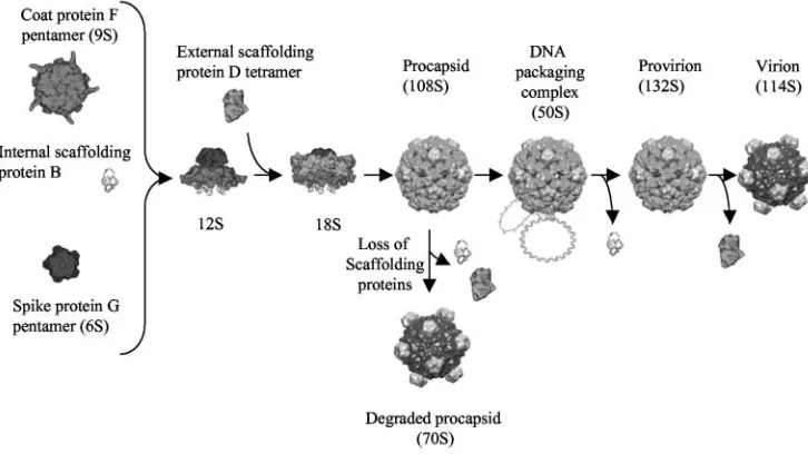
The microvirus assembly pathway is illustrated in Fig. 1. The first detectable intermediates are the 9S and 6S particles, re-spective pentamers of the major coat F and spike G proteins (22). Five internal scaffolding B proteins bind to the underside of the 9S particles, inducing a conformational change that enables interactions with major spike G, minor spike H, and
external scaffolding D proteins (5, 6, 18, 21, 22). While the identity of the next intermediate remains obscure, structural data suggest that it contains the F, G, H, B, and D proteins (5, 6). A particle sedimenting at 18S most likely represents this particle (4, 8). In a reaction mediated by the external scaffold-ing protein (10, 22), 12 18S particles associate to form the procapsid (108S). The preinitiation complex for single-stranded DNA synthesis and packaging binds to the procapsid, most likely along a twofold axis of symmetry, forming the 50S complex (7). Single-stranded genomic DNA is concurrently synthesized and packaged, presumably through a pore at the threefold axis of symmetry. There are no other openings large enough in the viral procapsid through which a DNA molecule could pass (1, 12).
The threefold-related pores are visible only in cryo-image reconstructions of the procapsids (1, 12). In the atomic struc-ture, this area is occupied by coat protein sequences in an arrangement similar to that observed in the mature virion (5, 6). There are other salient differences between the cryo-image and atomic models. For example, in the crystal structure, coat protein pentamers have a decreased radial distance and inter-act across the twofold axes of symmetry. In contrast, pentamers in the cryo-image reconstruction make little or no contact with each other and appear to be associated more tightly with the external scaffolding protein lattice. These differences suggest that the crystallized procapsids underwent a limited maturation event during sample preparation. The results of the genetic anal-yses presented here and in a previous report support this suppo-sition: coat-external scaffolding protein interactions are probably more extensive than the crystal structure indicates (2).
* Corresponding author. Mailing address: Department of Veteri-nary Sciences and Microbiology, Building 90, University of Arizona, Tucson AZ 85721-0090. Phone: (520) 626-6634. Fax: (520) 621-6366. E-mail: [email protected].
6751
on November 8, 2019 by guest
While the crystal structure may not divulge the full extent of coat-external scaffolding protein associations, it does reveal the intricate interactions of the four external scaffolding proteins in the asymmetric unit (Fig. 2A), which form asymmetric dimers (D1D2 and D3D4). Each monomer consists of seven
␣-helices connected by loop structures. There is a considerable variation between the subunits, which bears no resemblance to quasi-equivalence (5, 6). Each D protein makes a unique set of contacts with the neighboring D, major spike, and capsid pro-teins. The external scaffolding protein is the most conserved microvirus protein, sharing 70% identity on the amino acid level. Residues 27 to 128, comprising␣-helices 2 to 6, are 96% conserved. This region forms a structurally conserved core (5, 6) and mediates the vast majority of intra- and interdimer contacts. However, considerable divergence occurs in␣-helix 1, loop 6, and␣-helix 7 amino acid sequences.
The results of biochemical analyses conducted with an␣3/
X174 chimeric external scaffolding protein, in which the first
␣-helix of the ␣3 protein replaced theX174 structure, sug-gested that this region acts as a coat protein substrate
speci-ficity domain early in the morphogenetic pathway (2). However, those studies were limited. The chimeric protein exhibited little or no activity vis-a`-visX174 morphogenesis, andX174 mutants that could productively utilize the chimeric protein could not be obtained at frequencies indicative of point mutations. Low se-quence conservation between the two phages may have impeded the genetic analyses. In an effort to elucidate a more mechanistic model, a chimeric external scaffolding protein was constructed between the more closely related phages G4 andX174 (Fig. 2B). The results of the analyses suggest that␣-helix 1 mediates the initial interaction with the coat protein, identify the region of the coat protein with which␣-helix 1 most likely interacts, and provide evidence, albeit indirect, that the open pores at the three-fold axes of symmetry are the site of DNA entry during the packaging reaction.
MATERIALS AND METHODS
[image:2.585.108.471.73.281.2]Phage plating, media, buffers, stock preparation, generation of RF DNA, and DNA isolation.The reagents, media, buffers, and protocols for single-stranded and replicative-form (RF) DNA isolation have been described previously (4, 8). FIG. 1.X174 morphogenesis.
FIG. 2. (A) Four external scaffolding protein subunits associated with each asymmetric unit; (B) primary sequences of␣-helices 1 from bacteriophages G4,X174, and␣3.
on November 8, 2019 by guest
http://jvi.asm.org/
[image:2.585.103.483.564.701.2]with NcoI and PstI and ligated into pSE420 digested with the same enzymes.
Isolation of chimeric-protein utilizer mutants.TheX174nullDmutant was plated on BAF30 pG4/XD and incubated at 30 or 33°C until plaques appeared. Progeny were stabbed into indicator lawns seeded with C122, BAF30 pG4/XD, and BAF30 pXDJ. Utilizers were identified by the retention of the comple-mentation-dependent phenotype and their ability to grow at restrictive temper-atures on BAF30 pG4/XD.
Detection of virion and intermediate particles from infected cells.One hun-dred milliliters ofslyDpG4/XD cells was infected withX174 wild-type,nullD, orutT204I/nullDphage at a multiplicity of infection of 3.5 and incubated for 4 h at 30°C or 37°C. Cells were pelleted, resuspended in 2.0 ml BE buffer (8), and lysed. Extracts were prepared as described previously (11). Virions and soluble proteins were isolated in CsCl gradients (3). After dialysis, extracts were con-centrated to 200l, layered atop 5 to 30% sucrose gradients, and spun at 45,000 rpm for 1 h in an SW50.1 rotor. Material was detected by taking readings of the optical density at 280 nm (OD280) of each fraction. The position of infectious virion was determined by plaque assays.
RESULTS
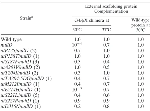
Biological activity of the G4/X174 chimeric external scaf-folding protein. A chimeric G4/X174 external scaffolding gene was constructed as described in Materials and Methods. The expressed protein contains␣-helix 1 from bacteriophage G4; the remainder of the protein is derived fromX174. To determine whether the chimeric protein could support virion morphogenesis, the cloned gene was assayed for the ability to complement aX174nullDmutant in plaque assays (Table 1). A clone of the wild-type protein was used as a control. Unlike the wild-type protein, which supported growth at all tempera-tures, the chimeric protein supported plaque formation only above 35°C. Although the plating efficiency was close to that of the positive control, the plaque size was considerably reduced, suggesting weak complementation (see below). Extragenic mu-tations that can utilize the chimeric external scaffolding protein between 30 and 35°C have been isolated (see below). There-fore, the cold-sensitive complementation phenotype most likely does not reflect protein folding or scaffolding protein dimerization defects.
Biochemical analysis of particles produced by the chimeric protein of G4/X D external scaffolding protein during assem-bly.To determine the morphogenetic steps not supported by the chimeric protein, the assembly intermediates synthesized in slyD cells expressing the chimeric protein were analyzed. The slyD mutation blocks X174 E protein-mediated lysis. After incubation, cells infected with either X174 nullD or wild-type phages were pelleted to remove unabsorbed virions.
After chemical lysis, 1.3 to 1.4 gm/cm3 material which con-tained soluble proteins and assembled particles was purified in CsCl gradients, concentrated, and analyzed by sucrose gradient sedimentation. After the fractionation, uninfectious assembled particles (procapsids, degraded procapsids, or nonviable viri-ons) were detected by spectroscopy (OD280); infectious parti-cles (virion or 114S virion) were detected by both spectroscopy and a direct plating assay.
Figure 3 presents the results of these experiments. Plaque assays are more sensitive than spectroscopy in detecting small amounts of virions that may be produced during infections. Therefore, the titer data for the fractions (1 to 10) that contain infectious particles (114S virions and 132S provirions) are pre-sented in the figure. The particles within fractions 1 to 10 that were detected by spectroscopy OD280(data not shown) were also detected by plaque assays. Thus, no uninfectious virion-like particles, such as 108S native procapsids, were detected in these assays. OD280 readings are presented for fractions 11 onward. Unlike the wild-type control, at the nonpermissive temperature of 30°C, X174 nullD-infected cells (Fig. 3A) produced no large particles, indicating a defect early in the assembly pathway. At 37°C, both 114S particles (infectious virions) and 70S particles (degraded procapsids; see below) were detected (Fig. 3B). However, unlike the products of the wild-type infection, 70S particles were more prevalent than virions, as determined by both titer and spectroscopy with the most infectious fraction. The OD280of the wild-type fraction was 0.2 (data not shown), while the corresponding OD280of the mutant fraction was not above background (0.02). These data indicate that the chimeric external scaffolding protein confers two assembly defects: procapsid formation, which is more pronounced at 30°C, and DNA packaging.
utE214H/nullD(1) 10 0.7 1.0
utS221L/nullD(5) 0.4 0.6 1.0
utS227P/nullD(1) 0.9 0.9 1.0
utD316N/nullD(1) 0.2 0.8 1.0
aComplementation is reported as the plating efficiency titer with the chimera/
titer with the wild-type protein.
bThe first letter and number in the name of each utilizer mutation indicate the
wild-type amino acid found at that position in the wild-type coat protein se-quence. The second letter indicates the substitution that confers the utilizer phenotype. Numbers within parentheses indicate the numbers of independent isolations of the mutants.
on November 8, 2019 by guest
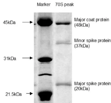
[image:3.585.300.541.96.275.2]Characterization of 70S particles.70S particles usually rep-resent degraded procapsids, which were never filled and which lost the scaffolding proteins (2, 18). To determine whether the 70S particles observed were degraded procapsids, spectroscopy and sodium dodecyl sulfate-polyacrylamide gel electrophoresis were performed. The OD280/OD260ratios for procapsids and virions were 0.90⫾0.05 and 1.55⫾0.05, respectively (12). The OD260/OD280ratio of the 70S particle was determined to be 0.93, which indicates protein particles containing little or no DNA. Sodium dodecyl sulfate-polyacrylamide gel electro-phoresis analysis (Fig. 4) of the 70S particles revealed three bands at 48 kDa, 37 kDa, and 20 kDa, which correspond to coat (F), minor spike (H), and major spike (G) proteins, re-spectively.
Isolation ofX174 mutants that can utilize the G4/X174 chimeric external scaffolding protein.As shown in Table 1, the chimeric protein supports morphogenesis only at elevated tem-peratures. To isolate mutants that can productively utilize the chimeric protein at lower temperatures, X174 nullD was
plated on cells expressing the chimeric protein at 30 and 33°C. Utilizers were identified by their ability to plate on BAF30 pG4/X D but retained a D protein complementation-depen-dent phenotype. The mutations are listed in Table 1 along with the number of independent isolations. Initially, genes D and F, which encode the external scaffolding protein and major coat protein, respectively, of the utilizers’ genomes, were se-quenced. The D gene sequencing confirmed the presence of thenullDbackground. All of the utilizer mutations conferred substitutions in the viral coat protein and formed a cluster in the tertiary structure (see Discussion). The entire genome of one of the utilizers, theutS221L/nullDmutant, was sequenced. No other mutations were found, indicating that the identified amino acid substitution was both necessary and sufficient for chimeric protein utilization.
Characterization of X174 mutants that can utilize the G4/X174 chimeric external scaffolding protein.The interme-diate particles synthesized in utT204I/nullD mutant-infected cells expressing the chimeric protein were also analyzed as described above (Fig. 3). UnlikeX174nullDinfections, in-fectious virions and 70S particles were isolated at both tem-peratures. Furthermore, the relative particle ratio did not dif-fer significantly from the wild-type infection, suggesting that the utilizer mutation suppresses both the procapsid assembly and the DNA-packaging defects and that the two defects may be fundamentally related. To determine whether the utilizer mutations circumvent the need for the D protein first␣-helix, a clone expressing an N-terminal deletion protein was assayed for the ability to complement the utilizer mutation/nullD strains. No complementation was detected, indicating that these mutants still require the first␣-helix for assembly.
DISCUSSION
In a previous study, an␣3/X chimeric D protein was con-structed (2, 3). This chimeric protein neither supportedX174 procapsid morphogenesis nor inhibited the wild-type protein in coinfections. However, this protein strongly inhibited wild-type
␣3 morphogenesis. From these data, a preliminary hypothesis
FIG. 3. (A) Particles produced in cells expressing the chimeric G4/X D protein at 30°C. (B) Particles produced in infected cells expressing the chimeric G4/X D protein at 37°C. Infectious particles within fractions 1 to 10 (114S virions and 132S provirions) were de-tected by plaque assays. OD280readings are presented for fractions 11
onward, which contain uninfectious particles (70S degraded procap-sids). Symbols: triangles, wild type; squares,nullDmutant; diamonds,
[image:4.585.325.512.68.241.2]utT204I/nullDmutant.
FIG. 4. Protein compositions of 70S particles (degraded procap-sids).
on November 8, 2019 by guest
http://jvi.asm.org/
was formed: the first helix is a substrate specificity domain, vis-a`-vis coat protein recognition, and mediates the initial coat protein interaction. However, attempts to probe this hypothe-sis further were limited. For example, X174 mutants that could utilize the chimera could not be isolated. Therefore, it was not possible to exclude other models involving defects, such as subunit dimerization, which would have produced sim-ilar experimental results. The inability to isolate utilizer mu-tants was probably due to the large amino acid divergence between the two phages. The first␣-helices share only 23% identity, while the G4 andX174 helices share 73% identity. Thus, the G4/X chimeric D protein described in this report was constructed.
Unlike the ␣3/X chimera, the G4/X chimera supported
X174 viral replication above 33°C, albeit poorly. Consistent with the hypothesis that␣-helix 1 mediates the first contact with the viral coat protein, the results of biochemical analyses demonstrated that procapsids were not formed below this tem-perature. While procapsids form at higher temperatures, DNA-packaging efficiency was severely reduced, suggesting that coat-scaffolding associations may be too weak to maintain the open pores at the threefold axis of symmetry, through which the genome is most likely translocated (see below).
Chimeric G4/X D protein utilizers were isolated. These utilizers suppress both the procapsid morphogenesis and DNA-packaging defects, indicating that these two phenomena may be related. The utilizer mutation/nullD double mutants were not rescued by the expression of a D protein lacking the first helix, suggesting that they do not confer an␣-helix 1-in-dependent phenotype. The utilizer substitutions cluster in
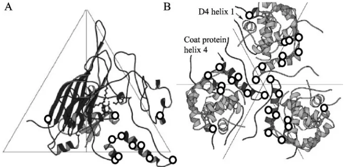
␣-helix 4 of the viral coat protein, in adjacent loop regions, which could affect the orientation of the helix, and in other threefold-related regions (Fig. 5). While it is impossible to ascertain which D subunit is involved in this interaction, three key structural and genetic observations suggest that it is the D4 subunit. (i) This subunit is located at the threefold axis of symmetry and is in the closest proximity to␣-helix 4 of the coat protein. (ii) Helix 1 points downward, toward the coat protein, only in this subunit. (iii) Genetic data indicate that the
inter-actions between D4and the underlying coat protein are more extensive than indicated by the crystal model, in which the coat protein has fallen inward (2).
Recently, the structure of the external scaffolding protein prior to its interaction with other structural proteins was solved (17). The asymmetric dimers consist of a DAand a DBsubunit, which resembles the structure of the dimers found in the pro-capsid. However, there are pronounced differences, which are localized to regions believed to mediate critical interactions with the viral coat protein, one of which is␣-helix 1. Those structural studies, together with the data presented here, sug-gest a more detailed assembly model. Before interacting with other viral proteins, the D protein exists as dimers and tetram-ers in solution (17). Thus, it is not unreasonable to assume that tetramers attach to pentamers of the coat protein. Several observations indicate that the initial D-F protein interaction involves␣-helix 1 of a DBsubunit in a tetramer recognizing threefold-related viral coat protein sequences in an adjacent fivefold-related asymmetric unit. For example, the G4/X and
␣3/X chimeric proteins (2) do not support large-particle for-mation under nonpermissive conditions. However, unlike other mutant D proteins that affect later morphogenetic stages, they do not inhibit wild-type external scaffolding protein for-mation in coinfected cells. The clustering of utilizer mutations and several structural models (1, 5, 6, 17) suggest that the interacting DBsubunit becomes the D4protein, as visualized in the procapsid. This, in turn, would bring the rest of the tet-ramer in contact with the underlying viral coat protein.␣-Helix 7 of D4then associates with the coat protein to locate tetram-ers in the right position (2, 5). The resulting pentameric par-ticles most likely associate via twofold-related D-D interactions mediated by loop 5 sequences (5, 17). After procapsid forma-tion, interactions between ␣-helix 1 of D4 and threefold-re-lated viral coat protein sequences must be maintained for successful DNA packaging, which provides more evidence, al-beit indirect, that a pore at the threefold axis of symmetry is the site of DNA entry.
FIG. 5. (A) Locations of utilizer mutations within the atomic structure of the viral coat protein. The utilizer mutations are identified by circles. (B) Locations of utilizer mutations in the atomic structure of a procapsid at the threefold axis of symmetry, containing both the viral coat and the D4external scaffolding proteins.
on November 8, 2019 by guest
[image:5.585.120.467.72.241.2]ACKNOWLEDGMENTS
We acknowledge the technical assistance of Min Chen. This work was supported by NSF grant MCB0234976 to B.A.F.
REFERENCES
1.Bernal, R. A., S. Hafenstein, N. H. Olson, V. D. Bowman, P. R. Chipman, T. S. Baker, B. A. Fane, and M. G. Rossmann.2003. Structural studies of bacteriophage␣3 assembly. J. Mol. Biol.325:11–24.
2.Burch, A. D., and B. A. Fane.2003. Genetic analyses of putative conforma-tion switching and cross-species inhibitory domains inMicroviridaeexternal scaffolding proteins. Virology310:64–71.
3.Burch, A. D., and B. A. Fane.2000. Foreign and chimeric external scaffolding proteins as inhibitors ofMicroviridaemorphogenesis. J. Virol.74:9347–9352. 4.Burch, A. D., J. Ta, and B. A. Fane.1999. Cross-functional analysis of the
Microviridaeinternal scaffolding protein. J. Mol. Biol.286:95–104. 5.Dokland, T., R. McKenna, L. L. Ilag, B. R. Bowman, N. L. Incardona, B. A.
Fane, and M. G. Rossmann.1997. Structure of a viral procapsid with mo-lecular scaffolding. Nature389:308–313.
6.Dokland, T., R. A. Bernal, A. Burch, S. Pletnev, B. A. Fane, and M. G. Rossmann.1999. The role of scaffolding proteins in the assembly of the small, single-stranded DNA virusX174. J. Mol. Biol.288:595–608. 7.Ekechukwu, M. C., D. J. Oberste, and B. A. Fane.1995. Host andX 174
mutations affecting the morphogenesis or stabilization of the 50S complex, a single-stranded DNA synthesizing intermediate. Genetics140:1167–1174. 8.Fane, B. A., and M. Hayashi. 1991. Second-site suppressors of a
cold-sensitive prohead accessory protein of bacteriophageX174. Genetics128:
663–671.
9.Fane, B. A., and P. E. Prevelige, Jr.2003. Mechanism of scaffolding-assisted viral assembly. Adv. Protein Chem.64:259–299.
10.Fujisawa, H., and M. Hayashi. 1977. Functions of gene C and gene D products of bacteriophageX174. J. Virol.21:506–515.
11.Hafenstein, S., and B. A. Fane.2002.X174 genome-capsid interactions influence the biophysical properties of the virion: evidence for a
scaffolding-like function for the genome during the final stages of morphogenesis. J. Virol.76:5350–5356.
12.Ilag, L. L., N. H. Olson, T. Dokland, C. L. Music, R. H. Cheng, Z. Bowen, R. McKenna, M. G. Rossmann, T. S. Baker, and N. L. Incardona.1995. DNA packaging intermediates of bacteriophageX174. Structure3:353–363. 13.King, J., and S. Casjens.1974. Catalytic head assembling protein in virus
morphogenesis. Nature251:112–119.
14.Liebowitz, J., and M. S. Horwitz.1975. Synthesis and assembly of adenovirus polypeptides. III. Reversible inhibition of hexon assembly in adenovirus type 5 temperature-sensitive mutants. Virology66:10–24.
15.McKenna, R., L. L. Ilag, and M. G. Rossmann.1994. Analysis of the single-stranded DNA bacteriophageX174, refined at a resolution of 3.0 Å. J. Mol. Biol.237:517–543.
16.McKenna, R., D. Xia, P. Willingmann, L. L. Ilag, S. Krishnaswamy, M. G. Rossmann, N. H. Olson, T. S. Baker, and N. L. Incardona.1992. Atomic structure of single-stranded DNA bacteriophageX174 and its functional implications. Nature355:137–143.
17.Morais, M. C., M. Fisher, S. Kanamaru, L. Przybyla, J. Burgner, B. A. Fane, and M. G. Rossmann.2004. Conformational switching by the scaffolding protein D directs the assembly of bacteriophageX174. Mol. Cell15:991– 997.
18.Novak, C. R., and B. A. Fane.2004. The functions of the N terminus of the X174 internal scaffolding protein, a protein encoded in an overlapping reading frame in a two scaffolding protein system. J. Mol. Biol.335:383–390. 19.Rixon, F. J.1993. Structure and assembly of herpes viruses. Semin. Virol.
4:135–144.
20.Roof, W. D., S. M. Horne, K. D. Young, and R. Young.1994.slyD, a host gene required forX174 lysis, is related to the FK506-binding protein family of peptidyl-prolylcis-trans-isomerases. J. Biol. Chem.269:2902–2910. 21.Siden, E. J., and M. Hayashi.1974. Role of the gene-product in
bacterio-phageX174 development. J. Mol. Biol.89:1–16.
22.Tonegawa, S., and M. Hayashi. 1970. Intermediates in the assembly of X174. J. Mol. Biol.48:219–242.