DEVELOPMENT AND VALIDATION OF RP-HPLC METHOD FOR QUANTIFICATION OF TAPENTADOL HYDROCHLORIDE IN
TABLET DOSAGE FORM
D
Diisssseerrttaattiioonn SSuubbmmiitttteedd ttoo
T
Thhee TTaammiillNNaadudu DDrr.. MM..GG..RR MMeeddiiccaall UUnniivveerrssiittyy, , CChheennnnaai i
I
Inn ppaarrttiiaall ffuullffiillllmmeenntt ffoorr tthhee aawwaarrdd oof f tthhe e DDeeggrreeee
M
MAASSTTEERR OOFF PPHHAARRMMAACCYY
(
(PPhhaarrmmaacceeuuttiiccaall AAnnaallyyssiiss))
A
APPRRIILL--22001122
Department of Pharmaceutical Analysis KMCH COLLEGE OF PHARMACY KOVAI ESTATE, KALAPPATTI ROAD,
Kovai Estate, Kalapatti Road, Coimbatore-641048
CERTIFICATE
This is to certify that the dissertation work entitled “DEVELOPMENT AND VALIDATION OF RP-HPLC METHOD FOR QUANTIFICATION OF TAPENTADOL HYDROCHLORIDE IN TABLET DOSAGE FORM” is a bonafide research work carried out by Mr. VENKANA BABU. MADETI (Reg. No: 26107229) , student in Master of Pharmacy, Department of Pharmaceutical Analysis, K.M.C.H.College of Pharmacy, Coimbatore, Tamilnadu, under the guidance of Mr. J.DHARUMAN, Professor, Head, Department of Pharmaceutical Analysis, K.M.C.H. College of Pharmacy during the academic year 2011-2012.
I wish for him best career,
Signature
Date: Dr. A.Rajasekaran M.Pharm, Ph.D.
Place: Principal
ACKNOWLEDGEMENT
My dissertation work entitled “DEVELOPMENT AND VALIDATION OF RP-HPLC METHOD FOR QUANTIFICATION OF TAPENTADOL HYDROCHLORIDE IN TABLET DOSAGE FORM” would not have been a feasible one without the grace of god almighty who gave me morale till the completion of my project.
I am extremely thankful to my Academic Guide Mr. J.Dharuman.M.Pharm.,(Ph.D.,)Professor, Head, Department of Pharmaceutical Analysis, KMCH. College of Pharmacy, for his constant insight, guidance, countless serenity, encouragement and painstaking efforts in my project work. I am indebted to his kindness and never failing co – operation.
To begin with I would like to thank Dr. A. Rajasekaran, M. Pharm, Ph.D., Principal, K.M.C.H. College of Pharmacy for his constant encouragement, support and the facilities provided.
I will always remain indebted to Dr Nalla G. Palanisamy, Chairman, and Dr. Thavamani D. Palanisamy, Managing Trustee, K.M.C.H. College of Pharmacy, Coimbatore for all the facilities, which have been provided to us at the institution, anabling me to do work of this magnitude.
My special thanks to all other staff members of KMCH College of Pharmacy, Coimbatore and Library faculties who directly or indirectly gave a helping hand during the course of study.
I express my sincere thanks to all my classmates, and friends for their timely help and co-operation.
Rambabu, T. Srikanth Reddy, J. K. Ghaharin, Dona Sara Kurian, A. V. S. Hemanth, Tinu Thomas, T. Manavalan, G. Arun Kumar) and my M.Pharm Ist year juniors (G. Venkatesh, Chaitanya kumar.T, N. Mahesh, M. Gowtham) and I take this opportunity to acknowledge them with thanks.
I also express my heartful thanks to J. S. M. Gupta, , S. Bugga Reddy of Dept. of pharmaceutical Chemistryand V. Chaithanya of Dept. of Pharmacology.
Finally I would like to express my sincere thanks to all those people who directly or indirectly helped me to complete this work successfully.
Above all I dedicate myself before the unfailing presence of GOD and constant love and encouragement given to me by my beloved Father, Mother, Sister and all of my family memberswho deserves the credit of success in whatever work I did.
Dept. of Pharmaceutical Analysis Introduction
KMCH college of pharmacy Page 1
INTRODUCTION
Analytical chemistry [1] is often described as the area of chemistry responsible for characterizing
the composition of matter, both qualitatively (what is present) and quantitatively (how much is
present).Analytical chemistry is not a separate branch of chemistry, but simply the application of
chemical knowledge.
Pharmaceutical Analysis [2] is the branch of chemistry involved in separating, identifying and
determining the relative amounts of the components making up a sample of matter. It is mainly
involved in the qualitative identification or detection of compounds and quantitative
measurements of the substances present in bulk and pharmaceutical preparation.
Quantitative analysis constitutes the largest part of analytical chemistry and is related to the
various methods and instrumentation employed in determining the amounts or concentration of
constituents in samples. It is also one of the basic criteria in the field of pharmacy where quality
is to be critically maintained. Analytical chemistry may be defined as the “Science and art of
determining the composition of materials in terms of the elements or compounds contained”.
Analytical method is a specific application of a technique to solve an analytical problem [3].
Physico-chemical methods [4, 5] are used to study the physical phenomenon that occurs as a result
of chemical reactions. Among the Physico-chemical methods, the most important are optical
(Refractometry, Polarimetry, Emission, Fluorescence methods of analysis, Photometry including
Photo-colorimetry and Spectrophotometry covering UV-Visible and IR regions and
Nephelometry or Turbidimetry) and chromatographic (Column, Paper, TLC, GLC, HPLC)
methods. Methods such as Nuclear Magnetic Resonance and Para Magnetic Resonance are
Chromatography and Liquid Chromatography are the most powerful tools available. The
chemical methods include the gravimetric and volumetric procedures which are based on
complex formation; acid-base, precipitation and redox reactions. Titrations in non-aqueous
media and complexometry have also been used in pharmaceutical analysis.
The number of new drugs is constantly growing. This requires new methods for controlling their
quality. Modern pharmaceutical analysis must need the following requirements.
I. The analysis should take a minimal time.
II. The accuracy of the analysis should meet the demands of Pharmacopoeia.
III. The analysis should be economical.
IV. The selected method should be precise and selective.
These requirements are met by the Physico-chemical methods of analysis, a merit of which is
their universal nature that can be employed for analyzing organic compounds with a diverse
structure. Of them, Visible Spectrophotometry is generally preferred especially by small scale
industries as the cost of the equipment is less and the maintenance problems are minimal.
Pharmaceutical analysis techniques are applied mainly in two areas [6]:
Traditionally, analytical chemistry has been split into two main types, Qualitative and
Quantitative
1. Qualitative: Qualitative analysis seeks to establish the presence of a given element or
compound in a sample.
2. Quantitative: Quantitative analysis seeks to establish the amount of a given element or
Dept. of Pharmaceutical Analysis Introduction
KMCH college of pharmacy Page 3
Instrumental methods of Chemical analysis:
Instrumental method is an exciting and fascinating part of chemical analysis that interacts with
all areas of chemistry and with many other areas of pure and applied sciences. Analytical
instrumentation plays an important role in the production and evaluation of new products and in
the protection of consumers and environment. This instrumentation provides lower detection
limits required to assure safe foods, drugs, water and air. Instrumental methods are widely used
by analytical chemists to save time, to avoid chemical separation and to obtain increased
accuracy. Different types of techniques are listed below:
A) Spectrometric techniques:
o Atomic Spectrometry (emission and absorption)
o Electron Spin Resonance Spectroscopy
o Fluorescence and Phosphorescence Spectrophotometry
o Infrared Spectrophotometry
o Nuclear Magnetic Resonance Spectroscopy
o Radiochemical Techniques including activation analysis
o Raman Spectroscopy
o Ultraviolet and Visible Spectrophotometry
o X-Ray Spectroscopy
B) Chromatographic Techniques:
o Gas Chromatography
o High Performance Liquid Chromatography
C) Miscellaneous Techniques:
o Kinetic Techniques
o Mass Spectrometry
o Thermal Analysis
D) Hyphenated Techniques:
o GC-MS (Gas Chromatography – Mass Spectrometry).
o ICP-MS (Inductivity Coupled Plasma- Mass Spectrometry).
o GC-IR (Gas Chromatography – Infrared Spectroscopy).
o MS-MS (Mass Spectrometry – Mass Spectrometry).
INTRODUCTION TO CHROMATOGRAPHY [7]:
Chromatography, by classical definition, is a separation process where resolution is achieved by
the distribution of the components of a mixture between two phases, a stationary phase and a
mobile phase. Those components held preferentially in the stationary phase are retained longer in
the system than those that are distributed in the mobile phase. As a consequence solutes are
eluted from the system in the order of their increasing distribution coefficients with respect to the
stationary phase. The mobile phase can be a gas or a liquid which gives rise to the two basic
forms of Chromatography, namely, Gas Chromatography (GC) and Liquid Chromatography
(LC). The stationary phase can also take two forms, solid and liquid, which provides two
subgroups of GC and LC, namely; Gas-Solid Chromatography (GSC) and Gas-Liquid
Chromatography (GLC), together with Liquid Solid Chromatography (LSC) and Liquid- Liquid
Dept. of Pharmaceutical Analysis Introduction
KMCH college of pharmacy Page 5
INTRODUCTION -HIGH PERFORMANCE LIQUID CHROMATOGRAPHY [8, 9]:
In the modern pharmaceutical industry, HPLC is a major analytical tool applied at all stages of
drug discovery, development and production. Fast and effective development of rugged
analytical HPLC methods is more efficiently undertaken with a thorough understanding of HPLC
principles, theory and instrumentation.
Principle [10]:
The principle of separation in normal phase mode and reverse phase mode is adsorption. When
mixtures of components are introduced in to a HPLC column, they travel according to their
relative affinities towards the stationary phase. The component which has more affinity towards
the adsorbent travels slower.
Types of HPLC [11]:
There are many ways to classify Liquid Column Chromatography. If classification is based on
the nature of the stationary phase and the separation process,
Three modes can be specified:
In Adsorption Chromatography:
The stationary phase is an adsorbent (like silica gel or any other silica based packing) and the
separation is based on repeated adsorption desorption steps.
In Ion-Exchange Chromatography:
1. The stationary bed has an ionically charged surface of opposite charge to the sample ions.
3. The stronger the charge on the sample, the stronger it will be attracted to the ionic surface and
thus, the longer it will take to elute.
4. The mobile phase is an aqueous buffer, where both pH and ionic strength are used to control
elution time.
In Size Exclusion Chromatography:
1. The column is filled with material having precisely controlled pore sizes, and the sample is
simply screened or filtered according to its solvated molecular size.
2. Larger molecules are rapidly washed through the column; smaller molecules penetrate inside
the porous of the packing particles and elute later.
3. Concerning the first type, two modes are defined depending on the relative polarity of the two
phases: Normal and Reversed-Phase Chromatography.
In Normal-Phase Chromatography:
1. The stationary bed is strongly polar in nature (e.g., silica gel), and the mobile phase is
non-polar (such as n-hexane or tetrahydrofuran).
2. Polar samples are thus retained on the polar surface of the column packing longer than less
polar materials.
In Reversed-Phase Chromatography:
The stationary bed is non-polar in nature, while the mobile phase is a polar liquid, such as
mixtures of Water and Methanol or Acetonitrile. Here the more non-polar the material is, the
Dept. of Pharmaceutical Analysis Introduction
KMCH college of pharmacy Page 7
travels faster. Since no two components have the same affinity towards the stationary phase, the
components are separated.
Liquid Chromatography (LC), which is one of the forms of Chromatography, is an
analytical technique that is used to separate a mixture in solution into its individual components.
The separation relies on the use of two different "phases" or "immiscible layers," one of which is
held stationary while the other moves over it. Liquid Chromatography is the generic name used
to describe any chromatographic procedure in which the mobile phase is a liquid. The separation
occurs because, under an optimum set of conditions, each component in a mixture will interact
with the two phases differently relative to the other components in the mixture. HPLC is the term
used to describe Liquid Chromatography in which the liquid mobile phase is mechanically
pumped through a column that contains the stationary phase.
An HPLC instrument, therefore, consists of an injector, a pump, a column, and a detector given
in the Fig: 1.
Liquid Chromatography has come a long way with regard to the practical development of HPLC
instrumentation and the theoretical understanding of different mechanisms involved in the
analyte retention as well as the development of adsorbents with different geometries and surface
chemistry.
In HPLC, a liquid sample or a solid sample dissolved in a suitable solvent is carried through a
Chromatographic column by a liquid mobile phase. Separation is determined by
solute/stationary-phase interactions, including liquid–solid adsorption, liquid–liquid partitioning,
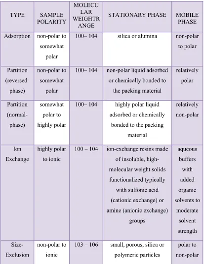
Table: 1 with their applications. In each case, however, the basic instrumentations are essentially
[image:12.612.100.513.161.694.2]same.
Table no: 1 Various Types of HPLC
TYPE SAMPLE
POLARITY
MOLECU LAR WEIGHTR
ANGE
STATIONARY PHASE MOBILE
PHASE
Adsorption non-polar to
somewhat polar
100– 104 silica or alumina non-polar
to polar Partition (reversed-phase) non-polar to somewhat polar
100– 104 non-polar liquid adsorbed
or chemically bonded to the packing material
relatively polar Partition (normal-phase) somewhat polar to highly polar
100– 104 highly polar liquid
adsorbed or chemically bonded to the packing
material relatively non-polar Ion Exchange highly polar to ionic
100 – 104 ion-exchange resins made
of insoluble, high-molecular weight solids functionalized typically
with sulfonic acid (cationic exchange) or amine (anionic exchange)
groups aqueous buffers with added organic solvents to moderate solvent strength Size-Exclusion non-polar to ionic
103 – 106 small, porous, silica or polymeric particles
Dept. of Pharmaceutical Analysis Introduction
[image:13.612.71.491.142.555.2]KMCH college of pharmacy Page 9
Various components involved in HPLC:
1. Solvent delivery systems:
The purpose of the pump, or solvent delivery system, is to ensure the delivery of a precise,
reproducible, constant, and pulse-free flow of mobile phase.
There are two classes of HPLC pumps:
1. Constant pressure pumps
2. Constant flow pumps
The most common type of HPLC constant flow pump is the reciprocating piston pump, in which
a piston is driven in and out of the solvent chamber by a gear. On the forward stroke, the inlet
check valve closes, the outlet check valve opens, and the mobile phase is pumped to the column.
On the return stroke, the check valves reverse and solvent is drawn into the chamber. In the
single head reciprocating pump, 50 % of the time the mobile phase flows to the column and 50
% of time the chamber is refilling. With the twin-head reciprocating pump the pump heads
operate simultaneously but 1800 out of phase with each other. As a result mobile phase flows to
the column 100 % of the time, providing an essentially pulse less flow.
Most separations can be done using isocratic elution which is the use of a single-solvent system
that does not change during the analysis. For more complex analysis gradient elution is required.
Gradient elution can be generated in three ways. In all cases a computer controlled pumping
system is required. In the first phase, controlled amounts of each eluent are metered into a
mixing chamber before reaching the high-pressure pump which sends the mixture to the column.
Dept. of Pharmaceutical Analysis Introduction
KMCH college of pharmacy Page 11
controlled by a microprocessor. The mixed solvent then enters the high pressure pump and flows
to the column. In the third case the delivery of high delivery multiple pumps is controlled
individually with a programming device and mixer is send to high-pressure mixing chamber
2. Columns [13]: The heart of the system is the column. The choice of common packing material and mobile phases depend on the physical properties of the drug. The column selection flow
chart can assist one in determining which columns to examine. Many different reverse phase
columns will provide excellent specificity for any particular separation. It is therefore best to
routinely attempt separations with a standard C-8 or C-18 column (e.g. Zorbax RX C-8) and
determine if it provides good separations. If this column does not provide good separation or the
mobile phase is unsatisfactory, alternate methods or columns should be explored. Reverse phase
column differ by the carbon chain lengths, degree of end capping and percent carbon loading.
Diol, Cyano and amino groups can be used for Reverse Phase Chromatography.The most
commonly used columns for HPLC are constructed from stainless steel with internal diameters
between 2.1 mm and 4.6 mm, and lengths ranging from approximately 30 mm to 300 mm. These
columns are packed with 3–10 mm porous silica particles that may have an irregular or spherical
shape. Typical column efficiencies are 40,000–60,000 theoretical plates/m. Micro columns use
less solvent and, because the sample is diluted to a lesser extent, produce larger signals at the
detector. These columns are made from fused silica capillaries with internal diameters of 44–200
mm and lengths of up to several meters. Micro columns packed with 3–5 mm particles have been
prepared with column efficiencies of up to 250,000 theoretical plates. Open tubular micro
columns also have been developed, with internal diameters of1–50 mm and lengths of
approximately 1 m. These columns, which contain no packing material, may be capable of
tubular columns, however, has been limited by the difficulty of preparing columns with internal
diameters less than 10 mm.
3. Stationary Phases [14]:
In Liquid–Liquid Chromatography the stationary phase is a liquid film coated on a packing
material consisting of 3–10 mm porous silica particles. The stationary phase may be partially
soluble in the mobile phase, causing it to “bleed” from the column over time. To prevent this loss
of stationary phase, it is covalently bound to the silica particles. Bonded stationary phases are
attached by reacting the silica particles with an organochlorosilane of the general form Si
(CH3)2RCl, where R is an alkyl or substituted alkyl group. To prevent unwanted interactions
between the solutes and any unreacted –SiOH groups, the silica frequently is “capped” by
reacting it with Si (CH3)3Cl; such columns are designated as end-capped. The properties of a
stationary phase are determined by the nature of theorganosilane’s alkyl group. If R is a polar
functional group, then the stationary phase will be polar. Since the stationary phase is polar, the
mobile phase is a non-polar or moderately polar solvent. The combination of a polar stationary
phase and a non-polar mobile phase is called normal phase Chromatography.
In reverse phase Chromatography, which is the more commonly encountered form of HPLC, the
stationary phase is non-polar and the mobile phase is polar. The most common non-polar
stationary phases use an organochlorosilane for which the R group is an octyl (C8) or
n-octyldecyl (C18) hydrocarbon chain. Most reverse phase separations are carried out using a
Dept. of Pharmaceutical Analysis Introduction
KMCH college of pharmacy Page 13
4. Mobile Phases (15)
The elution order of solutes in HPLC is governed by polarity. In a normal-phase separation the
least polar solute spends proportionally less time in the polar stationary phase and is the first
solute to elute from the column.
The mobile phases used in normal-phase chromatography are based on non-polar hydrocarbons,
such as hexane, heptane, or octane, to which is added a small amount of a more polar solvent,
such as 2-propanol. Solvent selectivity is controlled by the nature of the added solvent. Additives
with large dipole moments, such as methylene chloride and 1, 2-dichloroethane, interact
preferentially with solutes that have large dipole moments, such as nitro-compounds, nitriles,
amines, and sulfoxides. Good proton donors such as Chloroform, m-Cresol, and Water interact
preferentially with basic solutes such as amines and Sulfoxides, whereas good proton acceptors
such as alcohols, ethers, and amines tend to interact best with hydroxylated molecules such as
acids and phenols. A variety of solvents used as mobile phases in normal-phase Chromatography
are listed in Table: 1.2, some of which may need to be stabilized by addition of an antioxidant,
such as 3-5 % Ethanol, because of the propensity for peroxide formation.
In a reverse-phase separation the order of elution is reversed, with the most polar solute being the
first to elute. The mobile phases used in reversed-phase Chromatography are based on a polar
solvent, typically water, to which a less polar solvent such as Acetonitrile or Methanol is added.
Solvents with large dipole moments, such as methylene chloride and 1,2-dichloroethane, interact
preferentially with solutes that have large dipole moments, such as nitro- compounds, nitriles,
amines, and sulfoxides. Solvents that are good proton donors, such as Chloroform, m-Cresol, and
are good proton acceptors, such as alcohols, ethers, and amines tend to interact best with
hydroxylated molecules such as acids and phenols.
Table: 2 List of solvents used in HPLC
Solvent Adsorption energy(e0)
on Al2O3
Solvent Adsorption energy(e0)
on Al2O3
n-Pentane 0.00 Isooctane 0.01 Cyclohexane 0.04 Carbon Tetrachloride 0.18 Toluene 0.29 Benzene 0.32 Chloroform 0.40 Methyl Ethyl Ketone 0.51
Acetone 0.56 Ethyl Acetate 0.58 Dimethylamine 0.63 Acetonitrile 0.65 Ethanol 0.88 Methanol 0.95 Acetic Acid Large
Water Very large
5. Detectors [16]
The detection of UV light absorbance offers both convenience and sensitivity for molecules.
When a chromophore, the wavelength of detection for a drug should be based on its UV
spectrum in the mobile phase and not in pure solvents, the most selective wavelength for
detecting a drug is frequently the longest wavelength maximum to avoid interference from
solvents, buffers and excipients. Other method of detection can be useful are required in some
instances.
• Solute specific detectors (UV, Visible, Fluorescence, Electrochemical, IR, Radioactivity).
• Bulk property detectors (Refractive index, Viscometric, Conductivity).
[image:18.612.97.520.149.385.2]Dept. of Pharmaceutical Analysis Introduction
KMCH college of pharmacy Page 15
• LC-MS detectors.
• Reaction detectors.
SYSTEM SUITABILITY PARAMETERS [17]
System suitability tests are an integral part of Gas and Liquid Chromatography. They are used to
verify that the resolution and reproducibility of the Chromatographic system are adequate for the
analysis to be done. These tests are based on the concept that the equipment, electronics,
analytical operations and samples to be analyzed constitute an integral system that can be
evaluated as such.
There are numerous guidelines which detail the expected limits for typical Chromatographic
methods. In the current FDA guideline on “Validation of Chromatographic methods” the
following acceptance limits are proposed as initial criteria.
1. Capacity factor (k'):
k' = (t R- t0) / t 0
The capacity factor is a measure of the degree of retention of an analyte relative to an unretained
peak.
Where, tR- retention time for the sample peak.
t0- retention time for the unretained peak.
The peak should be well-resolved from other peaks and the void volume. Generally the value of
2. Backpressure:
The pressure required to pump the mobile phase through the column. It is related to mobile phase
viscosity (η), flow rate (F), column length (L), and diameter (dc), and particle size (dp) by the
following equation:
ΔP α FLη / dp2 dc2
3. Resolution (Rs):
Ability of a column to separate chromatographic peaks, Resolution can be improved by
increasing column length, decreasing particle size, increasing temperature, changing the eluent or
stationary phase. It can also be expressed in terms of the separation of the apex of two peaks
divided by the tangential width average of the peaks.
Rs = ΔtR / 0.5 (W1 + W2);
Where, ΔtR = t2 – t1.
For reliable quantization, well-separated peaks are essential for quantitation. Rs of > 2 between
the peak of interest and the closest potential interfering peak (impurity, excipients, degradation
product, internal standard, etc.) are desirable.
4. Theoretical plate number / Efficiency (N):
A measure of peak band spreading determined by various methods, some of which are sensitive
to peak asymmetry. The most common are shown here, with the ones most sensitive to peak
shape shown first.
Dept. of Pharmaceutical Analysis Introduction
KMCH college of pharmacy Page 17
N = 16 (tR / W) 2 = L / H
Half height
N = 5.54(tR/ W) 2 = L / H
Theoretical plate number is a measure of column efficiency, that is, how many peaks can be
located per unit run-time of the chromatogram.
Where, tR - Retention time for the sample peak.
W - Peak width.
N is fairly constant for each peak on a chromatogram with a fixed set of operating
conditions. H (height), or HETP (height equivalent of a theoretical plate), measures the column
efficiency per unit length (L) of the column. Parameters which can affect N or H include.
Peak position, particle size in column, flow-rate of mobile phase, column temperature, viscosity
of mobile phase, and molecular weight of the analyte.
The theoretical plate number depends on elution time but in general should be > 2000.
5. Tailing factor (T):
It is the measure of the symmetry of a peak.
T = W0.05 / 2f
Where, W0.05 - Peak width at 5% height
The accuracy of quantitation decreases with increase in peak tailing because of the difficulties
encountered by the integrator in determining where/when the peak ends and hence the
calculation of the area under the peak.
Limits -T</= 2.
Advantages of HPLC [18]:
The HPLC is the method of choice in the field of analytical chemistry, since this method is
Specific, Robust, Linear, Precise and Accurate and the Limit of detection is low and also it offers
the following advantages.
1. Speed (many analysis can be accomplished in 20 min or less).
2. Greater sensitivity (various detectors can be employed).
3. Improved resolution (wide variety of stationary phases).
4. Reusable columns (expensive columns but can be used for many analysis).
5. Easy sample recovery, handling and maintenance.
6. Instrumentation leads itself to automation and quantification (less time & less labour).
Dept. of P KMCH co INTROD Validatio Validatio studies, intended Validatio
1. Food a
Pharmaceutica
ollege of pharm DUCTION T
on:
on of an an
that the pe
analytical ap
on is define
and Drug ad
al Analysis
macy
TO VALID
nalytical met
erformance c
pplications.
d as follows
dministrati
DATION [19‐2
thod is the
characteristi
s by differen
on (FDA):
5]:
process by
ics of the m
nt agencies:
y which it i
method mee
s establishe
et the requi
Introdu
Pa ed, by labor
irements fo
uction
age 19 ratory
r the
Establishing documentation evidence, which provides a high degree of assurance that a specific
process, will consistently produce a product meeting its predetermined specifications and quality
attributes.
2. World Health Organization (WHO):
Action of providing that, any procedure, process, equipment, material, activity, or system
actually leads to the expected results.
3. European Committee (EC):
Action of providing in accordance with the principles of good manufacturing practice that any
procedure, process, equipment, material, activity or system actually leads to the expected results.
In brief validation is a key process for effective Quality Assurance.
Analytical method validation:
Method validation is the process to confirm that the analytical procedure employed for a specific
test is suitable for its intended use. Methods need to be validated or revalidated.
Before their introduction into routine use
• Whenever the conditions change for which the method has been validated, e.g., instruments
with different characteristics.
• Whenever the method is changed, and the change is outside the original scope of the method.
The International Conference on Harmonization (ICH) of Technical Requirements for the
Registration of Pharmaceutical for human use has developed a consensus text on the
validation of analytical procedures. The document includes definitions for validation
Dept. of Pharmaceutical Analysis Introduction
KMCH college of pharmacy Page 21
The parameters are:
i. Precision
ii. Specificity
iii. Accuracy
iv. Linearity
v. Range
vi. Limit of Detection
vii. Limit of Quantitation
viii. Robustness
ix. Ruggedness
The parameters as defined by the ICH and by other organizations are;
Precision:
“The precision of an analytical procedure expresses the closeness of agreement (degree of
scatter) between a series of measurements obtained from multiple sampling of the same
homogeneous sample under the prescribed conditions. Precision may be considered at three
levels; repeatability, intermediate precision and reproducibility.”
Precision should be obtained preferably using authentic samples. As parameters, the standard
deviation (SD), the relative standard deviation (coefficient of variation) and the confidence
interval should be calculated for each level of precision.
Repeatability expresses the analytical variability under the same operating conditions over a
short interval of time (within-assay, intra-assay). At least nine determinations covering the
Intermediate precision includes the influence of additional random effects within laboratories,
according to the intended use of the procedure, for example, different days, analysts or
equipment, etc.
Reproducibility, i.e., the precision between laboratories (collaborative or inter-laboratory
Studies), is not required for submission, but can be taken into account for standardization of
analytical procedures.
Specificity:
“Specificity is the ability to assess unequivocally the analyte in the presence of components
which may be expected to be present. Typically these might include impurities, degradants,
matrix, etc. Lack of specificity of an individual procedure may be compensated by other
supporting analytical procedure(s)”.
With respect to identification, discrimination between closely related compounds likely to be
present should be demonstrated by positive and negative samples. In the case of chromatographic
assay and impurity tests, available impurities/degradants can be spiked at appropriate levels to
the corresponding matrix or else degraded samples can be used. For assay, it can be
demonstrated that the result is unaffected by the spiked material. Impurities should be separated
individually and/or from other matrix components. Specificity can also be demonstrated by
verification of the result with an independent. In the case of chromatographic separation,
resolution factors should be obtained for critical separation. Tests for peak homogeneity, for
Dept. of Pharmaceutical Analysis Introduction
KMCH college of pharmacy Page 23
Accuracy:
“The accuracy of an analytical procedure expresses the closeness of agreement between the
value which is accepted either as a conventional true value or an accepted reference value and
the value found”.
Accuracy can be demonstrated by the following approaches:
• Inferred from precision, linearity and specificity
• Comparison of the results with those of a well characterized, independent procedure
• Application to a reference material (for drug substance)
• Recovery of drug substance spiked to placebo or drug product (for drug product).
• Recovery of the impurity spiked to drug substance or drug product (for impurities).
For the quantitative approaches, at least nine determinations across the specified range should be
obtained, for example, three replicates at three concentration levels each. The percentage
recovery or the difference between the mean and the accepted true value together with the
confidence intervals are recommended.
It is important to use the same quantitation method (calibration model) in the accuracy studies as
used in the control test procedure. Sometimes in the literature, the data from linearity studies are
simply used to calculate the content of spiked samples. However, the validation linearity study is
usually not identical to the calibration applied in routine analysis. Again, validation has to
demonstrate the suitability of the routine analytical procedure. Deviations from the theoretical
recovery values, while performing a calibration with a drug substance alone, may indicate
interferences between the analyte and placebo components, incomplete extraction, etc. In such a
standard. Such interferences will also be detected by comparing the linearity’s of diluted drug
substance and of spiked placebo, but the evaluation is more complex. In contrast, recovery
studies usually concentrate directly on the working range and are simpler (but not always easy)
to evaluate.
Linearity:
“The linearity of an analytical procedure is its ability (within a given range) to obtain test results
which are directly proportional to the concentration (amount) of analyte in the sample”.
It may be demonstrated directly on the analyte, or on spiked samples using at least five
concentrations over the whole working range. Besides a visual evaluation of the analyte signal as
a function of the concentration, appropriate statistical calculations are recommended, such as a
linear regression. The parameters slope and intercept, residual sum of squares and the coefficient
of correlation should reported. A graphical presentation of the data and the residuals is
recommended.
Range:
“The range of an analytical procedure is the interval between the upper and lower concentration
(amounts) of analyte in the sample (including these concentrations) for which it has been
demonstrated that the analytical procedure has a suitable level of precision, accuracy and
Dept. of Pharmaceutical Analysis Introduction
KMCH college of pharmacy Page 25
Limit of detection (LOD):
“The detection limit of an individual analytical procedure is the lowest amount of analyte in a
sample which can be detected but not necessarily quantitated as an exact value. The quantitation
limit of an individual analytical procedure is the lowest concentration of analyte in a sample
which can be quantitatively determined with suitable precision and accuracy.”
Various approaches can be applied:
Visual definition
Calculation from the signal-to-noise ratio (LOD and LOQ correspond to 3 or2 and 10
times the noise level, respectively)
Calculation from the standard deviation of the blank
Calculation from the calibration line at low concentrations
LOD; LOQ ¼ F_SDb (2.6-1)
F: factor of 3.3 and 10 for LOD and LOQ, respectively
SD: standard deviation of the blank, standard deviation of the ordinate intercept, or residual
standard deviation of the linear regression
b: slope of the regression line
The estimated limits should be verified by analyzing a suitable number of samples containing the
analyte at the corresponding concentrations. The LOD or LOQ and the procedure used for
Limit of Quantitation (LOQ):
The quantitation limit is the lowest level of analyte that can be accurately and precisely
measured. This limit is required only for impurity methods and is determined by reducing the
analyte concentration until a level is reached where the precision of the method is unacceptable.
If not determined experimentally, the quantitation limit is often calculated as the analyte
concentration that gives S/N = 10. An example of quantitation limit criteria is that the limit will
be defined as the lowest concentration level for which an RSD 20% is obtained when an
intra-assay precision study is performed.
Robustness:
According to ICH Q2A [1a] “the robustness of an analytical procedure is a measure of its
capacity to remain unaffected by small, but deliberate variations in method parameters and
provides an indication of its reliability during normal usage”.
Furthermore, it is stated in ICH Q2B [1b], “The evaluation of robustness should be considered
during the development phase and depends on the type of procedure understudy. It should show
the reliability of an analysis with respect to deliberate variations in method parameters. If
measurements are susceptible to variations in analytical conditions, the analytical conditions
should be suitably controlled or a precautionary statement should be included in the procedure.
One consequence of the evaluation of robustness should be that a series of system suitability
parameters (e.g., resolution test) is established to ensure that the validity of the analytical
Dept. of Pharmaceutical Analysis Introduction
KMCH college of pharmacy Page 27
Ruggedness:
“The ruggedness of an analytical method is the degree of reproducibility of test results obtained
by the analysis of the same samples under a variety of conditions, such as different laboratories,
different analysts, different instruments, different days, etc. Ruggedness is normally expressed as
the lack of influence on test results of operational and environmental variables of the analytical
method. Ruggedness is a measure of reproducibility of test results under the variation in
conditions normally expected from laboratory to laboratory and from analyst to analyst”. The
degree of reproducibility is then evaluated by comparison of the results obtained under varied
conditions with those under standard conditions.
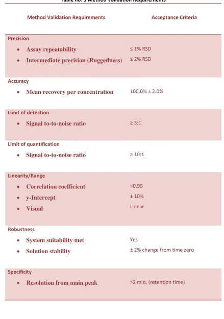
Table no: 3 Method Validation Requirements
Method Validation Requirements Acceptance Criteria Precision
• Assay repeatability
• Intermediate precision (Ruggedness)
≤ 1% RSD
≤ 2% RSD
Accuracy
• Mean recovery per concentration
100.0% ± 2.0%
Limit of detection
• Signal to-to-noise ratio
≥ 3:1
Limit of quantification
• Signal to-to-noise ratio
≥ 10:1
Linearity/Range
• Correlation coefficient
• y-Intercept • Visual >0.99 ± 10% Linear Robustness
• System suitability met
• Solution stability
Yes
± 2% change from time zero
Specificity
• Resolution from main peak
[image:32.612.84.523.93.718.2]
Dept. of Pharmaceutical Analysis Literature Review
KMCH college of pharmacy Page 29
LITERATURE REVIEW
JIN Yi et al(2011) [26] developed a sensitive and rapid LC-MS/MS method to determine
the concentration of tapentadol in rat plasma. Methods After the extraction from plasma by
protein precipitation, analytes and internal standards were separated by a Diamonsil C18 column.
Methanol-5 mmol·L-1 ammonium acetate-acetic acid (V V V=58 42 0.5) were used as the mobile
phase. The multiple reactions monitoring (MRM) was used for quantitative determination in
positive mode. The transitions were m/z: 222.2→107.1 for tapentadol ([M+H]+) , m/z:
307.1→220.1 for fluconazol (IS, [M+H]+). Results No significant interferences for the detection
of tapentadol and fluconazol from endogenous substances in plasma were observed in the present
study. The calibration curve was linear over the range of 2.0-200 μg·L-1 for tapentadol. The
accuracy was in the range of 96.2%-97.5% and the inter-day and intra-day precision was less
than 15%. Conclusions The LC-MS/MS method is simple, rapid and sensitive enough for the
pharmacokinetic study of Tapentadol in rats
Thomas M. Tzschentke et al[27] (_)-(1R,2R
)-3-(3-Dimethylamino-1-ethyl-2-methyl-propyl)-phenol hydrochloride (Tapentadol HCl) is a novel _-opioid receptor (MOR)
agonist (Ki _ 0.1 _M; relative efficacy compared with morphine 88% in a [35S]guanosine
5_-3-O-(thio)triphosphate binding assay) and NE reuptake inhibitor (Ki _ 0.5 _M for synaptosomal
reuptake inhibition). In vivo intracerebral microdialysis showed that tapentadol, in contrast to
morphine, produces large increases in extracellular levels of NE (_450% at 10 mg/kg i.p.).
Tapentadol exhibited analgesic effects in a wide range of animal models of acute and chronic
pain [hot plate, tail-flick, writhing, Randall-Selitto, mustard oil colitis, chronic constriction
injury (CCI), and spinal nerve ligation (SNL)], with ED50 values ranging from 8.2 to 13 mg/kg
potency of tapentadol was only two to three times lower than that of morphine, suggesting that
the dual mode of action of tapentadol may result in an opiate-sparing effect. A role of NE in the
analgesic efficacy of tapentadol was directly demonstrated in the SNL model, where the
analgesic effect of tapentadol was strongly reduced by theα2-adrenoceptor antagonist yohimbine
but only moderately attenuated by the MOR antagonist naloxone, whereas the opposite was seen
for morphine. Tolerance development to the analgesic effect of tapentadol in the CCI model was
twice as slow as that of morphine. It is suggested that the broad analgesic profile of tapentadol
and its relative resistance to tolerance development may be due to a dual mode of action
consisting of both MOR activation and NE reuptake inhibition.
Rolf terlinden et al [28] proposed Tapentadol is a novel, centrally acting oral analgesic with a dual mode of action that has demonstrated efficacy in preclinical and clinical models of
pain relief. The present study investigated and characterized the absorption, metabolism, and
excretion of tapentadol in humans. Four healthy male subjects received a single 100-mg oral
dose of 3-[14C]-labeled tapentadol HCl for evaluation of the pharmacokinetics of the drug and
the excretion balance of radiocarbon. The concentration-time profiles of radiocarbon in whole
blood and serum and radiocarbon excretion in the urine and feces, and the expired C02 were
determined. The serum pharmacokinetics and excretion kinetics of tapentadol and its conjugates
were assessed, as was its tolerability. Absorption was rapid (with a mean maximum serum
concentration [Cmax], 2.45 μg-eq/m]; a time to Cmax, 1.25–1.5 h), and the drug was present
primarily in the form of conjugated metabolites (conjugated: unconjugated metabolites = 24 1).
Excretion of radiocarbon was rapid and complete (>95% within 24 h; 99.9% within 5 days) and
almost exclusively renal (99% 69% conjugates; 27% other metabolites; 3% in unchanged form).
Dept. of Pharmaceutical Analysis Literature Review
KMCH college of pharmacy Page 31
electrocardiogram recording, or physical examination findings were reported. In our study group,
it was found that a single oral dose of tapentadol was rapidly absorbed, then excreted into the
urine, primarily in the form of conjugated metabolites, and was well tolerated.
Thomas M Tzschentke et al(2009) [29] stated that Tapentadol exerts its analgesic effects through micro opioid receptor agonism and nor-adrenaline reuptake inhibition in the central
nervous system. Preclinical studies demonstrated that tapentadol is effective in a broad range of
pain models, including nociceptive, inflammatory, visceral, mono- and polyneuropathic models.
Moreover, clinical studies showed that tapentadol effectively relieves moderate to severe pain in
various pain care settings. In addition, it was reported to be associated with significantly fewer
treatment discontinuations due to a significantly lower incidence of gastrointestinal-related
adverse events compared with equivalent doses of oxycodone. The combination of these reduced
treatment discontinuation rates and tapentadol efficacy for the relief of moderate to severe
nociceptive and neuropathic pain may offer an improvement in pain therapy by increasing
patient compliance with their treatment regimen.
Xu Steven Xu et al(2010) [30] Background Tapentadol is a new, centrally active
analgesic agent with two modes of action - μ opioid receptor agonism and nor-epinephrine
reuptake inhibition - and the immediate-release (IR) formulation is approved in the US for the
relief of moderate to severe acute pain. The aims of this analysis were to develop a population
pharmacokinetic model to facilitate the understanding of the pharmacokinetics of tapentadol IR
in healthy subjects and patients following single and multiple dosing, and to identify covariates
that might explain variability in exposure following oral administration.
Methods: The analysis included pooled data from 11 385 serum pharmacokinetic samples from
modeling was conducted using nonlinear mixed-effects modeling (NONMEM®) software to
estimate population pharmacokinetic parameters and the influence of the subject's demographic
characteristics, clinical laboratory chemistry values and disease status on these parameters.
Simulations were performed to assess the clinical relevance of the covariate effects on tapentadol
exposure.
Results: A two-compartment model with zero-order release followed by first-order absorption
and first-order elimination best described the pharmacokinetics of tapentadol IR following oral
administration. The inter-individual variability (coefficient of variation) in apparent oral
clearance (CL/F) and the apparent central volume of distribution after oral administration were
30% and 29%, respectively. An additive error model was used to describe the residual variability
in the log-transformed data, and the standard deviation values were 0.308 and 0.314 for
intensively and sparsely sampled data, respectively. Covariate analysis showed that sex, age,
bodyweight, race, body fat, hepatic function (using total bilirubin and total protein as surrogate
markers), health status and creatinine clearance were statistically significant factors influencing
the pharmacokinetics of tapentadol. Total bilirubin was a particularly important factor that
influenced CL/F, which decreased by more than 60% in subjects with total bilirubin greater than
50 μmol/L.
Conclusions: The population pharmacokinetic model for tapentadol IR identified the
relationship between pharmacokinetic parameters and a wide range of covariates. The
simulations of tapentadol exposure with identified, statistically significant covariates
demonstrated that only hepatic function (as characterized by total bilirubin and total protein) may
be considered a clinically relevant factor that warrants dose adjustment. None of the other
Dept. of Pharmaceutical Analysis Literature Review
KMCH college of pharmacy Page 33
Cooper et al [31] developed a sensitive LC-MS/MS method for the quantification of oxycodone, oxymorphone and noroxycodone which has been used to analyze clinical samples.
The analytes are well separated on a typical C18, 5 micron HPLC column, but the cycle time of 9
minutes is a hindrance to maximizing throughput. BASi has sought to improve cycle time while
also improving resolution by converting the method to utilize an ultra small particle size C18
column with a conventional HPLC.
Methods: The range of the validated assay is 0.1-100 ng for oxycodone and oxymorphone, and 0.5-100 ng/ml for noroxycodone. Detection is by positive Turbo Ion spray on a Sciex API-4000.
The HPLC column is a 2.1 X 50 mm, 5 micron, Waters XBridge C18. Typical retention times of
the analytes are oxycodone 5.2 minutes, oxymorphone 3.5 minutes, and noroxycodone 2.4
minutes. The cycle time is approximately 9 minutes. For the new, faster method, the
chromatography has been altered to accommodate the small particle column [2 x 30 mm, 1.5
micron, Grace Vison HT C18] but the extraction and mass spectrometer conditions remain the
same. The analytes are eluted on a steep gradient from 10 to 100% organic with a 1 minute hold
at 5 minutes and a flow rate of 0.2 mL/min.
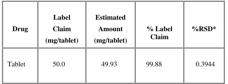
Arunadevi S. Birajdar(2009) [32] developed a high-performance liquid chromatographic method has been developed for the simultaneous analysis of paracetamol and
tramadol in combined solid dosage form. The mobile phase consisting of Acetonitrile- 0.26 %
Triethylamine buffer (pH 7.3) in ratio of (45:55 % v/v) was delivered at the flow rate of 1.0
mL/min and UV detection was carried out at 264 nm. The separation was achieved using C18
reverse-phase column (250 X 4.6 mm I.D., particle size 5µm). The method was linear over the
concentration range of 1.0-12.0 µg/mL for Paracetmol and 0.1-1.2 µg/mL for tramadol.
99.88%. The validation of method carried out as per ICH guidelines. The described HPLC
method was successfully employed for the analysis of pharmaceutical formulations containing
combined dosage form and can be employed for bioequivalence study in future for the same
formulations.
Markus G Gebauer et al(2001) [33] An HPLC method for the quantification of oxycodone and lidocaine in a gel matrix is described. The mobile phase consisted of methanol–
water–acetic acid (35:15:1 v/v/v) and was delivered at 1.5 ml/min through a 4.6×250 mm
Zorbax® SB-C8 column. Oxycodone was detected at 285 nm and lidocaine at 264 nm. Linear
calibration curves were obtained for oxycodone in the range of 0.05–1.5% (w/w) and for
lidocaine in the range of 0.1–5.0% (w/w). Oxycodone and lidocaine were treated with hydrogen
peroxide and the oxidation products were readily separated on the column. The method was
applied to assess the stability of a gel containing oxycodone hydrochloride (0.3% w/w) and
lidocaine (1.5% w/w). The gel was stored under refrigeration in ready-to-use syringes and under
these conditions oxycodone and lidocaine were stable for at least 1 year. The gel is useful in the
management of tenesmus in rectal cancer.
Lambropoulos J et al(1999) [34]The stability indicating properties of the USP method for the assay of fentanyl in fentanyl citrate injection were evaluated [1] by analyzing fentanyl drug
substance and product after acid, hydrogen peroxide, heat, and light treatment.
N-phenyl-N-(4-piperidinyl) propionamide (PPA), which is a known degradation product/process impurity of
fentanyl, was not adequately resolved from the fentanyl peak, and mobile phase adjustments did
not improve the resolution (Fig. 1). Therefore, the USP method did not meet the requirements for
Dept. of Pharmaceutical Analysis Literature Review
KMCH college of pharmacy Page 35
nm) to provide adequate levels for the quantitation of the related substances of fentanyl and, in
addition, the acetate ions in the mobile phase could interfere with a lower wavelength detection.
An isocratic, reversed phase, stability indicating, high performance liquid chromatographic
(HPLC) method for the assay of fentanyl and related substances in fentanyl citrate injection, USP
has been developed and validated. The chromatographic conditions employed an Inertsil C8, 5
column (25 cm x 4.6 mm), a mobile phase of aqueous perchloric acid [0.23%, w/v]-Acetonitrile
[65:35, v/v], and ultraviolet (UV) detection at 206 nm. Under the chromatographic conditions of
the method, PPA and seven other known process impurities were separated from the active.
Degradation studies showed that the active eluted as a spectrally pure peak resolved from its
degradation products.
John Lambropoulos et al(2000) [35] A high performance liquid chromatography (HPLC) method for the assay of fentanyl citrate, alfentanil hydrochloride, and sufentanil citrate swab
samples was developed and validated in order to control a cleaning procedure. The swabbing
procedure involved Super POLX 1200 wipers moistened with water. The assay employed
extraction of swabs with water and analysis by isocratic, reversed-phase, HPLC with varying
ultraviolet (UV) detection for desired sensitivity, depending on the analyte. The method was
shown to be selective and linear from the limits of quantitation (0. 10, 0. 20, and 0.15 μg/swab
for fentanyl citrate, alfentanil, and sufentanil, respectively) to over three times these
concentrations. The assay limits (detection levels) per swab area were set at least at 0.2% of the
concentrations of the actives in the drug products (0.02, 0. 10, and 0. 10 μg/swab or
approximately 0.03, 0.02, and 0.2% for fentanyl citrate, alfentanil, and sufentanil, respectively).
It should be noted that all active concentrations listed in this work were calculated based on the
sufentanil. No reference standard was available for alfentanil hydrochloride and sufentanil
citrate. Drug product was used instead throughout this study.
J Lambropoulos et al(1999)[36] proposed the stability indicating properties of the USP method for the assay of fentanyl in fentanyl citrate injection were evaluated [1] by analyzing
fentanyl drug substance and product after acid, hydrogen peroxide, heat, and light treatment.
N-phenyl-N-(4-piperidinyl) propionamide (PPA), which is a known degradation product/process
impurity of fentanyl, was not adequately resolved from the fentanyl peak, and mobile phase
adjustments did not improve the resolution (Fig. 1). Therefore, the USP method did not meet the
requirements for a stability-indicating assay. In addition, the wavelength in the USP method was
too high (230 nm) to provide adequate levels for the quantitation of the related substances of
fentanyl and, in addition, the acetate ions in the mobile phase could interfere with a lower
wavelength detection. An isocratic, reversed phase, stability indicating, high performance liquid
chromatographic (HPLC) method for the assay of fentanyl and related substances in fentanyl
citrate injection, USP has been developed and validated. The chromatographic conditions
employed an Inertsil C8, 5 column (25 cm x 4.6 mm), a mobile phase of aqueous perchloric acid
[0.23%, w/v]-Acetonitrile [65:35, v/v], and ultraviolet (UV) detection at 206 nm. Under the
chromatographic conditions of the method, PPA and seven other known process impurities were
separated from the active. Degradation studies showed that the active eluted as a spectrally pure
peak resolved from its degradation products.
Amir mehdizadeh et al(2005) [37] developed a simple, sensitive and specific HPLC
method and also a simple and fast extraction procedure were developed for quantitative analysis
of fentanyl transdermal patches. Chloroform, methanol and ethanol were used as extracting
Dept. of Pharmaceutical Analysis Literature Review
KMCH college of pharmacy Page 37
ethanol and the eluted fentanyl through the C18 column was monitored by UV detection at 230
nm. The linearity was at the range of 0.5-10 μg/mL with correlation coefficient (r2) of 0.9992.
Both intra and inter-day accuracy and precision were within acceptable limits. The detection
limit (DL) and quantitation limit (QL) were 0.15 and 0.5 μg/mL, respectively. Other validation
characteristics such as selectivity, robustness and ruggedness were evaluated. Following method
validation, a system suitability test (SST) including capacity factor (k´), plate number (N), tailing
factor (T), and RSD was defined for routine test.
Finally after searching of the all literature no method was developed for estimation of Tapentadol Hydrochloride by RP-HPLC. So by this, I have decided to do method
TAPENT 5.1 Chem Brand n Nu Chemica 3 Molecula C14 Mol. ma Structur Fig Pharmac Bioa TADOL HY mical profile ame: ucynta, Nuc al name/IUP 3-[(1R, 2R)-ar formula:
4H23NO.HC
ass: 221.339
re:
g: 2 MOLE
cokinetic da
availability
YDROCHL
e [38, 39]
ynta ER, Ni
PAC NAME 3-(Dimethyl : CL g/mo CULAR ST ata:
: 31.9 ± 6.
DRU ORIDE ap E: lamine)-1-et TRUCTURE 8% (oral) G PROFIL thyl-2-methy
Dept. of Pharmaceutical Analysis Drug Profile
KMCH college of pharmacy Page 39
Metabolism : Hepatic glucuronidation and sulfate conjugation
Half-life : 4 hrs
Excretion : Renal (>95%) and feca
Routes : Oral, Oral, Other ROA Unknown
Description:
White or off white powder
Category [40]:
Centrally acting analgesic, agonist at the m-opioid receptor,
Dose [41]:
Immediate release:
50 mg, 75 mg, or 100 mg orally every 4 to 6
Extended release:
Opioid naive: Initial: 50 mg twice daily (recommended interval: 12 hours); titrate in increments
of 50 mg no more frequently than twice daily every 3 days to effective dose (therapeutic range:
100 to 250 mg twice daily) (maximum dose: 500 mg/day)
5.2 Pharmacological profile [42]
Mechanism of action:
Tapentadol is a centrally-acting synthetic analgesic. Although its exact mechanism is
unknown, analgesic efficacy is thought to be due to mu-opioid agonist activity and the inhibition
Pharmac
Tapentad
binding t
animal m
rats resu
activity d
selective
is sensiti
pharmaco
Effects o
doses of
codynamics
dol is a cent
to the human
models. Tape
lting in incr
due to the
mu-opioid a
ive to nor-e
ologically ac
on the cardio
[image:44.612.201.410.75.328.2]f tapentadol
Fig: 3 M
s
trally-acting
n mu-opioid
entadol has b
reased nor-e
mu-opioid r
antagonists (
epinephrine
ctive metabo
ovascular sy
on the QT
M.O.A of T
synthetic an
d receptor an
been shown epinephrine receptor ago (e.g., naloxo modulators. olite. ystem: There interval. In Tapentadol H
nalgesic. It i
nd is 2–3 tim
n to inhibit n
concentratio
onist activity
one), wherea
. Tapentado
e was no eff
a randomize
Hydrochlor
is 18 times l
mes less pote
nor-epinephr
ons. In precl
y of tapenta
as the nor-ep
ol exerts its
fect of thera
ed, double-b ride
less potent t
ent in produ
rine reuptake
linical mode
adol can be
inephrine re
analgesic e
apeutic and s
blind, placeb
than morphi
ucing analges
e in the brai
els, the anal
e antagonize
euptake inhib
effects witho
supra therap
itive-Dept. of Pharmaceutical Analysis Drug Profile
KMCH college of pharmacy Page 41
controlled crossover study, healthy subjects were administered five consecutive doses of
Nucynta® 100 mg every 6 hours, Nucynta® 150 mg every 6 hours, placebo and a single oral
dose of moxifloxacin. Similarly, Nucynta® had no relevant effect on other ECG parameters
(heart rate, PR interval, QRS duration, and T-wave or U-wave morphology).
Pharmacokinetics
Absorption
Mean absolute bioavailability after single-dose administration (fasting) is approximately 32%
due to extensive first-pass metabolism. Maximum serum concentrations of tapentadol are
typically observed at around 1.25 hours after dosing.
Dose-proportional increases in the Cmax and AUC values of tapentadol have been observed over
the 50 to 150 mg dose range.
A multiple (every 6 hour) dose study with doses ranging from 75 to 175 mg tapentadol showed a
mean accumulation factor of 1.6 for the parent drug and 1.8 for the major metabolite
tapentadol-O-glucuronide, which are primarily determined by the dosing interval and apparent half-life of
tapentadol and its metabolite.
Food Effect
The AUC and Cmax increased by 25% and 16%, respectively, when Nucynta® was administered
after a high-fat, high-calorie breakfast. Nucynta® may be given with or without food.
Tapentadol is widely distributed throughout the body. Following intravenous administration, the
volume of distribution (Vz) for tapentadol is 540 +/- 98 L. The plasma protein binding is low and
amounts to approximately 20%.
Metabolism and Elimination
In humans, the metabolism of Tapentadol Hydrochloride is extensive. About 97% of the parent
compound is metabolized. Tapentadol is mainly metabolized via Phase 2 pathways, and only a
small amount is metabolized by Phase 1 oxidative pathways. The major pathway of tapentadol
metabolism is conjugation with glucuronic acid to produce glucuronide. After oral administration
approximately 70% (55% O-glucuronide and 15% sulfate of tapentadol) of the dose is excreted
in urine in the conjugated form. A total of 3% of drug was excreted in urine as unchanged drug.
Tapentadol is additionally metabolized to N-desmethyl tapentadol (13%) by CYP2C9 and
CYP2C19 and to hydroxyl tapentadol (2%) by CYP2D6, which are further metabolized by
conjugation. Therefore, drug metabolism mediated by cytochrome P450 system is of less
importance than phase 2 conjugation.
None of the metabolites contributes to the analgesic activity.
Tapentadol and its metabolites are excreted almost exclusively (99%) via the kidneys. The
terminal half-life is on average 4 hours after oral administration. The total clearance is 1530 +/-
177 ml/min.
Special Populations
Dept. of Pharmaceutical Analysis Drug Profile
KMCH college of pharmacy Page 43
The mean exposure (AUC) to tapentadol was similar in elderly subjects compared to young
adults, with a 16% lower mean Cmax observed in the elderly subject group compared to young
adult subjects.
Renal Impairment
AUC and Cmax of tapentadol were comparable in subjects with varying degrees of renal
function (from normal to severely impaired). In contrast, increasing exposure (AUC) to
tapentadol-O-glucuronide was observed with increasing degree of renal impairment. In subjects
with mild, moderate, and severe renal impairment, the AUC of tapentadol-O-glucuronide are
1.5-, 2.5-1.5-, and 5.5-fold higher compared with normal renal function1.5-, respectively.
Hepatic Impairment
Administration of Nucynta® resulted in higher exposures and serum levels to tapentadol in
subjects with impaired hepatic function compared to subjects with normal hepatic function. The
ratio of tapentadol pharmacokinetic parameters for the mild and moderate hepatic impairment
groups in comparison to the normal hepatic function group were 1.7 and 4.2, respectively, for
AUC; 1.4 and 2.5, respectively, for Cmax; and 1.2 and 1.4, respectively, for t1/2. The rate of
formation of tapentadol-O-glucuronide was lower in subjects with increased liver impairment.
Pharmacokinetic Drug Interactions
Tapentadol is mainly metabolized by Phase 2 glucuronidation, a high capacity/low affinity
system; therefore, clinically relevant interactions caused by Phase 2 metabolism are unlikely to
respectively. These changes are not considered clinically relevant and no change in dose is
required.
No changes in the pharmacokinetic parameters of tapentadol were observed when
acetaminophen and acetylsalicylic acid were given concomitantly.
In vitro studies did not reveal any potential of tapentadol to either inhibit or induce cytochrome
P450 enzymes. Thus, clinically relevant interactions mediated by the cytochrome P450 system
are unlikely to occur.
The pharmacokinetics of tapentadol were not affected when gastric pH or gastrointestinal
motility were increased by omeprazole and metoclopramide, respectively.
Plasma protein binding of tapentadol is low (approximately 20%). Therefore, the likelihood of
pharmacokinetic drug-drug interactions by displacement from the protein binding site is low.
![Fig: 1 HPLC BASIC INSTRUMENT [12]](https://thumb-us.123doks.com/thumbv2/123dok_us/549153.73445/13.612.71.491.142.555/fig-hplc-basic-instrument.webp)