Advancing biomedical imaging
The Harvard community has made this
article openly available.
Please share
how
this access benefits you. Your story matters
Citation
Weissleder, Ralph, and Matthias Nahrendorf. 2015. “Advancing
Biomedical Imaging.” Proceedings of the National Academy
of Sciences 112 (47): 14424–28. https://doi.org/10.1073/
pnas.1508524112.
Citable link
http://nrs.harvard.edu/urn-3:HUL.InstRepos:41384303
Terms of Use
This article was downloaded from Harvard University’s DASH
repository, and is made available under the terms and conditions
applicable to Other Posted Material, as set forth at
http://
Advancing biomedical imaging
Ralph Weissleder1and Matthias Nahrendorf
Center for Systems Biology, Massachusetts General Hospital and Harvard Medical School, Boston, MA 02114
Edited by Mark E. Davis, California Institute of Technology, Pasadena, CA, and approved May 28, 2015 (received for review May 4, 2015)
Imaging reveals complex structures and dynamic interactive processes, located deep inside the body, that are otherwise difficult to decipher. Numerous imaging modalities harness every last inch of the energy spectrum. Clinical modalities include magnetic resonance imaging (MRI), X-ray computed tomography (CT), ultrasound, and light-based methods [endoscopy and optical coherence tomography (OCT)]. Research modalities include various light microscopy techniques (confocal, multiphoton, total internal reflection, superresolution fluorescence microscopy), electron microscopy, mass spectrometry imaging, fluorescence tomography, bioluminescence, variations of OCT, and optoacoustic imaging, among a few others. Although clinical imaging and research microscopy are often isolated from one another, we argue that their combination and integration is not only informative but also essential to discovering new biology and interpreting clinical datasets in which signals invariably originate from hundreds to thousands of cells per voxel.
imaging
|
intravital microscopy|
inflammationEngineering sciences have played a major role in advancing biomedical imaging by improv-ing and miniaturizimprov-ing detectors, enhancimprov-ing system design, increasing speed, sensitivity and resolution, accelerating computational analysis, and developing methods to minimize the side effects of applied energy. Additionally, chemical engineering has produced advanced imaging probes (nanomaterials, labeled small and large molecules, and fluorescent proteins) to improve tissue, cell, and molecular specific-ity. Currently, imaging is evolving rapidly in three distinct biomedical areas: (i) imaging molecular biomarkers or contributing to biomarker analysis, (ii) single cell imaging, and (iii) imaging therapeutics. Each area has highly significant potential for accelerating progress, as we will discuss below after an overview of available tools.
Engineering Advances Have Yielded Impressive Tools
Clinical Imaging Systems. Modern
imag-ing systems have made great progress since the first devices were developed more than 100 y ago. X-rays, for example, introduced by Wilhelm Röntgen’s images of his wife’s hand, are now used in sophisticated three-dimensional computed tomography (CT) scans that can detect millimeter-sized pul-monary nodules in high-risk populations (1), among many other applications. The ex-plosion of imaging technologies has also pro-duced complementary information because the energy–matter interaction generates dif-ferent contrast mechanisms (e.g., magnetic relaxivity, susceptibility, diffusion, temperature, elasticity, electrical impedance, radiation ab-sorption, scattering, and fluorescence) (2). Imaging systems can be grouped according to energy type (e.g., X-rays, positrons, photons,
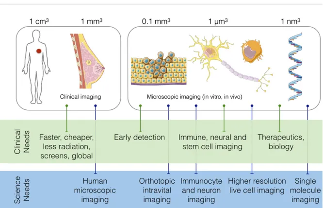
or sound waves), spatial resolution (e.g., macroscopic or microscopic), or obtained information type (anatomical, physiological, cellular, or molecular) (Fig. 1) (2). Macro-scopic imaging systems that provide ana-tomical and physiological information are now in widespread clinical and preclinical use. By contrast, systems that provide mi-croscopic resolution are widely used in basic science (Fig. 1). Imaging modality selection is largely determined by the scientific or medical question at hand. Although cur-rent imaging technologies’technical capabil-ities are often amazing, their future potential is equally exciting. Examples include mi-croscopy performed in live subjects (3, 4) and imaging at extreme resolutions in live cells (5). At these resolutions, real-time observations will provide spectacular insight into the mammalian cells’inner workings.
Research Imaging.Microscopes that allow
imaging in live animals have been indispens-able in discovering cancer biology (6–9), im-munology (10–14), and brain function (15– 17). Research microscopy systems are often based on confocal or multiphoton scopes with long working distance objectives, special lasers, and unique motion compensation techniques. In addition to advances in optics and detector technology, imaging’s research contributions have been enabled by fluores-cent proteins and exponentially expanded computational power. The discovery of fluorescent proteins, for which the Nobel prize was awarded in 2008 (Shimomura, Chalfie, and Tsien), allowed researchers to visualize a broad range of specific proteins or cells for the first time. One stunning ex-ample is the combinatorial color-labeling method based on the stochastic expression
of several fluorescent proteins (Brainbow) (16). Lichtman and coworkers (16) were able to mark individual neurons with over 100 distinct colors and subsequently trace and reconstruct entire connectome brain maps. Similarly, fluorescent proteins facili-tated the development of ultraresolution mi-croscopy techniques for which the Nobel prize was awarded in 2014 (Betzig, Hell, and Moerner). Reporter genes have also been described for other imaging technologies such as MRI (18, 19), nuclear imaging (20, 21), and ultrasound (22). Although computational advances have greatly contributed to new imag-ing techniques, much work remains to be done, particularly with regard to automated image analysis (23), data mining, integrating complex datasets into multiscale models, and developing new visualization tools.
Chemical Tools for Biomolecular Imag-ing. Chemical tools are increasingly impor-tant in both clinical and research imaging because they can add molecular and cellular specificity and/or enhance physiological data extraction. Additionally, chemical imaging agents have two major advantages over fluorescent proteins although the two are often used complementarily: chemical tools enable imaging in humans and obviate the need for genetically engineered mouse mod-els. A considerable number of imaging agents have been developed over the last decade
Author contributions: R.W. and M.N. analyzed data and wrote the paper.
The authors declare no conflict of interest.
This article is a PNAS Direct Submission.
This article is part of the special series of PNAS 100th Anniversary articles to commemorate exceptional research published in PNAS over the last century.
1To whom correspondence should be addressed. Email: rweissleder@
[Molecular Imaging and Contrast Agent Database (MICAD); www.ncbi.nlm.nih.gov/ books/NBK5330/], and some agents are com-mercially available or even FDA-approved (24). Nanoparticles are particularly prom-ising because they tend to accumulate in innate immunocytes, which are often
“first responders” in pathologic processes (25). Further, nanoparticles have unique pharmacokinetics: i.e., they circulate longer and are not immediately cleared renally and can be targeted to specific organs, cells, or proteins. Magnetic nanoparticles, which are detected by magnetic resonance imaging (MRI), are perhaps the best-studied nano-particle type. Ferumoxytol, for example, is an FDA-approved nanomaterial for iron replacement in treating anemia but has been used to enhance MRI (26); when tagged with fluorochromes, ferumoxytol also dou-bles as an MR and/or optical imaging agent (12). Quantum dots have been es-sential in certain microscopic imaging exper-iments (27, 28), especially in conjunction with environmentally sensitive particles (29), tar-geted particles, and short wave infrared parti-cles that can be detected much deeper in tissue (30). Labeled antibodies and antibody frag-ments have long been used for targeted im-aging, and the introduction of long-lived imaging isotopes (89Zr, 68Ga, 64Cu,124I) has resulted in some spectacular clinical results (31–33). Newer alpaca-derived antibody fragments currently being developed offer several advantages over traditional anti-bodies (34, 35). Specifically, single chain camelid antibody fragments lack an Fc portion and are much smaller (∼15 kDa) than immunoglobulins (∼150 kDa),“diabody”
antibody derivatives (∼60 kDa), Fab fragments (∼50 kDa), or single-chain variable fragments (ScFvs) (∼25 kDa). Other important chemical imaging tools now in routine use include a large number of isotope, fluorochrome, or metal-labeled small molecules (24). Finally, there are a number of hyperpolarized C13 metabolites being developed for metabolic MR imaging (36).
Imaging Molecular and Cellular Biomarkers
In vivo imaging of molecular and cellular biomarkers is most useful for studying organs not readily biopsied (such as at the brain), finding early cancers, and mapping disease severity and location. Molecular biomarker development has largely been guided by
“omics”techniques and immunopathological studies. Emerging multiplexed imaging (37) and cytometry (38–41) approaches will likely play an important role in defining new im-aging targets. Finally, clinical imim-aging can enhance biomarker information by providing complementary information (42, 43).
Imaging Receptors.Applying imaging tech-nologies to receptors has expanded our knowl-edge of human biology and improved treat-ments for numerous conditions. For example, receptor imaging has been used to study the dopamine reward pathway in people with at-tention deficit hyperactivity disorder (ADHD). One study found that adults with ADHD had fewer D2 and D3 receptors (mea-sured via11C-raclopride and11C-cocaine) in their reward circuits and that receptor levels were proportional to inattention symptoms (44). Additionally, ADHD patients’reward
circuits were less sensitive. Receptor imaging is also influencing cancer diagnostics. Tumor receptors play an important role in carci-nogenesis and tumor growth: relevant re-ceptors include steroid rere-ceptors (estrogen receptor in breast cancer and androgen ceptor in prostate cancer), somatostatin re-ceptors (SSTR2), and growth factor rere-ceptors (EGFR, HER2) among others (e.g., trans-ferrin, folate, and asialoglycoprotein recep-tors). Tumor receptor imaging has been used to spot cancers (45, 46), understand cancer biology (47), and quantitate the effects of receptor inhibition on tumor growth (48).
Finding Smaller Cancers.Cancer remains
[image:3.585.35.368.48.262.2]the second most common cause of death in the United States. In 2014, there were 1,665,540 new cancer cases diagnosed and 585,720 cancer deaths in the United States. However, the vast majority of cancers are curable when detected early (>90% in stage 1). Most clinical imaging technologies can easily visualize cancers when they approach 1 cm3, which is equivalent to∼3 billion cells. Through recent advances in image resolution and chemical agents, the detection size boundary is being pushed toward 1mm3, which cor-responds to∼3 million cancer cells. Hope-fully even smaller sized cancer lesions will likely be detectable in the future. To achieve this goal, we will need tools to determine which properties of precancerous lesions predict the likelihood of progression to ma-lignant metastatic disease. There are extra-ordinary opportunities in pushing these boundaries: (i) exploring new imaging tech-nologies, sensors, and agents through en-gineering advances, (ii) combining blood biomarkers with imaging, and (iii) devel-oping microscopic imaging tools that can be used intraoperatively or during minimally invasive procedures (i.e., microendoscopy) (49). Combining imaging and blood bio-marker analysis may be particularly helpful in increasing the accuracy of screening pro-cedures. For example, in addition to iden-tifying easily confirmed lung cancers, low-dose CT scans invariably discover many harmless lesions that require further work-up at high costs. Blood tests for circulating tumor DNA, microvesicles, circulating can-cer cells, and/or other makers may increase the accuracy of CT screening (42, 50, 51). Intraoperative imaging with fluorescent affinity ligands or antibodies (52, 53) is now a clinical reality, and reports from the first clin-ical trials are very promising (49, 54). These approaches will ultimately change cancer surgery standards by giving surgeons real-time feedback about tumor margins and whether any cancer remains. Imaging-facilitated
Fig. 1. Overview of clinical and basic science imaging needs.
Weissleder and Nahrendorf PNAS | November 24, 2015 | vol. 112 | no. 47 | 14425
SPE
CIAL
FEATURE
:
PERSPE
Imaging Physiology. Imaging physiology has long been the mainstay of clinical di-agnostics. Using a sensitive contrast agent that informs on vascular parameters (density, per-meability, etc.) often reveals disease processes. Because many physiological processes simply do not happen ex vivo, the only way to learn about them is to watch them in vivo. A stunning example is certain leukocytes’ ca-pacity to crawl along the endothelial surface of small vessels, sometimes even against the flow of blood (55). This patrolling behavior was discovered only because newly de-veloped imaging tools had the sensitivity and resolution to follow cell group in-teractions distinguished by specific re-porter genes. Noninvasive imaging, even clinical imaging, will likely adopt the advan-tages of spectrally resolving several targets. Key aspects of complex physiology and dis-ease process, including those that seem to conflict one another, occur simultaneously. Integrating comprehensive imaging data can provide unprecedented insight into pathol-ogy. For instance, near infrared fluorescence imaging of macrophage presence, angio-genesis, and protease activity in ischemic mouse hearts linked these healing bio-markers to cellular function in the setting of heart failure (56). Translatable PET/MR imaging may enable multispectral imaging, as recently shown in mice with heart failure (57) and Alzheimer’s disease (58).
Single Cell Imaging
Intravital microscopy can reveal cells’ 3D morphology and interactions with neighbor-ing cells in their native microenvironment. Some emerging discoveries have direct im-plications for understanding clinical findings.
Immune Cell Imaging. For the most part,
immunology is still studied via flow cytom-etry and genomics (59). Nevertheless, single cell immunocyte imaging has tremendous po-tential for deciphering cells’in vivo spatial distribution, dynamics, lineage, and behavior in disease. High resolution imaging has re-cently lead to surprising discoveries. For example, new mouse models with bright fluorescence reporter genes (e.g., Cx3cr1GFP and others) show that macrophages are much more widely distributed than pre-viously thought, that macrophages have projecting dendrites that facilitate sensing (60), and that these cells display remarkable dy-namics and effector functions. The heart, for instance, contains a dense network of macrophages whose delicate far-reaching
diseased organs, and imaging facilitates ex-ploration of these networks’ functions in normal and diseased tissues especially in cancer, myocardial infarction, type 1 diabetes, and autoimmune diseases. Taking cancer as an example, the following are some out-standing questions: (i) What is the respective role of tissue-resident (i.e., yolk sac-derived) tissue macrophages versus those recruited from hematopoietic sources during tumor initiation and metastases? (ii) Can tumor-associated macrophages be used therapeuti-cally to enhance tumor killing? (iii) Why do some patients respond much better than others to emerging immune checkpoint blockades? and (iv) Why do some patients experience extraordinary toxicities with im-munotherapies whereas others do not ?
Beyond imaging at single cell resolution, reporting on the immune cell populations is becoming feasible in patients. Recent MRI and PET studies in patients with atheroscle-rosis and acute myocardial infarction relied on macrophage-avid iron oxide nanoparticles or the glucose analog 18F-FDG to study in-flammatory responses (61–63). These imag-ing trials described a systemic activation of the immune system, with accumulation of leukocytes in the ischemic tissue and in re-mote atherosclerotic plaques. In addition, increased activity was observed in the spleen and the bone marrow. Taken together, the imaging data imply that acute organ ischemia triggers increased bone marrow and splenic production of myeloid cells, which migrate to the ischemic organ but also to remote ath-erosclerotic plaques, thus promoting disease progression. Translating these insights from mouse to man would likely be impossible without imaging, which, unlike biopsies, can sample the entire human body noninvasively. Newer approaches of cell labeling, reporter gene strategies (64), immune cell imaging (35), and checkpoint blockade imaging [e.g., programmed death-1 (PD1) and cytotoxic T-lymphocyte antigen (CTLA-4)] are also being explored clinically.
Stem Cell Imaging. Important questions
that microscopic imaging can help to answer are those related to survival, proliferation, and differentiation of stem and progenitor cells. In vivo microscopy can follow individual fluo-rescently tagged hematopoietic stem cells over several days and report on their propensity to divide or migrate as a function of their local-ization in the hematopoietic niche and as a function of disease: for instance, in mice with increased sympathetic tone after ischemic
whole body level, bioluminescence and PET imaging of reporter gene expression in the tracked cells are leading the field because they can be quite sensitive and tracking labels do not dilute with cell division. In addition, the imaging signal ceases when the cells die. These techniques have been used for tracking cells transplanted into failing mouse hearts, where imaging provided the sobering but important feedback of limited stem cell sur-vival. Clinically, cells have been tagged with magnetic materials (70) and isotopes to monitor their in vivo distribution (64). Ex-cellent reviews exist on these topics (71–73).
Imaging in Drug Therapy
Imaging has the potential to play a leading role in the routine use of therapeutics, par-ticularly in oncology where drug resistance develops over time and targeted therapies can be extremely expensive. Similarly, therapeutic intervention in Alzheimer’s disease may ben-efit from clinical imaging. The US Food and Drug Administration (FDA) recently ap-proved three PET imaging agents (florbe-taben, florbetabir, and flutemetamol) that target amyloid. Unfortunately, because the Centers for Medicare and Medicaid Ser-vices (CMS) often does not reimburse use of these and other PET ligands, their use is ironically limited in favor of more costly alternatives. Consequently, most PET im-aging is currently performed during clinical trials. Here, imaging is used to enroll pa-tients into specific trials: test drug distri-bution in phase 1 trials via PET imaging (often11C-labeled rather than the18F com-panion imaging drugs); guide biopsy of spe-cific tissues for pathological analysis, and deliver drugs by image guidance or as a readout of efficacy in therapeutic trials (e.g., tumor shrinkage or change in metabolic ac-tivity in target tissue). Imaging can also be used as a companion diagnostic to provide information essential to safe and effective use of a corresponding therapeutic prod-uct. In reality, however, the FDA-approved list of companion diagnostics relies much more heavily on in vitro diagnostic devices to interrogate tissue samples (www.fda.gov/ MedicalDevices/ProductsandMedicalProcedures/ InVitroDiagnostics/ucm301431.htm).
In the research setting, intravital microscopy has been used to study new drugs’ pharma-cokinetics and pharmacodynamics (3). A growing list of fluorescent companion imag-ing drugs enables these advances (9, 75–82) and immobilization techniques allow ortho-topic imaging (83–85) and methods to study drug/target binding (86). These advances al-low detailed insight into when and why drugs fail. Until now, most research on the thera-peutics’mechanisms of action and failures has been performed in cell culture, rather than at the cellular level in vivo. Reliance on cell culture leaves unanswered a num-ber of questions regarding delivery to tar-get cells and whether or not the assumed mechanism of drug action occurs in vivo. For example, what are the drug concentra-tions inside cellular compartments (nucleus vs. cytoplasm)? Is the drug mechanism the same for every cell within the tumor, or is there heterogeneity? Do response mecha-nisms differ within tumor classes (i.e., dif-ferent models of ovarian cancer)? How, when, and where does resistance develop? These questions are exemplified by a recent study
of eribulin (9), which was developed and FDA-approved as a potent microtubule-tar-geting cytotoxic agent to treat taxane-resistant cancers. However, recent clinical trials showed that this drug eventually fails in many patient subpopulations for unclear reasons. To in-vestigate eribulin’s resistance mechanisms, researchers developed a fluorescent analog, with sufficiently similar pharmacokinetic (PK) properties and cytotoxic activity across a human cell line panel, to study the parent drug’s cellular PK and tissue distribution. Results showed that resistance to eribulin and its fluorescent analog depended directly on the multidrug resistance protein 1 (MDR1). In vivo, MDR1-mediated drug efflux and 3D tumor vascular architecture critically de-termined drug accumulation in tumor cells. Also, standard i.v.-administered third-gener-ation MDR1 inhibitor failed to rescue drug accumulation; however, encapsulating the same MDR1 inhibitor within a nanoparticle delivery system reversed the multidrug-resistant phenotype and potentiated the eribulin effect in vitro and in vivo in mice. This study is just one example of how in vivo imaging of an anticancer drug’s cellular PK is a powerful strategy for elucidating drug re-sistance mechanisms in heterogeneous tu-mors and evaluating strategies to overcome this resistance. This type of essential in-formation is hard to obtain without imaging.
The Future
If the last decade’s rapid advances in imaging and engineering are a good harbinger, then the future looks bright indeed. There are extraordinary opportunities in further ad-vancing imaging capabilities to support basic science and translational and clinical mis-sions (Fig. 1). We argue that these new tools will ultimately allow new types of measure-ments. The most useful techniques will
quantitatively and comprehensively access the cellular and/or subcellular/molecular levels in vivo. In the following, we list some of the current technological challenges (Fig. 1): (i) How do we improve clinical detection of earlier forms of cancers to<1 mm3(e.g., 0.1 mm3consisting of∼103 cells)? (ii) Can we develop single cell imaging techniques to image beyond the current depth capabilities (i.e., deeper than∼200–500μm)? (iii) Can we develop methods to characterize in-dividual cells’functional states within tissues and tumors? (iv) How can we vastly increase data acquisition speeds to accelerate imaging or enable broader coverage (field of view) and how do we increase the spatial resolution by 10- to 100-fold without increasing acquisition times? (v) How can we push multiplexing: i.e., simultaneously imaging 10–100 targets? (vi) Can we develop advanced chemical tools: e.g., brighter, small footprint fluorochromes that are biocompatible and/or can be used as sensors? (vii) Can we develop a complete set of mouse models with genetically encoded fluorescent proteins in all relevant classes of immune cells in addition to lineage tracers for each of these cells? (viii) How do we ac-celerate the development of human micro-scopic imaging through endoscopes and probes? (ix) Can we harness technological advances to develop miniaturized sensors and implantable microscopes for long-term im-aging on the scale of days to months?
In summary, imaging has critically con-tributed to all aspects of basic science, trans-lational studies, and clinical medicine. A world without imaging is clearly not imaginable. We anticipate that future developments will allow us to push the boundaries of what can be measured and detected.
1Church TR, et al.; National Lung Screening Trial Research Team (2013) Results of initial low-dose computed tomographic screening for lung cancer.N Engl J Med368(21):1980–1991.
2Weissleder R, Pittet MJ (2008) Imaging in the era of molecular oncology.Nature452(7187):580–589.
3Pittet MJ, Weissleder R (2011) Intravital imaging.Cell147(5): 983–991.
4Chen BC, et al. (2014) Lattice light-sheet microscopy: Imaging molecules to embryos at high spatiotemporal resolution.Science
346(6208):1257998.
5Gao L, Shao L, Chen BC, Betzig E (2014) 3D live fluorescence imaging of cellular dynamics using Bessel beam plane illumination microscopy.Nat Protoc9(5):1083–1101.
6Lohela M, et al. (2014) Intravital imaging reveals distinct responses of depleting dynamic tumor-associated macrophage and dendritic cell subpopulations.Proc Natl Acad Sci USA111(47):E5086–E5095.
7Carmeliet P, Jain RK (2011) Molecular mechanisms and clinical applications of angiogenesis.Nature473(7347):298–307.
8Pignatelli J, et al. (2014) Invasive breast carcinoma cells from patients exhibit MenaINV- and macrophage-dependent transendothelial migration.Sci Signal7(353):ra112.
9Laughney AM, et al. (2014) Single-cell pharmacokinetic imaging reveals a therapeutic strategy to overcome drug resistance to the microtubule inhibitor eribulin.Sci Transl Med6:261ra152.
10Clatworthy MR, et al. (2014) Immune complexes stimulate CCR7-dependent dendritic cell migration to lymph nodes.Nat Med
20(12):1458–1463.
11Lämmermann T, et al. (2013) Neutrophil swarms require LTB4 and integrins at sites of cell death in vivo.Nature498(7454): 371–375.
12Fu W, Wojtkiewicz G, Weissleder R, Benoist C, Mathis D (2012) Early window of diabetes determinism in NOD mice, dependent on the complement receptor CRIg, identified by noninvasive imaging.
Nat Immunol13(4):361–368.
13Abtin A, et al. (2014) Perivascular macrophages mediate neutrophil recruitment during bacterial skin infection.Nat Immunol
15(1):45–53.
14Murooka TT, et al. (2012) HIV-infected T cells are migratory vehicles for viral dissemination.Nature490(7419):283–287.
15Kuchibhotla KV, Lattarulo CR, Hyman BT, Bacskai BJ (2009) Synchronous hyperactivity and intercellular calcium waves in astrocytes in Alzheimer mice.Science323(5918):1211–1215.
Weissleder and Nahrendorf PNAS | November 24, 2015 | vol. 112 | no. 47 | 14427
SPE
CIAL
FEATURE
:
PERSPE
brain imaging in behaving mammals: An engineering approach.
Neuron86(1):140–159.
18Weissleder R, et al. (2000) In vivo magnetic resonance imaging of transgene expression.Nat Med6(3):351–355.
19Genove G, DeMarco U, Xu H, Goins WF, Ahrens ET (2005) A new transgene reporter for in vivo magnetic resonance imaging.Nat Med
11(4):450–454.
20Gambhir SS, et al. (2000) A mutant herpes simplex virus type 1 thymidine kinase reporter gene shows improved sensitivity for imaging reporter gene expression with positron emission tomography.Proc Natl Acad Sci USA97(6):2785–2790.
21Chung JK (2002) Sodium iodide symporter: Its role in nuclear medicine.J Nucl Med43(9):1188–1200.
22Shapiro MG, et al. (2014) Biogenic gas nanostructures as ultrasonic molecular reporters.Nat Nanotechnol9(4):311–316.
23Chittajallu DR, et al. (2015) In vivo cell-cycle profiling in xenograft tumors by quantitative intravital microscopy.Nat Methods12(6): 577–585.
24Chopra A, et al. (2012) Molecular Imaging and Contrast Agent Database (MICAD): Evolution and progress.Mol Imaging Biol14(1): 4–13.
25Weissleder R, Nahrendorf M, Pittet MJ (2014) Imaging macrophages with nanoparticles.Nat Mater13(2):125–138.
26Gaglia JL, et al. (2015) Noninvasive mapping of pancreatic inflammation in recent-onset type-1 diabetes patients.Proc Natl Acad Sci USA112(7):2139–2144.
27Chen O, et al. (2013) Compact high-quality CdSe-CdS core-shell nanocrystals with narrow emission linewidths and suppressed blinking.Nat Mater12(5):445–451.
28Kim S, et al. (2004) Near-infrared fluorescent type II quantum dots for sentinel lymph node mapping.Nat Biotechnol22(1):93–97.
29Lemon CM, et al. (2014) Metabolic tumor profiling with pH, oxygen, and glucose chemosensors on a quantum dot scaffold.Inorg Chem53(4):1900–1915.
30Hong G, et al. (2012) Multifunctional in vivo vascular imaging using near-infrared II fluorescence.Nat Med18(12):1841–1846.
31Holland JP, et al. (2012) Annotating MYC status with 89Zr-transferrin imaging.Nat Med18(10):1586–1591.
32Ho AL, et al. (2013) Selumetinib-enhanced radioiodine uptake in advanced thyroid cancer.N Engl J Med368(7):623–632.
33Pandit-Taskar N, et al. (2014)⁸⁹Zr-huJ591 immuno-PET imaging in patients with advanced metastatic prostate cancer.Eur J Nucl Med Mol Imaging41(11):2093–2105.
34Ashour J, et al. (2015) Intracellular expression of camelid single-domain antibodies specific for influenza virus nucleoprotein uncovers distinct features of its nuclear localization.J Virol89(5):2792–2800.
35Rashidian M, et al. (2015) Noninvasive imaging of immune responses.Proc Natl Acad Sci USA112(19):6146–6151.
36Nelson SJ, et al. (2013) Metabolic imaging of patients with prostate cancer using hyperpolarized [1-13C]pyruvate.Sci Transl Med
5:198ra108.
37Angelo M, et al. (2014) Multiplexed ion beam imaging of human breast tumors.Nat Med20(4):436–442.
38Bendall SC, Nolan GP (2012) From single cells to deep phenotypes in cancer.Nat Biotechnol30(7):639–647.
39Bodenmiller B, et al. (2012) Multiplexed mass cytometry profiling of cellular states perturbed by small-molecule regulators.Nat Biotechnol30(9):858–867.
40Fienberg HG, Nolan GP (2014) Mass cytometry to decipher the mechanism of nongenetic drug resistance in cancer.Curr Top Microbiol Immunol377:85–94.
diagnostics for patients with non-small-cell lung cancer.J Thorac Oncol9(8):1111–1119.
43Hasan N, Kumar R, Kavuru MS (2014) Lung cancer screening beyond low-dose computed tomography: The role of novel biomarkers.Lung192(5):639–648.
44Volkow ND, et al. (2011) Motivation deficit in ADHD is associated with dysfunction of the dopamine reward pathway.Mol Psychiatry16(11):1147–1154.
45Mankoff DA, Link JM, Linden HM, Sundararajan L, Krohn KA (2008) Tumor receptor imaging.J Nucl Med49(Suppl 2):149S–163S.
46Maurer AH, et al. (2014) Imaging the folate receptor on cancer cells with 99mTc-etarfolatide: Properties, clinical use, and future potential of folate receptor imaging.J Nucl Med55(5):701–704.
47Wang Y, Fruhwirth G, Cai E, Ng T, Selvin PR (2013) 3D super-resolution imaging with blinking quantum dots.Nano Lett13(11): 5233–5241.
48van Dijk LK, et al. (2013) Imaging of epidermal growth factor receptor expression in head and neck cancer with SPECT/CT and 111In-labeled cetuximab-F(ab’)2.J Nucl Med54(12):2118–2124.
49Pan Y, et al. (2014) Endoscopic molecular imaging of human bladder cancer using a CD47 antibody.Sci Transl Med6:260ra148.
50Ilie M, et al. (2014)“Sentinel”circulating tumor cells allow early diagnosis of lung cancer in patients with chronic obstructive pulmonary disease.PLoS ONE9(10):e111597.
51Ghazani AA, et al. (2014) Molecular characterization of scant lung tumor cells using iron-oxide nanoparticles and micro-nuclear magnetic resonance.Nanomedicine (Lond Print)10(3):661–668.
52Thurber GM, Figueiredo JL, Weissleder R (2010) Detection limits of intraoperative near infrared imaging for tumor resection.J Surg Oncol102(7):758–764.
53Kirsch DG, et al. (2007) A spatially and temporally restricted mouse model of soft tissue sarcoma.Nat Med13(8):992–997.
54van Dam GM, et al. (2011) Intraoperative tumor-specific fluorescence imaging in ovarian cancer by folate receptor-α targeting: First in-human results.Nat Med17(10):1315–1319.
55Auffray C, et al. (2007) Monitoring of blood vessels and tissues by a population of monocytes with patrolling behavior.Science
317(5838):666–670.
56Leuschner F, et al. (2012) Rapid monocyte kinetics in acute myocardial infarction are sustained by extramedullary monocytopoiesis.J Exp Med209(1):123–137.
57Majmudar MD, et al. (2013) Monocyte-directed RNAi targeting CCR2 improves infarct healing in atherosclerosis-prone mice.
Circulation127(20):2038–2046.
58Maier FC, et al. (2014) Longitudinal PET-MRI revealsβ-amyloid deposition and rCBF dynamics and connects vascular amyloidosis to quantitative loss of perfusion.Nat Med20(12):1485–1492.
59Benoist C, Lanier L, Merad M, Mathis D; Immunological Genome Project (2012) Consortium biology in immunology: The perspective from the Immunological Genome Project.Nat Rev Immunol12(10): 734–740.
60Da Silva N, et al. (2011) A dense network of dendritic cells populates the murine epididymis.Reproduction141(5):653–663.
61Alam SR, et al. (2012) Ultrasmall superparamagnetic particles of iron oxide in patients with acute myocardial infarction: Early clinical experience.Circ Cardiovasc Imaging5(5):559–565.
62Wollenweber T, et al. (2014) Characterizing the inflammatory tissue response to acute myocardial infarction by clinical multimodality noninvasive imaging.Circ Cardiovasc Imaging7(5): 811–818.
64Kurtz DM, Gambhir SS (2014) Tracking cellular and immune therapies in cancer.Adv Cancer Res124:257–296.
65Courties G, et al. (2015) Ischemic stroke activates hematopoietic bone marrow stem cells.Circ Res116(3):407–417.
66Spencer JA, et al. (2014) Direct measurement of local oxygen concentration in the bone marrow of live animals.Nature508(7495): 269–273.
67Ferraro F, et al. (2011) Diabetes impairs hematopoietic stem cell mobilization by altering niche function.Sci Transl Med3:104ra101.
68Fujisaki J, et al. (2011) In vivo imaging of Treg cells providing immune privilege to the haematopoietic stem-cell niche.Nature
474(7350):216–219.
69Krause DS, et al. (2013) Differential regulation of myeloid leukemias by the bone marrow microenvironment.Nat Med19(11): 1513–1517.
70Bulte JW (2013) Science to practice: Can stem cells be labeled inside the body instead of outside?Radiology269(1):1–3.
71Nguyen PK, Riegler J, Wu JC (2014) Stem cell imaging: From bench to bedside.Cell Stem Cell14(4):431–444.
72Cromer Berman SM, Walczak P, Bulte JW (2011) Tracking stem cells using magnetic nanoparticles.Wiley Interdiscip Rev Nanomed Nanobiotechnol3(4):343–355.
73Srivastava AK, Bulte JW (2014) Seeing stem cells at work in vivo.
Stem Cell Rev10(1):127–144.
74Chung K, et al. (2013) Structural and molecular interrogation of intact biological systems.Nature497(7449):332–337.
75Kim E, Yang KS, Weissleder R (2013) Bioorthogonal small molecule imaging agents allow single-cell imaging of MET.PLoS ONE
8(11):e81275.
76Kim E, Yang KS, Giedt RJ, Weissleder R (2014) Red Si-rhodamine drug conjugates enable imaging in GFP cells.Chem Commun (Camb)
50(34):4504–4507.
77Budin G, Yang KS, Reiner T, Weissleder R (2011) Bioorthogonal probes for polo-like kinase 1 imaging and quantification.Angew Chem Int Ed Engl50(40):9378–9381.
78Reiner T, et al. (2011) Accurate measurement of pancreatic islet beta-cell mass using a second-generation fluorescent exendin-4 analog.Proc Natl Acad Sci USA108(31):12815–12820.
79Reiner T, et al. (2012) Imaging therapeutic PARP inhibition in vivo through bioorthogonally developed companion imaging agents.
Neoplasia14(3):169–177.
80Thurber GM, et al. (2013) Single-cell and subcellular pharmacokinetic imaging allows insight into drug action in vivo.Nat Commun4:1504.
81Yang KS, Budin G, Reiner T, Vinegoni C, Weissleder R (2012) Bioorthogonal imaging of aurora kinase A in live cells.Angew Chem Int Ed Engl51(27):6598–6603.
82Miller MA, Askevold B, Yang KS, Kohler RH, Weissleder R (2014) Platinum compounds for high-resolution in vivo cancer imaging.
ChemMedChem9(6):1131–1135.
83Lee S, et al. (2012) Real-time in vivo imaging of the beating mouse heart at microscopic resolution.Nat Commun3:1054.
84Giedt RJ, Koch PD, Weissleder R (2013) Single cell analysis of drug distribution by intravital imaging.PLoS ONE8(4):e60988.
85Aguirre AD, Vinegoni C, Sebas M, Weissleder R (2014) Intravital imaging of cardiac function at the single-cell level.Proc Natl Acad Sci USA111(31):11257–11262.