
This is a repository copy of Cathasept Line Lock and Microbial Colonization of Tunneled Hemodialysis Catheters: A Multicenter Randomized Controlled Trial.
White Rose Research Online URL for this paper: http://eprints.whiterose.ac.uk/89843/
Version: Accepted Version
Article:
Kanaa, M, Wright, MJ, Akbani, H et al. (3 more authors) (2016) Cathasept Line Lock and Microbial Colonization of Tunneled Hemodialysis Catheters: A Multicenter Randomized Controlled Trial. American Journal of Kidney Diseases, 66 (6). 1015 - 1023. ISSN 0272-6386
https://doi.org/10.1053/j.ajkd.2015.04.047
© 2015. This manuscript version is made available under the CC-BY-NC-ND 4.0 license http://creativecommons.org/licenses/by-nc-nd/4.0/
[email protected] https://eprints.whiterose.ac.uk/
Reuse
Unless indicated otherwise, fulltext items are protected by copyright with all rights reserved. The copyright exception in section 29 of the Copyright, Designs and Patents Act 1988 allows the making of a single copy solely for the purpose of non-commercial research or private study within the limits of fair dealing. The publisher or other rights-holder may allow further reproduction and re-use of this version - refer to the White Rose Research Online record for this item. Where records identify the publisher as the copyright holder, users can verify any specific terms of use on the publisher’s website.
Takedown
If you consider content in White Rose Research Online to be in breach of UK law, please notify us by
1
Cathasept Line Lock and Microbial Colonization of Tunneled Hemodialysis
Catheters: A Multicenter Randomized Controlled Trial
Authors:
1. Dr Muhammad Kanaa, MD (Corresponding author)
Consultant Nephrologist
Doncaster & Bassetlaw Hospitals NHS Foundation Trust
Doncaster Royal Infirmary
Armthorpe Road
Doncaster, South Yorkshire
DN2 5LT
UK
Tel: 0044-1302 366666 Ext: 4750
Email: [email protected]
2. Dr Mark J Wright, MD
Consultant Nephrologist
Leeds Teaching Hospitals NHS Trust
3. Dr Habib Akbani, FRCP
Consultant Nephrologist
Bradford Teaching Hospitals NHS Foundation Trust
4. Dr Paul Laboi, MD
Consultant Nephrologist
York Teaching Hospital NHS Foundation Trust
5. Professor Sunil Bhandari, PhD
Consultant Nephrologist/ Honorary Clinical Professor
Hull and East Yorkshire Hospitals NHS Trust and Hull York Medical School
2
Consultant Microbiologist/ Associate Clinical Professor
Leeds Teaching Hospitals NHS Trust and University of Leeds
Word Counts:
Abstract: 317
Manuscript: 3566
Short title: Cathasept for prevention of catheter colonization
Keywords: Hemodialysis, Colonization, Central venous catheters, Catheter-related
3
Abstract
Background
Catheter-related bloodstream infections (CRBSI) are associated with increased
morbidity and mortality in hemodialysis patients (HD). Cathasept (Tetra-sodium
EDTA) solution has antimicrobial and anticoagulant activities.
Study Design
Multi-center, prospective, randomized, and controlled study.
Setting & Participants
117 maintenance HD patients with confirmed uncolonised tunneled hemodialysis
catheters (t-HDC) were recruited from four hemodialysis centers.
Intervention
Patients were randomly assigned to remain on heparin 5000 units/ml locks (Heparin
Group - HG) or to receive Cathasept 4% locks (Cathasept Group - CG) thrice weekly
according to the catheter lumen volume and were followed up until their catheter was
removed or for a maximum of 8 months.
Outcomes
Primary outcome was the incidence rate of clinically significant microbial
colonization of t-HDC defined as through-catheter quantitative blood culture
(TCQBC) yielding ≥1000 CFU/ml of bacteria or yeast. Secondary outcomes included
CRBSI rate, catheter patency, and biomarkers of inflammation and anemia.
Measurements
Weekly TCQBC. hs-CRP was measured fortnightly, and FBC and Ferritin monthly.
Results
Catheter colonization rate was 0.14/1000 catheter-days in the CG and 1.08/1000
catheter-days in the HG [Incidence rate ratio (IRR) 0.13; 95% CI, 0.003- 0.94;
4
days in the HG [IRR 0.40; 95% CI, 0.08- 2.09; p=0.3]. The proportion of dialysis
sessions with achieved prescribed blood flow rate was significantly lower in the CG
(66.8% vs 75.3%; p<0.001), with more patients required thrombolytic locks or
infusion to maintain catheter patency (22 vs 9; p=0.01). On average, hs-CRP was 11.6
(SE ±5.3) mg/l lower for patients in the HG (p=0.03). Anemia markers were
comparable in both groups.
Limitations
The study was underpowered to assess the effect on CRBSI, was terminated early due
to slow recruitment, and was not double-blinded.
Conclusion
Cathasept significantly reduced t-HDC colonization but the reduction in CRBSI was
not statistically significant, and it was associated with more thrombotic complications.
5
Introduction
Infection is the second most common cause of death in the dialysis population in the
UK and the western world, accounting for 18%of deaths in the prevalent dialysis
population in the UK alone1. Patients receiving hemodialysis (HD) have a higher
incidence of bloodstream infections (BSI) compared to patients on peritoneal dialysis
(PD) 2-4, with central venous catheters (CVCs) being a major and independent risk
factor for developing BSI 3, 5-8. Although discouraged as a permanent form of vascular
access, the use of tunneled HD catheters (t-HDC) has remained steady and even
increased in most countries participating in the Dialysis Outcomes and Practice
Patterns (DOPPS) study 9-11. BSI are associated with a significant cardiovascular
morbidity 4 and intra-luminal colonization of catheters which usually precedes BSI12
is reported to cause subclinical inflammation in the HD population 3, 13-17 contributing
to anemia 18-20, malnutrition and atherosclerosis 3, 21-23. Heparin, which is commonly
used to maintain catheter patency, has been implicated in the growth of biofilms on
abiotic surfaces 24, 25 and is associated with an increased risk of bleeding, episodes of
hyperkalemia, hair loss and heparin-induced thrombocytopenia 26, 27. Alternative
catheter lock solutions have been used to prevent intravascular catheter related
bloodstream infection (CRBSI) and avoid complications associated with heparin.
Several meta-analyses of randomized controlled studies (RCTs) have demonstrated
significant reduction in CRBSI rates when various antimicrobial lock solutions were
used to prevent CRBSI in HD patients, but there was a significant heterogeneity
between the studies 28-34. Concerns remain over the risk of bacterial resistance
developing when antibiotic solutions are used and the potential side effects of some
antibiotics 35. One randomized controlled study in 110 HD patients showed that
6
any cause with t-HDC 36. A randomized trial of a novel catheter lock containing
sodium citrate 7%, methylene blue 0.15%, methylparaben 0.15%, and propylparabe
0.015%, showed a significant reduction in CRBSI compared to heparin locks37.
Cathasept is composed of 4% tetra-sodium ethylene diamine tetra-acetic acid
(EDTA) and has both anticoagulant 38, 39 and antimicrobial effects 40 and is therefore
a potentially useful line lock solution.
Methods
Study Design
Multi-center, prospective, randomized (1:1), controlled, part-blinded (laboratory
personnel were blinded), comparative study. The study was approved by the Leeds
(West) Medical Research Ethics Committee, UK (Reference: 05/Q1205/241) and was
registered and regulated by the Medicines and Healthcare Products Regulatory
Agency (MHRA, UK; Reference: CI/2006/0003).
The study was conducted at four hemodialysis centers in the Yorkshire region in the
UK between August 2006 and October 2008.
Study Participants
Eligible participants were adult HD patients aged 18 to 85 years who met all inclusion
and had no exclusion criteria.
Inclusion criteria
Participants had to be able to provide informed consent, with a history of established
renal failure (ERF) and were starting or undergoing hemodialysis using a t-HDC in an
internal jugular or subclavian vein.
7
Any medical, social or psychological condition that would compromise
participation and follow-up in study.
Females who were pregnant or lactating.
Patients who had a tunneled catheter with an expected duration of placement
or use of less than 60 days.
Patients enrolled in another clinical study, or had participated in the study.
Life expectancy of less than three months.
Patients with existing tunneled central venous catheters who had positive
blood cultures or received antimicrobial therapy, including antibiotic lock
solution and/or antimicrobial catheters, for documented or suspected CRBSI
within 14 days prior to enrolment.
Evidence of systemic infection or catheter exit site infection at the time of
enrolment.
Patients with colonized catheters (screening quantitative through catheter
blood cultures (QTCBC) yielding >20 cfu/ml bacteria or yeasts).
Patients whose catheters demonstrated signs of dysfunction in two or more
dialysis sessions during the last two weeks prior to enrolment. These signs
were defined as:
a) Blood flow rate < 200 ml/min, or prescribed blood flow rate was not
achieved, OR
b) Elevated venous pressure (>250 mmHg), or negative arterial
pressure of greater than (-250 mmHg),
OR
c) Line reversal (using the arterial port to aspirate and the venous port to return
8
A known sensitivity to heparin, disodium EDTA, or natural rubber latex.
Outcomes
Primary outcome:
Incidence rate of clinically significant microbial colonization of t-HDC per 1000
catheter-days, defined as TCQBC yielding ≥1000 CFU/ml of bacteria or yeast.
Secondary outcomes:
Incidence rate of CRBSI; number of dialysis sessions where the prescribed blood flow
was achieved; incidence rate of interventions to improve catheter patency including
intra-catheter lock or infusion of thrombolytic agents; and difference between both
groups for hemoglobin, hs-CRP, serum ferritin, Kt/V, and iron and
erythropoietin-stimulating requirements. Kt/V is a measurement used to quantify dialysis treatment
adequacy (K is the dialyzer clearance of urea, t is the time spent on dialysis that day
and not the prescribed time on dialysis, and V is the volume of distribution of urea
which is approximately equal to total body water). The Kt/V was calculated monthly
using the following formula from the national Kidney Foundation’s Kidney Disease
Outcomes Quality Initiative (KDOQI) guidelines:
Kt/V = -Ln(R-0.008t) + (4 – 3.5R) ( body weight / end dialysis body weight)
where R is the post-dialysis urea to pre-dialysis urea ratio, and denotes the change
in body weight before and after dialysis.
Interventions
Eligible patients underwent a through-catheter quantitative blood culture (TCQBC) to
determine the colonization status of their catheter. After discarding the existing lock
solution, 1 ml of blood was obtained from each catheter lumen and cultured for
9
as blood culture from one or both lumens yielding ≥1000 colony forming units
(CFU)/mL bacteria or yeasts. Patients with TCQBC yielded ≤20 CFU/ml were
randomized to either continue heparin lock solution 5000 units/ml (Control Group) or
to receive Cathasept 4% (Intervention Group). The amount of lock solution was
determined by the catheter lumen volume. Participants were followed up until their
catheter was removed or for eight months if the catheter was not removed.
Measurements
Baseline characteristics:
The following demographic, clinical and laboratory data were collected: age, gender,
height, weight, Davies comorbidity score 41, drug history (including erythropoietin
stimulating agents and iron supplements), allergies, cause of ERF, start of
hemodialysis treatment, site of central venous catheter insertion, type and brand of
catheter used, date of catheter insertion, current prescribed blood flow rate in
ml/minute, dialysis adequacy as measured using Kt/V, Full blood count (FBC), high
sensitive C-reactive protein (hs-CRP), and albumin.
Study Data:
All episodes of CRBSI; episodes of catheter exit-site infections; maximum blood flow
rate achieved during every dialysis session; catheter interventions aimed at improving
blood flow rate (for example, treatment with thrombolytic agents, flushing with 0.9%
saline, reversal of lumens, or catheter replacement); monthly Kt/V, and the monthly
iron and erythropoietin requirements.
Treatment Evaluations
Clinical assessment
At the start of each dialysis session tympanic body temperature was taken and patients
10
had clinical signs of a possible exit site infection, or presented with (or developed)
fever ≥ 38°C during dialysis, or had rigors, were assessed by their medical teams for a
source of infection and appropriate investigations including blood culture, exit site
swab, urine culture and chest x-ray if clinically indicated were performed.
Laboratory assessments
Catheter colonization was assessed by weekly TCQBC from each catheter lumen.
hs-CRP was performed every two weeks to check for sub-clinical inflammation. Monthly
blood samples were obtained for FBC and serum ferritin levels.
Catheter care procedure
Catheters had been inserted by the nephrology team responsible for the patient’s care
at each participating center. The Patient’s skin was cleaned with 2% chlorhexidine
gluconate and 70% isopropyl alcohol solution in all centers. Heparin 5000 units/ml
had been instilled into each catheter lumen, according to the lumen volume, after
catheter insertion and after each dialysis session until enrolment onto the study. The
exit site was covered with a transparent polyurethane dressing and was inspected for
signs of infection and loss of dressing integrity at each dialysis session. The exit site
was cleaned with sterile 0.9% saline followed by using 2% chlorhexidine gluconate
and 70% isopropyl alcohol solution, and dressings were changed every week unless
inspection revealed loss of the dressing integrity, which resulted in an earlier dressing
change. Catheter hubs were cleaned with 2% chlorhexidine gluconate and 70%
isopropyl alcohol solution.
Microbiology Methods
TCQBC for the diagnosis of microbial colonization
TCQBC has been previously used for detecting microbial colonization of central
11
Once a week, at the start of a dialysis session, the lock solution in each catheter lumen
(arterial and venous) was aspirated and discarded using aseptic technique.
Subsequently, 1ml of blood was aspirated from each catheter lumen, collected into a
1.3ml pediatric heparin tube (UK-Greiner Bio-one Ltd, Ref. LH/1.3 Greiner), and sent
for quantitative culture of bacteria and yeasts as follows. Three 50 microliter samples
of blood were plated on three separate pre-poured
Cystine-Lactose-Electrolyte-Deficient (CLED) agar plates (E&O Laboratories Ltd, UK) using the Whitley
Automated Spiral Plater (WASP) (Don Whitley Scientific Ltd, UK) to make a 3-fold
log dilution across the plate. After overnight (18-24 hrs) incubation at 37°C, each
plate was read using the ProtoCol counter (Don Whitley Scientific Ltd, UK) to
calculate the colony counts per ml. The average colony count of the three samples
was calculated and used to determine the catheter colonization status. Catheters with
an average colony count of ≥ 1000 CFU/ml from one or both lumens on at least one
occasion were considered to have reached clinically significant colonization. Standard
microbiological methods were applied to identify the cultured organism.
Diagnosis of intravascular catheter-related bloodstream infection (CRBSI)
Patients with clinical signs suggestive of CRBSI had 10ml of blood obtained from
each catheter lumen and, where possible, from a peripheral vein, and all samples were
split equally between the anaerobic and aerobic blood collection bottles. Qualitative
blood culture with differential time to positivity (DTP) was performed according to
the local microbiology protocol46, 47. The criteria outlined by the Centres for Disease
Control and Prevention (http://www.cdc.gov/) for the definite diagnosis of CRBSI
were used. “Definite” diagnosis of CRBSI was made if a growth of microbes from
blood aspirated through the catheter hub was detected at least 2 hours before it was
detected from a blood sample obtained from a peripheral vein; or if the same microbe
12
(≥15 CFU by semi-quantitative, roll-plate culture); or if the same microbe was
cultured from a peripheral blood culture and exit site swab when signs of exit site
infection were present 48-50. Where the above criteria could not be satisfied, the
diagnosis of CRBSI was regarded as “Probable” when: 1) pyrexia resolved after
removal of the catheter and appropriate antibiotic treatment AND blood cultures or
catheter tip cultures were positive, but microbiological criteria for definite CRBSI
were not met; or 2) after appropriate antibiotic treatment when only blood cultures
were positive in a symptomatic patient with no other apparent source of infection.
Episodes were regarded as “Possible” CRBSI when pyrexia resolved after removal of
catheter with or without antibiotic treatment in the absence of laboratory confirmation
of bloodstream infection in a symptomatic patient with no other apparent source of
infection51, 52.
Statistical Methods
Sample size
A previous eight month audit by the Yorkshire Research Group who performed
fortnightly QTCBC on HD patients with tunneled catheters estimated the prevalence
of catheter colonization to be of the order of 25% 53. It was hypothesized that a
reduction in colonization rate of 75% would be clinically significant. A two-tailed
chi-squared test with a 0.05 significance level and a power of 80% would require two
groups of 70 catheters (140 total), allowing 10% for dropouts.
Randomization
Randomization was performed using a random number table and blocking factor of 10
in a 1:1 ratio. Treatment allocation was concealed in a sealed opaque envelope.
13
Only microbiology laboratory personnel were blinded to the study lock solution used.
Medical teams treating study subjects were not informed of the randomized lock
solution unless it was specifically requested for patient care purposes.
Analysis
Baseline characteristics were summarized with descriptive statistics using SPSS
version 14. Normally distributed data were reported as means (± SD) and
non-normally distributed data as medians and range. Categorical data were compared
using Chi-square test or Fisher’s exact test. Numerical data were compared using
Mann-Whitney U for non-parametric data and independent samples t test for
parametric data. To compare incidence rates in the HG and CG, we calculated the
incidence rate ratio (IRR) and 95% Confidence Intervals (CI)54. Time to first
colonization was assessed by Kaplan-Meier curves with the log-rank test used for
comparison between groups. Odds ratio was used to compare catheter blood flow
rates. All other secondary outcomes were assessed for change over time by the
repeated measures (random-effects) regression model using Stata 9 software and were
expressed as means ± SE. All analyses were performed on an intention-to-treat basis.
Results
A total of 308 patients with t-HDC were assessed for eligibility to participate in the
study, of whom, 117 patients were randomized to either continue heparin lock
solution [Heparin Group (HG); 58 patients] or Cathasept lock solution [Cathasept
Group (CG); 59 patients). Two patients randomized into the HG died before they
received the first dose on the study and therefore were excluded from the
intention-to-treat analysis. Enrolment and allocation procedures and reasons for withdrawal from
study are outlined in figure 1. The study was terminated early by the sponsors due to
14
access service and consequent reduction in the number of patients receiving HD by
t-HDC.
There was a total of 14693 catheter days (7306 in the CG and 7387 in the HG,).The
median number of catheter days was 91 (7- 248 days) in the CG and 117 (range 1-
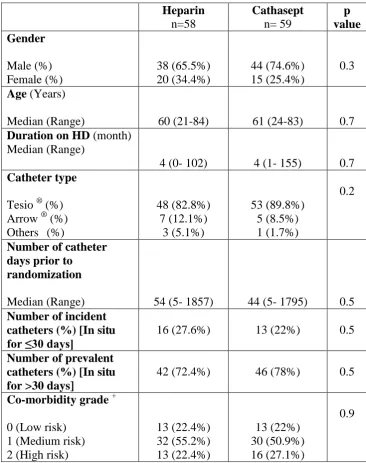
249) in the HG (p=0.5). Patients and catheter baseline characteristics are summarized
in table 1.
Primary outcome
Clinically significant catheter colonization occurred in nine catheters (1 in the CG and
8 in the HG and). The catheter colonization rate was statistically significantly
different in the two study groups: 0.14 per 1000 catheter-days in the CG and 1.08 per
1000 catheter-days in the HG (IRR 0.13; 95% CI, 0.003-0.94; p=0.02). Time to
colonization (catheter survival without microbial colonization) was also statistically
significantly longer in the CG (Figure 2).
Five catheters became colonized with coagulase negative staphylococci and four
catheters were colonized with gram-negative bacteria [one grew multiple organisms
(Serratia liquefaciens, Pseudomonas aeruginosa, Pseudomonas luteola, and Serratia
marcescens); one was colonized with an Acinetobacter species; one was colonized
with a coliform and one with a Pseudomonas species.].
Secondary outcomes
CRBSI
Four episodes (1 in the CG and 3 in the HG) met the criteria for “definite” CRBSI,
and further three episodes (1 in the CG and 2 in the HG) met the criteria for
15
Responsible organisms were methicillin-sensitive Staphylococcus aureus (3),
coagulase negative staphylococci (2), methicillin-resistant Staphylococcus aureus (1)
and Proteus species (1).
Weekly TCQBC were positive in only one case of CRBSI in the heparin group. Two
cases of CRBSI had a positive culture for the same organism by endoluminal brushing
and blood culture, one of which also showed endoluminal bacteria by SEM imaging
(Results reported elsewhere) 55.
Catheter patency
Data on blood flow rate were available for 114 patients (59 in the CG and 55 in the
HG). Prescribed blood flow rate was achieved in 1861 out of 2788 (66.8%) dialysis
sessions in the CG compared to 2178 out of 2893 (75.3%) of dialysis sessions in the
HG (odds ratio 1.94; 95% CI, 1.03 to 3.6; p=0.04), indicating that blood flow rate was
almost twice as likely to be achieved in the HG.
The number of patients who required any intervention to improve catheter patency
was not statistically different in the two groups (CG, 44 patients vs HG, 34; p=0.2).
However, the number of patients who received thrombolytic locks or infusions was
significantly higher in the CG (22 vs 9 patients; p=0.01).
Interventions used to improve catheter patency are summarized in table 3.
Laboratory data
Monthly hemoglobin remained stable during the study in both groups and was 0.4 (SE
± 0.23) g/dl higher in the HG but this was not statistically significant (p=0.09).
Ferritin level was on average 87.8 (SE±79.8) ng/ml lower in the HG but with no
statistically significant difference (p= 0.3). Fortnightly hs-CRP was significantly
16
the HG (p=0.03). Monthly Kt/V measurements were similar in both groups with
patients in the HG achieving slightly higher Kt/V values at 0.08 (SE±0.07) per month
(p=0.3).
Darbepoetin and iron saccharate dose
There was no significant difference in the monthly requirements of darbepoetin alfa or
iron saccharate between both groups. Patients in the CG received on average 16.2
(SE±21.2) mcg of darbepoetin alfa per month more than patients in the HG (p=0.4).
Monthly iron requirements were on average 27 (SE±34.7) mg lower in the CG
(p=0.4).
Adverse/Serious adverse events
The number of reported adverse events was 224 in the HG and 233 in the CG
(Independent sample t-test; p=0.9). The number of reported serious adverse events
was 79 in the HG and 66 in the CG (Independent sample t-test; p=0.9) and the number
of reported serious adverse device effects was four in the HG and six in the CG
(Independent sample t-test; p=0.8). There were no safety concerns and in particular
there were no episodes of hypocalcaemia.
Discussion
In this randomized controlled study, Cathasept reduced clinically significant microbial
colonization of t-HDC by 87% (p=0.02) compared to heparin. Although this reduction
was statistically significant, it should be pointed out that the 95% confidence interval
was wide (0.003- 0.94) indicating reduced precision when estimating the risk
reduction. Despite fewer episodes of CRBSI in the Cathasept group, this was not
statistically significant. This may represent a type 2 error as the sample size may not
17
colonization rate in our institution before 2006, which was used to calculate the
sample size, was much higher than that observed in this study (25% vs 7.8%). The
observed reduction in CRBSI rates may have been the result of the “saving lives”
campaign and other UK-wide to reduce methicillin-resistant Staphylococcus aureus
(MRSA) bacteremia and healthcare associated infection.
Cathasept was not as effective as heparin in maintaining catheter patency. Prescribed
blood flow rate was more likely to be achieved in the HG than in the CG group (Odds
ratio 1.94; p=0.04) and the number of patients who required thrombolytic locks or
infusions was also significantly lower in the HG group (9 vs 22 patients; p=0.01).
However, the number of catheters removed because of clotting or luminal occlusion
was similar in both groups (3 in each group). This suggests that catheter dysfunction
may have been due to extra-luminal formation of fibrin sheaths or non-occlusive
clots, rather than luminal problems. Due to a significant leakage of the instilled lock
solution over time, as evident from experimental work and the well documented
systemic anticoagulation effect of heparin locks 27, 56-58, it is possible that catheters
locked with heparin are less likely to develop extra-luminal occlusion. This might
explain the differences seen between catheters locked with Cathasept and heparin in
this study, but confirmatory work would be required.
High-sensitive CRP was significantly higher in the CG despite having lower catheter
colonization rate and this finding might be explained by the higher number of
infections unrelated to the catheter lumen occurring in the CG. There were 68
infective complications (including Exit site infection, respiratory infections, and
others) in the CG, and 48 infections in the HG, with corresponding incidence rates of
18
Although it did not reach statistical significance, this difference in infection events
might be high enough to account for CRP differences.
Haemoglobin level, serum ferritin concentration, and Darbepoetin and iron
requirements were similar for both groups despite the differences in hs-CRP.
There are several limitations to this study. Firstly, it was stopped earlier than planned
(as explained earlier) with 117 patients recruited, when the recruitment target was
140. This did not impact on the primary outcome of this study, which reached
statistical significance in favor of the Cathasept lock solution. Secondly, the study was
not double-blinded which could have introduced bias. Double blinding was not
possible because Cathasept could not be manufactured in glass vials like heparin due
to its high pH at around 10.5. Thirdly, the study was not powered to show a
significant effect on CRBSI rate or other secondary outcomes as discussed above. The
incidence of “Definite” and “Probable” CRBSI was 8.9% in the HG and 3.4% in the
CG. A sample size of 300 patients in each group would be required to detect a
significant difference with a 0.05 significance and a power of 80%.
This study has confirmed the effectiveness of Cathasept in reducing microbial
colonization of t-HDC and would be applicable to other hemodialysis populations as
well as other CVC applications in non-dialysis patients. However, a larger study with
enough power to assess catheter dysfunction and CRBSI rate is recommended.
Acknowledgements
Statistical assistance was provided by Mr. Andy Scally; Faculty of Health Studies,
19
Support: We are grateful to the study sponsors, Tyco Healthcare Group LP d/b/a
Covidien, Mansfield, MA, 02048, USA for funding the study and contributing to the
study design and data collection; and to the Yorkshire Kidney Research Fund for their
educational grant to support MK. The sponsors had no role in analysis or
interpretation of data, writing the report, or the decision to submit the report for
publications. We are also indebted to a group of research nurses at the participating
centers for their support and invaluable input in data collection and day-to-day
running of the study.
Financial Disclosure: The authors declare that they have no relevant financial
interests.
Contributions: research idea and study design: MK, MJW, PL, SB, JATS; data
acquisition: MJW, SB, HA, PL; data analysis: MK; results interpretation: MK, MJW,
JATS; supervision or mentorship: MJW, JATS. Each author contributed important
intellectual content during manuscript drafting or revision and accepts accountability
for the overall work by ensuring that questions pertaining to the accuracy or integrity
of any portion of the work are appropriately investigated and resolved. MK takes
responsibility that this study has been reported honestly, accurately, and transparently;
that no important aspects of the study have been omitted, and that any discrepancies
20
References
1. Ansell D, Roderick P, Hodsman A, Ford D, Steenkamp R, Tomson C.
Survival and causes of death of UK adult patients on Renal Replacement Therapy in
2007: national and centre-specific analyses: UK Renal Registry, 2008 report, Chapter
7; 2008.
2. Aslam N, Bernardini J, Fried L, Burr R, Piraino B. Comparison of Infectious
Complications between Incident Hemodialysis and Peritoneal Dialysis Patients. Clin J
Am Soc Nephrol. November 1, 2006 2006;1(6):1226-1233.
3. Ishani A, Collins AJ, Herzog CA, Foley RN. Septicemia, access and
cardiovascular disease in dialysis patients: The USRDS Wave 2 Study. Kidney
International. Jul 2005;68(1):311-318.
4. Foley RN, Guo H, Snyder JJ, Gilbertson DT, Collins AJ. Septicemia in the
United States Dialysis Population, 1991 to 1999. J Am Soc Nephrol. April 1, 2004
2004;15(4):1038-1045.
5. Hoen B, Paul-Dauphin A, Hestin D, Kessler M. EPIBACDIAL: A multicenter
prospective study of risk factors for bacteremia in chronic hemodialysis patients.
Journal of the American Society of Nephrology. May 1998;9(5):869-876.
6. Powe NR, Jaar B, Furth SL, Hermann J, Briggs W. Septicemia in dialysis
patients: Incidence, risk factors, and prognosis. Kidney International. Mar
1999;55(3):1081-1090.
7. Tokars JI, Miller ER, Stein G. New national surveillance system for
hemodialysis-associated infections: Initial results. American Journal of Infection
21
8. Astor BC, Eustace JA, Powe NR, et al. Type of vascular access and survival
among incident hemodialysis patients: The choices for healthy outcomes in caring for
ESRD (CHOICE) study. Journal of the American Society of Nephrology. May
2005;16(5):1449-1455.
9. Pisoni RL, Young EW, Dykstra DM, et al. Vascular access use in Europe and
the United States: Results from the DOPPS. Kidney International. Jan
2002;61(1):305-316.
10. Rayner HC, Besarab A, Brown WW, Disney A, Saito A, Pisoni RL. Vascular
access results from the Dialysis Outcomes and Practice Patterns Study (DOPPS):
Performance against Kidney Disease Outcomes Quality Initiative (K/DOQI) clinical
practice guidelines. American Journal of Kidney Diseases. Nov 2004;44(5):S22-S26.
11. Ethier J, Mendelssohn DC, Elder SJ, et al. Vascular access use and outcomes:
an international perspective from the dialysis outcomes and practice patterns study.
Nephrology Dialysis Transplantation. Oct 2008;23(10):3219-3226.
12. Sherertz RJ. Pathogenesis of Vascular Catheter Infection. In: Waldvogel FA,
Bisno AL, eds. Infections Associated with Indwelling Medical Devices. Third ed.
Washington, D. C.: ASM Press; 2000:111- 125.
13. Costerton JW, Stewart, P.S., Greenberg, E.P. Bacterial biofilms: A common
cause of persistent infections. Science. 1999(284):1318- 1322.
14. Cappelli G, Ballestri M, Perrone S, Ciuffreda A, Inguaggiato P, Albertazzi A.
Biofilms invade nephrology: Effects in hemodialysis. Blood Purification.
22
15. Schindler R. Causes and therapy of microinflammation in renal failure.
Nephrol. Dial. Transplant. August 1, 2004 2004;19(suppl_5):v34-40.
16. Tsirpanlis G, Chatzipanagiotou S, Nicolaou C. Microinflammation versus
inflammation in chronic renal failure patients. Kidney International. Nov
2004;66(5):2093-2094.
17. Cappelli G, Tetta C, Canaud B. Is biofilm a cause of silent chronic
inflammation in haemodialysis patients? A fascinating working hypothesis. Nephrol.
Dial. Transplant. February 1, 2005 2005;20(2):266-270.
18. Macdougall IC, Cooper AC. Erythropoietin resistance: the role of
inflammation and pro-inflammatory cytokines. Nephrology Dialysis Transplantation.
2002;17:39-43.
19. Stenvinkel P, Barany P. Anaemia, rHuEPO resistance, and cardiovascular
disease in end-stage renal failure: links to inflammation and oxidative stress.
Nephrology Dialysis Transplantation. 2002;17:32-37.
20. Horl WH, Vanrenterghem Y. Optimal Treatment of Renal Anaemia (OPTA):
improving the efficacy and efficiency of renal anaemia therapy in haemodialysis
patients receiving intravenous epoetin. Nephrology Dialysis Transplantation. May
2005;20:III25-III32.
21. Stenvinkel P, Alvestrand A. Inflammation in end-stage renal disease: Sources,
consequences, and therapy. Seminars in Dialysis. Sep-Oct 2002;15(5):329-337.
22. Phanish MK, Marcora SM, Lemmey AB. Malnutrition, chronic inflammation
and atherosclerosis in dialysis patients. Nephrology Dialysis Transplantation. Feb
23
23. Wanner C, Metzger T. C-reactive protein a marker for all-cause and
cardiovascular mortality in haemodialysis patients. Nephrology Dialysis
Transplantation. 2002;17:29-32.
24. Shanks RMQ, Donegan NP, Graber ML, et al. Heparin stimulates
Staphylococcus aureus biofilm formation. Infection and Immunity. Aug
2005;73(8):4596-4606.
25. Shanks RMQ, Sargent JL, Martinez RM, Graber ML, O'Toole GA. Catheter
lock solutions influence staphylococcal biofilm formation on abiotic surfaces.
Nephrol. Dial. Transplant. August 1, 2006 2006;21(8):2247-2255.
26. Karaaslan H, Peyronnet P, Benevent D, Lagarde C, Rince M, Leroux-Robert
C. Risk of heparin lock-related bleeding when using indwelling venous catheter in
haemodialysis. Nephrol. Dial. Transplant. October 1, 2001 2001;16(10):2072-2074.
27. Agharazii M, Plamondon I, Lebel M, Douville P, Desmeules S. Estimation of
heparin leak into the systemic circulation after central venous catheter heparin lock.
Nephrol. Dial. Transplant. June 1, 2005 2005;20(6):1238-1240.
28. James MT, Conley J, Tonelli M, et al. Meta-analysis: Antibiotics for
prophylaxis against hemodialysis catheter-related infections. Annals of Internal
Medicine. 2008;148(8):596-605.
29. Jaffer Y, Selby NM, Taal MW, Fluck RJ, McIntyre CW. A meta-analysis of
hemodialysis catheter locking solutions in the prevention of catheter-related infection.
24
30. Labriola L, Crott R, Jadoul M. Preventing haemodialysis catheter-related
bacteraemia with an antimicrobial lock solution: a meta-analysis of prospective
randomized trials. Nephrol. Dial. Transplant. May 1, 2008 2008;23(5):1666-1672.
31. Yahav D, Rozen-Zvi B, Gafter-Gvili A, Leibovici L, Gafter U, Paul M.
Antimicrobial lock solutions for the prevention of infections associated with
intravascular catheters in patients undergoing hemodialysis: Systematic review and
meta-analysis of randomized, controlled trials. Clinical Infectious Diseases.
2008;47(1):83-93.
32. Rabindranath KS, Bansal T, Adams J, et al. Systematic review of
antimicrobials for the prevention of haemodialysis catheter-related infections.
Nephrol. Dial. Transplant. December 1, 2009 2009;24(12):3763-3774.
33. Snaterse M, Rüger W, Scholte op Reimer WJM, Lucas C. Antibiotic-based
catheter lock solutions for prevention of catheter-related bloodstream infection: a
systematic review of randomised controlled trials. Journal of Hospital Infection.
2010;75(1):1-11.
34. Zhao Y, Li Z, Zhang L, et al. Citrate Versus Heparin Lock for Hemodialysis
Catheters: A Systematic Review and Meta-analysis of Randomized Controlled Trials.
American Journal of Kidney Diseases. 2015/03/04 2014;63(3):479-490.
35. Landry DL, Braden GL, Gobeille SL, Haessler SD, Vaidya CK, Sweet SJ.
Emergence of Gentamicin-Resistant Bacteremia in Hemodialysis Patients Receiving
Gentamicin Lock Catheter Prophylaxis. Clinical Journal of the American Society of
25
36. Solomon LR, Cheesbrough JS, Ebah L, et al. A Randomized Double-Blind
Controlled Trial of Taurolidine-Citrate Catheter Locks for the Prevention of
Bacteremia in Patients Treated With Hemodialysis. American Journal of Kidney
Diseases. 2010;55(6):1060-1068.
37. Maki DG, Ash SR, Winger RK, Lavin P, for the ATI. A novel antimicrobial
and antithrombotic lock solution for hemodialysis catheters: A multi-center,
controlled, randomized trial*. Critical Care Medicine. 2011;39(4):613-620.
38. Zucker M, B., . Divalent cations in blood clotting. In: Seve M, J., Johnson,
L.,A., ed. Metal-Binding in Medicine. Philadelphia: PA: J B Lippincott;
1960:137-142.
39. Godal H, C., Brosstad, F., Kierulf, P. Influence of Na2-EDTA and some
compounds used for dissolution of fibrin clots on the physiochemical properties of
fibrinogen and fibrin. Biblthca Haemat. 1978;44:151- 155.
40. Kite P, Eastwood K, Sugden S, Percival SL. Use of in vivo-generated biofilms
from hemodialysis catheters to test the efficacy of a novel antimicrobial catheter lock
for biofilm eradication in vitro. Journal of Clinical Microbiology. Jul
2004;42(7):3073-3076.
41. Davies SJ, Phillips L, Naish PF, Russell GI. Quantifying comorbidity in
peritoneal dialysis patients and its relationship to other predictors of survival.
Nephrol. Dial. Transplant. June 1, 2002 2002;17(6):1085-1092.
42. Kite P, Eastwood K, Sugden S, et al. Prevalence of Intravascular Catheter
26
Interscience Conference of Antimicrobial Agents and Chemotherapy. San Diego,
California: National Library of Medicine; 2002.
43. Kite P, Dobbins BM, Wilcox MH, McMahon MJ. Rapid diagnosis of
central-venous-catheter-related bloodstream infection without catheter removal. Lancet.
10/30/ 1999;354(9189):1504-1507.
44. Catton J, Wood J., Dobbins, B. M., Burke, D., Kite, P., Eastwood, K., Sugden,
S.A., Wilcox, M.H. & McMahon, M.J. Quantitative culture of through line blood is
an accurate method for the diagnosis of central venous
catheter-related-bloodstream-infection(CRBSI) without catheter removal. Nutrition in Clinical Practice.
2002;17:71.
45. Catton JA, Dobbins BM, Kite P, et al. In situ diagnosis of intravascular
catheter-related bloodstream infection: a comparison of quantitative culture,
differential time to positivity, and endoluminal brushing. Crit Care Med. 04
2005;33(4):787-791.
46. Raad I, Hanna HA, Alakech B, Chatzinikolaou I, Johnson MM, Tarrand J.
Differential Time to Positivity: A Useful Method for Diagnosing Catheter-Related
Bloodstream Infections. Annals of Internal Medicine. 01/06/ 2004;140(1):18-25.
47. Safdar N, Fine JP, Maki DG. Meta-Analysis: Methods for Diagnosing
Intravascular Device-Related Bloodstream Infection. Annals of Internal Medicine.
03/15/ 2005;142(6):451-466.
48. Pearson ML. Guideline for Prevention of Intravascular-Device-Related
27
49. O’Grady NP, Alexander M, Dellinger EP, et al. Guidelines for the Prevention
of Intravascular Catheter-related Infections. Morbidity and Mortality Weekly Report.
Centers for Disease Control and Prevention.
http://www.cdc.gov/mmwr/PDF/rr/rr5110.pdf. Vol 51 (RR-10); 2002:1-29.
50. Mermel LA, Farr BM, Sherertz RJ, et al. Guidelines for the management of
Intra-vascular catheter-related infections. Clinical Infectious Diseases. 2001
2001;32:1249- 1272.
51. Little MA, O'Riordan A, Lucey B, et al. A prospective study of complications
associated with cuffed, tunnelled haemodialysis catheters. Nephrology Dialysis
Transplantation. Nov 2001;16(11):2194-2200.
52. Dogra GK, Herson H, Hutchison B, et al. Prevention of tunneled hemodialysis
catheter-related infections using catheter-restricted filling with gentamicin and citrate:
a randomized controlled study. Journal of the American Society of Nephrology. Aug
2002;13(8):2133-2139.
53. Wheatley RE, Higginbotham, M., Jones, C., Laboi, P.,Taylor, G.,Than, N.
&Turney,J. An audit on the use of chlorhexidine gluconate in the prevention of
dialysis catheter colonisation and infection. Paper presented at: British Renal Society
Conference 20- 22 May, 2004; Harrogate, UK, May 20-22.
54. Kirkwood B, R. & Sterne, J.,A.,C. Comparing rates. Essential Medical
Statistics. Massachusetts, USA: Blackwell Science; 2003:240-248.
55. Kanaa M, Wright MJ, Sandoe JAT. Examination of tunnelled haemodialysis
catheters using scanning electron microscopy. Clinical Microbiology and Infection.
28
56. Besarab A, Pandey R. Catheter Management in Hemodialysis Patients:
Delivering Adequate Flow. Clinical Journal of the American Society of Nephrology.
January 1, 2011 2011;6(1):227-234.
57. Yevzlin AS, Sanchez RJ, Hiatt JG, et al. Concentrated Heparin Lock Is
Associated with Major Bleeding Complications after Tunneled Hemodialysis Catheter
Placement. Seminars in Dialysis. 2007;20(4):351-354.
58. Pepper RJ, Gale DP, Wajed J, et al. Inadvertent postdialysis anticoagulation
29
Table 1: Baseline characteristics
(+: Davies Co-morbidity score: grade 0 (No Co-morbidity), grade 1 (1-2), grade 2 (≥3)
Heparin n=58 Cathasept n= 59 p value Gender Male (%) Female (%) 38 (65.5%) 20 (34.4%) 44 (74.6%) 15 (25.4%) 0.3 Age (Years)
Median (Range) 60 (21-84) 61 (24-83) 0.7
Duration on HD (month)
Median (Range)
4 (0- 102) 4 (1- 155) 0.7
Catheter type
Tesio ® (%) Arrow ® (%) Others (%)
48 (82.8%) 7 (12.1%) 3 (5.1%) 53 (89.8%) 5 (8.5%) 1 (1.7%) 0.2
Number of catheter days prior to
randomization
Median (Range) 54 (5- 1857) 44 (5- 1795) 0.5
Number of incident catheters (%) [In situ
for ≤30 days] 16 (27.6%) 13 (22%) 0.5
Number of prevalent catheters (%) [In situ for >30 days]
42 (72.4%) 46 (78%) 0.5
Co-morbidity grade +
0 (Low risk) 1 (Medium risk) 2 (High risk)
30
Table 2: Incidence rates for “definite” and “probable” catheter-related blood stream infections (CRBSI) in the Cathasept Group (CG) and Heparin Group (HG) [N, number of episodes; IR,
Incidence Rate/1000 catheter-days; IRR, Incidence Rate Ratio (HG is the reference group)]
CRBSI
Cathasept Heparin
N IR N IR IRR (95% CI) P value
Definite
1 0.14 3 0.41 0.34 (0.035- 3.24) 0.3
Probable
1 0.14 2 0.27 0.51 (0.046- 5.58) 0.6
31
Table 3: Number of interventions required to improve catheter patency [N, number of episodes; IR, Incidence Rate/1000 catheter-days; IRR, Incidence Rate Ratio (HG is the reference group)]
(+: Saline flushes and reversal of dialysis circuit connection to the catheter)
N IR IRR p
value Thrombolytic
locks
Cathasept Heparin
64 23
8.76
3.11 2.8 (1.7- 4.7) <0.001
Thrombolytic infusions
Cathasept Heparin
16 5
2.19
0.68 3.2 (1.13- 11.29) 0.02
Other +
Cathasept Heparin
495 279
67.7
32
Figure 1: Screening, randomization, and follow-up flow chart (ITT; Intention-to-treat)
![Table 2: Incidence rates for “definite” and “probable” catheter-related blood stream infections (CRBSI) in the Cathasept Group (CG) and Heparin Group (HG) [N, number of episodes; IR, Incidence Rate/1000 catheter-days; IRR, Incidence Rate Ratio (HG is the reference group)]](https://thumb-us.123doks.com/thumbv2/123dok_us/7853498.178694/31.595.134.459.145.275/incidence-definite-infections-cathasept-heparin-incidence-incidence-reference.webp)
![Table 3: Number of interventions required to improve catheter patency [N, number of episodes; IR, Incidence Rate/1000 catheter-days; IRR, Incidence Rate Ratio (HG is the reference group)]](https://thumb-us.123doks.com/thumbv2/123dok_us/7853498.178694/32.595.95.504.151.377/interventions-required-catheter-episodes-incidence-catheter-incidence-reference.webp)
