Open Access
Short Report
DNA watermarks in non-coding regulatory sequences
Dominik Heider
1,3, Martin Pyka
2and Angelika Barnekow*
1Address: 1Department of Experimental Tumorbiology, University of Münster, Badestr. 9, D-48149 Münster, Germany, 2Interdisciplinary Center
for Clinical Research, University Hospital Münster, Domagkstr. 3, D-48143 Münster, Germany and 3Department of Bioinformatics, Center for
Medical Biotechnology, University of Duisburg-Essen, Universitätsstr. 2, D-45117 Essen, Germany
Email: Dominik Heider - [email protected]; Martin Pyka - [email protected]; Angelika Barnekow* - [email protected]
* Corresponding author
Abstract
Background: DNA watermarks can be applied to identify the unauthorized use of genetically modified organisms. It has been shown that coding regions can be used to encrypt information into living organisms by using the DNA-Crypt algorithm. Yet, if the sequence of interest presents a non-coding DNA sequence, either the function of a resulting functional RNA molecule or a regulatory sequence, such as a promoter, could be affected. For our studies we used the small cytoplasmic RNA 1 in yeast and the lac promoter region of Escherichia coli.
Findings: The lac promoter was deactivated by the integrated watermark. In addition, the RNA molecules displayed altered configurations after introducing a watermark, but surprisingly were functionally intact, which has been verified by analyzing the growth characteristics of both wild type and watermarked scR1 transformed yeast cells. In a third approach we introduced a second overlapping watermark into the lac promoter, which did not affect the promoter activity.
Conclusion: Even though the watermarked RNA and one of the watermarked promoters did not show any significant differences compared to the wild type RNA and wild type promoter region, respectively, it cannot be generalized that other RNA molecules or regulatory sequences behave accordingly. Therefore, we do not recommend integrating watermark sequences into regulatory regions.
Background
DNA watermarks can be used for authenticating geneti-cally modified organisms or in future for labeling animals in breeding [1,2]. It has already been shown in silico and
in vivo that these watermarks do not affect the translation of proteins [1-4]. These assumptions only apply to coding regions, thus the insertion of watermarks into regulatory sequences like promoter regions or regulatory RNA mole-cules, had to be examined. In our studies we integrated watermark sequences into a widely known promoter region of bacteria. Watermarks integrated into regulatory
regions like promoter or enhancer sequences can affect their functionality. We integrated watermark sequences into the lac promoter of Escherichia coli to examine, whether the watermarks affect the promoter activity. The lac operon of Escherichia coli consists of a promoter, three operators and three genes (lacY, lacZ and lacA), coding for the β-galactoside permease, β-galactosidase and β -galacto-side transacetylase, which are required for the lactose metabolism in Escherichia coli [5]. The β-galactosidase cleaves the lactose into glucose and galactose [5]. The pro-moter sequence of the lac operon consists of two highly
Published: 7 July 2009
BMC Research Notes 2009, 2:125 doi:10.1186/1756-0500-2-125
Received: 4 February 2009 Accepted: 7 July 2009
This article is available from: http://www.biomedcentral.com/1756-0500/2/125 © 2009 Barnekow et al; licensee BioMed Central Ltd.
conserved regions called the -35 (TTTACA) and -10 (TAT-GTT) regions, upstream from the transcription start. It has already been shown that single point mutations within conserved regions can affect or even stop promoter activ-ity of different promoters (Additional file 1) [6-10].
In a second approach we integrated a watermark sequence into a regulatory RNA molecule called small cytoplasmic RNA 1 (scR1) from yeast (scR1 gene on chromosome V of
Saccharomyces cerevisiae, http://www.yeastgenome.org/).
The scR1 consists of 519 nucleotides and represents the homologous RNA molecule in Saccharomyces cerevisiae to the mammalian 7SL RNA, which is part of the signal rec-ognition particle (SRP), necessary for the targeting of pro-teins into the endoplasmic reticulum (ER) [11-13]. The SRP complex is a ribonucleoprotein consisting of six polypeptides and the 7SL RNA. In Saccharomyces cerevisiae
the six polypeptides are SRP14, SRP19, SRP21, SRP54, SRP68 and SRP72 [14]. The SRP displays a GTPase activity [15]. The 7SL molecule is a conserved functional RNA constituting the backbone of the SRP complex and has been shown to be essential for all organisms tested, except
Saccharomyces cerevisiae cells. Saccharomyces cerevisiae cells are viable without scR1, but have a reduced reproduction rate [11,12,14,16-21].
Results and discussion
The analyses of the watermarked DNA sequences of both, the scR1 and the lac promoter sequence with the DNA-Crypt fuzzy controller, reveal that the use of any correc-tion code is not recommended.
We inserted a watermark sequence starting at position 898 in the lac promoter of pBluescriptII KS+ plasmid. The watermark sequence deactivated the lac promoter, which was verified by qualitative β-galactosidase assay using X-Gal (5-bromo-4-chloro-3-indolyl-β -D-galactopyrano-side) as a substrate (Table 1).
In 2001, Dieci et al. showed that after transformation of YRA130 ΔscR1 yeast cells with the wild type scR1 Yep352
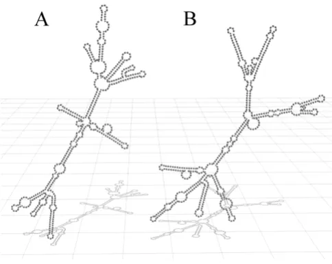
plasmid, a normal division rate was achieved and the wild type phenotype could be rescued [12]. For our studies we transformed the YRA130 ΔscR1 yeast cells with the Yep352 plasmid containing a watermarked scR1 gene (Yep352-SCR1-TB). The watermark sequence was inte-grated into the wild type scR1 gene starting at position 471 (see mutagenic primer sequences). The secondary structure predictions of the watermarked scR1 using the ViennaRNA-1.5 web interface revealed significant changes within the secondary structure compared to the structure of the wild type scR1 (Figure 1) [22]. The 3D model was created using Blender http://www.blender.org.
Although the predicted secondary structure of the water-marked scR1 differs from the wild type structure, the resulting RNA molecule proved to be functional active indirectly demonstrated by the YRA130 cells transformed either with the Yep352-SCR1 or the Yep352-SCR1-TB, which displayed no significant differences in their growth characteristics (Figure 2).
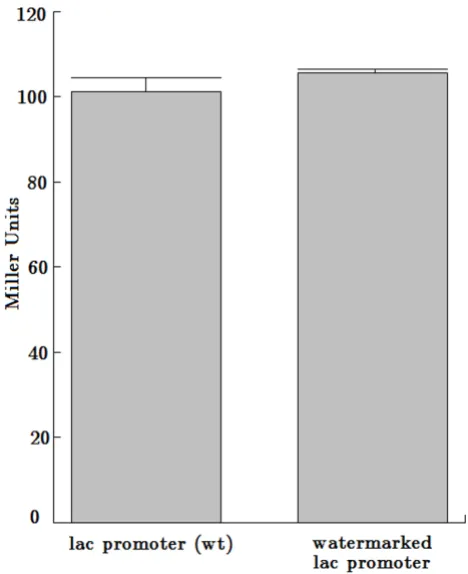
In addition, we inserted a second overlapping watermark sequence into the lac promoter region. The watermark sequence was integrated into the wild type lac promoter sequence starting at position 903 in the pBluescriptII KS+ plasmid and overlapped with the aforementioned deacti-vating watermark sequence. The watermarked lac pro-moter was proved to be active, verified by the qualitative
β-galactosidase assay using X-Gal. Furthermore, the
water-Table 1: Qualitative β-galactosidase assays
promoter qualitative β-galactosidase assay
lac +
lac K5 + lac II -lac wt: the wild type -lac promoter
lac K5: the lac promoter containing the watermark sequence WWU lac II: the lac promoter containing the watermark sequence 42 +: promoter is active
Secondary structure of the wild type scR1 and the water-marked scR1
Figure 1
[image:2.612.313.555.449.642.2]Growth characteristics
Figure 2
marked lac promoter did not display any significant dif-ferences compared to the wild type lac promoter using the quantitative β-galactosidase assay with ONPG (ortho-Nitrophenyl-β-galactoside) (Table 1, Figure 3).
Conclusion
Our results show that the integration of watermarks is not suitable for regulatory regions like promoter or regulatory RNA molecules. We have shown that these watermarks can deactivate promoter regions and further affect the sec-ondary structure of regulatory RNA molecules. Although the watermarked scR1 was not deactivated by the integra-tion of a watermark sequence, the secondary structure pre-diction displayed an altered structure. These results cannot be generalized for other RNA molecules.
The affect of integrated watermarks in non-coding regula-tory regions cannot be generalized and has to be tested in every single case. Therefore, we propose not to integrate watermark sequences into regulatory sequences.
Methods
Watermark design
We wanted to integrate a watermark into the lac promoter containing the answer to life, the universe, and everything
[23]. Further we wanted to encrypt the watermark 'TB' into the scR1 gene and 'WWU' into the lac promoter sequence, respectively. To demonstrate the flexibility of our algorithm we used different translation codes. The first one only used the binary representation. The second translation code used for the scR1 experiments just slightly differs from the standard one used in DNA-Crypt [1]. The third one imitates the official international Morse code. But identical binary encoding tables described in Heider and Barnekow 2007 were used [1]. The inserted DNA watermark sequences are
42 → 1010102 → CCC
TB → 10011000012 → CGCTG
and
WWU → .--.--..- → 01101100112 → GCATA
We created artificial reading frames within the scR1 gene and the lac promoter sequence that do not exist in non-coding regions, to apply the DNA-Crypt algorithm. Because of cost-benefit equation, we scanned manually for the best location within the DNA sequences of the scR1 gene and the lac promoter for integrating authenti-cating watermarks. The two watermark sequences in the lac promoter region overlap.
DNA-Crypt fuzzy controller
The watermark sequences were analyzed with the DNA-Crypt fuzzy controller with standard settings. The life time was set at 1000 cycles [1]. Based on the three input dimen-sions, the length of the watermark sequence, individual mutation rate and life time, the DNA-Crypt fuzzy control-ler recommends, based on the rule base, whether to use a specific mutation correction code or not [1].
Secondary structure prediction
The secondary structure predictions of the RNA molecules were performed using the ViennaRNA-1.5 web interface with rescaled energy parameters to 30°C [22]. The three dimensional structure predictions were created with Blender.
Site-directed mutagenesis
The site-directed mutagenesis was used to integrate the watermark sequences into the wild type DNA. We used a modified QuikChange(R) Site-Directed Mutagenesis Kit protocol from Stratagene (Stratagene, Amsterdam, The Netherlands) described in Heider and Barnekow 2008 [4]. For the scR1 studies we used the Yep352-SCR1 plasmid carrying the wild type sequence of the scR1 gene (a kind gift of Giorgio Dieci, Universita di Parma, Italy) [12]. For
[image:4.612.56.290.393.680.2]Quantitative β-galactosidase assay with ONPG
Figure 3
plasmid (Stratagene, Amsterdam, The Netherlands). The site-directed mutagenesis was performed with the follow-ing primer sequences:
scR1 – forward primer:
5'-GCACATTGTGGCCGTGCCCTCTGGG ATGGAGTGT-GTC-3'
scR1 – reverse primer:
5'-GACACACTCCATCCCAGAGGGCACGGCCACAAT-GTGC-3'
lac – forward primer:
5'-GTTAGCTCACTCATTTGGGACCCCAGGCTTTACA-3'
lac – reverse primer:
5'-TGTAAAGCCTGGGGTCCCAAATGAGTGAGCTAAC-3'
lac2 – forward primer:
5'-CTCACTCATTAGGCCCCCCAGGCC TTACACTT-TATGC-3'
lac2 – reverse primer:
5'-GCATAAAGTGTAAGGCCTGGGGGGCCTAATGAGT-GAG-3'
The bold letters represent the watermark sequences. The mutagenesis was confirmed by sequencing with the M13 primer 5'-GTAAAACGACGGCCAGT-3' and the lac-SEQ primer 5'-CCGCCTCTCCCCGCG-3', respectively.
Transformation of bacteria
For our lac promoter studies we used the Escherichia coli
DH5αstrain. 20 ml 2YT were inoculated with 200 μl over-night culture of DH5αbacteria and incubated at 37°C to an OD600 of 0.3 to 0.5. The cells were centrifuged for 10 minutes at 2000 × g and mixed with 10 ml ice-cold sterile 0.1 M CaCl2. After incubation for 30 minutes on ice, the cells were centrifuged for 10 minutes at 4°C 2000 × g and resuspended in 2 ml ice-cold 0.1 M CaCl2. 200 μl of the cells were mixed up with 50 ng DNA. After incubation for 30 minutes on ice, the cells were incubated for 45 seconds at 42°C and cooled down for 2 minutes on ice. After mix-ing with 500 μl 2YT the cells were incubated for 45 min-utes at 37°C and 220 rpm. Finally 200 μl of the cell suspension were plated and grown overnight on 2YT plates containing ampicillin.
Transformation of yeast
We used the YRA130 ΔscR1::His3 yeast strain for our scR1 experiments that has a reduced reproduction rate (a kind gift of Peter Walter, University of California, San Fran-cisco, USA). The yeast strain YRA130 was transformed using the lithium acetate method and grown on SD-ura plates [24].
β-galactosidase assay
The activity of the lac promoter was verified by qualitative and quantitative β-galactosidase assays, using 5-bromo-4-chloro-3-indolyl-β-D-galactopyranoside (X-Gal, Carl Roth GmbH, Karlsruhe, Germany) and ONPG (Carl Roth GmbH, Karlsruhe, Germany), respectively. For the quali-tative β-galactosidase assay we pipetted 40 μl 100 mM IPTG (Carl Roth GmbH, Karlsruhe, Germany) and 40 μl X-Gal (20 mg/ml) onto a 2YT agar plate containing amp-icillin. After drying the plates for one hour, 20 μl of over-night cultures were plated and incubated overover-night at 37°C. The quantitative analyses we performed using the method established by Miller in 1972, except some mod-ifications published by Zhang and Bremer and some addi-tional modifications [25,26]. We used 100 μl of IPTG induced overnight culture for our assays and froze them for 30 minutes at 20°C before adding the permealization buffer. In addition we used a substrate buffer containing 3 mg/ml instead of 1 mg/ml ONPG to guarantee saturation for the β-galactosidase.
Growth characteristics
The growth characteristics of YRA130, and the Yep352-SCR1 and Yep352-Yep352-SCR1-TB transformed YRA130 cells were analyzed by measuring optical densities at 600 nm every 60 minutes for nine hours (Pharmacia LKB Novaspec II).
Competing interests
The authors declare that they have no competing interests.
Authors' contributions
Publish with BioMed Central and every scientist can read your work free of charge "BioMed Central will be the most significant development for disseminating the results of biomedical researc h in our lifetime."
Sir Paul Nurse, Cancer Research UK
Your research papers will be:
available free of charge to the entire biomedical community
peer reviewed and published immediately upon acceptance
cited in PubMed and archived on PubMed Central
yours — you keep the copyright
Additional material
Acknowledgements
The authors want to thank Peter Walter (University of California, San Fran-cisco, USA) for the YRA130 strain, Giorgio Dieci (Universita di Parma, Italy) for the Yep352-SCR1 plasmid and Sarah-Jane Barnard for critically reading the manuscript. This work is part of the PhD thesis of D.H.
References
1. Heider D, Barnekow A: DNA-based watermarks using the DNA-Crypt algorithm. BMC Bioinformatics 2007, 8:176. 2. Heider D, Kessler D, Barnekow A: Watermarking sexually
reproducing diploid organisms. Bioinformatics 2008,
24:1961-1962.
3. Heider D, Barnekow A: DNA watermarks and sexual transmis-sion. Europ J Cell Biol 2008, 87S1:58.
4. Heider D, Barnekow A: DNA watermarks: A proof of concept.
BMC Molecular Biology 2008, 9:40.
5. Beckwith JR: Regulation of the lac operon. Recent studies on the regulation of lactose metabolism in Escherichia coli sup-port the operon model. Science 1967, 156(3775):597-604. 6. Rothmel RK, LeClerc JE: Mutational analysis of the lac
regula-tory region: second-site changes that activate mutant pro-moters. Nucleic Acids Research 1989, 17(10):3909-3925.
7. Depto AS, Stenberg RM: Regulated Expression of the Human Cytomegalovirus pp65 Gene: Octamer Sequence in the Pro-moter Is Required for Activation by Viral Gene Products.
Journal of Virology 1989, 63(3):1232-1238.
8. Bentley K, Deacon N, Sonza S, Zeichner S, Churchill M: Mutational analysis of the HIV-1 LTR as a promoter of negative sense transcription. Arch Virol 2004, 149:2277-2294.
9. Romano G, Suon S, Jin H, Donaldson AE, Iacovitti L: Characteriza-tion of Five EvoluCharacteriza-tionary Conserved Regions of the Human Tyrosine Hydroxylase (TH) Promoter: Implications for the Engineering of a Human TH Minimal Promoter Assembled in a Self-Inactivating Lentiviral Vector System. J Cell Physiol
2005, 204(2):666-677.
10. Almeida AM, Murakami Y, Layton DM, Hillmen P, Sellick GS, Maeda Y, Richards S, Patterson S, Kotsianidis I, Crawford LMDH, Baker A, Ferguson M, Roberts I, Houlston R, Kinoshita T, Karadimitris A:
Hypomorphic promoter mutation in PIGM causes inherited glycosylphosphatidylinositol deficiency. Nature Medicine 2006,
12:846-851.
11. Felici F, Cesareni G G, Hughes JM: The Most Abundant Small Cytoplasmic RNA of Saccharomyces cerevisiae Has an Important Function Required for Normal Cell Growth. Mol Cell Biol 1989, 9(8):3260-3268.
12. Dieci G, Giuliodori S, Catellani M, Percudani R, Ottonello S: Intra-genic Promoter Adaptation and Facili-tated RNA Polymer-ase III Recycling in the Transcription of SCR1, the 7SL RNA Gene of Saccharomyces cerevisiae. J Biol Chem 2001,
277:6903-6914.
13. Wild K, Weichenrieder O, Strub K, Sinning I, Cusack S: Towards the structure of the mammalian signal recognition particle.
Curr Opin Struct Biol 2002, 12:72-81.
14. Rosenblad AM, Gorodkin J, Knudsen B, Zwieb C, Samuelsson T:
SRPDB (Signal Recognition Particle Database). Nucleic Acids
15. Borschbach M, Hauke S, Pyka M, Heider D: Opportunities and lim-itations of a principle component analysis optimized machine learning approach for the identification and classifi-cation of cancer involved proteins. Communications of the SIWN
2009, 6:85-89.
16. Hann BC, Walter P: The signal sequence recognition particle in S.cerevisiae. Cell 1991, 67:131-144.
17. Stirling CJ, Hewitt EW: The S. cerevisiae SEC65 gene encodes a component of yeast signal recognition particle with homol-ogy to human SRP19. Nature 1992, 356:534-537.
18. Ogg SC, Poritz MA, Walter P: Signal sequence recognition par-ticle receptor is important for cell growth and protein secre-tion in Saccharomyces cerevisiae. Mol Biol Cell 1992, 3:895-911. 19. Brown J, Hann BC, Medzihradszky KF, Niwa M, Burlingame AL, Wal-ter P: Subunits of the Saccharomyces cerevisiae signal recog-nition particle required for itsfunctional expression. EMBO J
1994, 13:4390-4400.
20. Thompson JD, Higgins DG, Gibson TJ: CLUSTAL W: improving the sensitivity of progressive multiple sequence alignment through sequence weighting, position-specific gap penalties and weight matrix choice. Nucleic Acids Res 1994, 22:4673-4680. 21. Kin T, et al.: fRNAdb: a platform for mining/annotating func-tional RNA candidates from non-coding RNA sequences.
Nucleic Acids Res 2007, 35(Database issue):D145-D148.
22. Vienna RNA Secondary Structure Prediction, A web inter-face to the RNAfold programm version 1.5 [http://rna.tbi.uni vie.ac.at/cgi-bin/RNAfold.cgi]
23. Adams D: The Hitchhiker's Guide to the Galaxy Ballantine Books; 1979. 24. Kail M, Barnekow A: Identification and characterization of interacting partners of Rab GTPases by yeast two-hybrid analyses. Methods Mol Biol 2008, 440:111-125.
25. Miller JH: J.H. Miller Experiments in Molecular Genetics Cold Spring Har-bor LaHar-boratory 1972 chap. J.H. Miller Assay of β -Galactosi-dase:352-355.
26. Zhang X, Bremer H: Control of the Escherichia coli rrnB P1 Promoter Strength by ppGpp. J Biol Chem 1995,
270(19):11181-11189.
Additional file 1
Table S1 – Conserved promoter regions. lac: the lac promoter; hexoki-nase: Rat type III hexokinase promoter; H2B: histone H2B promoter; AD4: adenovirus type 4; AD2: adenovirus type 2; SV40: SV40 enhancer Click here for file